Article Text

Abstract
Neurological symptoms occur in approximately 20% of patients with Sjögren's syndrome, and may be the presenting manifestations of the disease. Here, we review several neurological conditions that can occur in Sjögren's syndrome: sensory ganglionopathy, painful small fibre neuropathy, and transverse myelitis (independently or as part of neuromyelitis optica). We present the symptoms, signs, differential diagnoses, recommended diagnostic evaluation, and treatment of each of these, highlighting the features that should alert neurologists to consider Sjögren's syndrome.
- Sjogren Syndrome
- Peripheral Neuropathy
- Transverse Myelitis
- Neuromyelitis Optica
Statistics from Altmetric.com
Introduction
Sjögren's syndrome affects the nervous system in approximately 20% of cases.1 ,2 Neurological symptoms may precede the onset of dry eyes and dry mouth (sicca symptoms) in 25–92%3 ,4 of patients. In such cases, the first presentation is often to a neurologist. Several reviews1 ,2 and case series5–8 describe the diverse neurological manifestations of Sjögren's syndrome. Here, we focus on three of the most common: sensory ganglionopathy (also known as sensory neuronopathy or sensory ataxic neuropathy), painful small fibre neuropathy, and transverse myelitis. We review when to consider Sjögren's syndrome in the differential diagnosis of neuropathy and transverse myelitis, and how to approach the diagnostic evaluation for Sjögren's syndrome as the cause of neurological disease.
In a series of 82 patients with neurological manifestations of Sjögren's syndrome, over half (57%) had neurological symptoms at presentation, and half of these patients (36% of total) had isolated neurological symptoms.8 Only 44% presented with concurrent sicca symptoms, and neurological symptoms preceded sicca symptoms in nearly half (47%), beginning on average 6 years before. Neurological symptoms preceded the ultimate diagnosis of Sjögren's syndrome in 81% of patients; when this occurred, the time to diagnosis of Sjögren's syndrome increased from 3 years to 5 years. Moreover, only 21% of patients with Sjögren's syndrome-associated neurological disease had anti-SSA (anti-Ro) or anti-SSB (anti-La) antibodies at presentation, and fewer than half (43%) ultimately developed positive serologies over the next 7 years. These data underscore the importance of considering Sjögren's syndrome in the differential diagnosis of the neurological disorders discussed below.
Peripheral nervous system manifestations
Prevalence
Neuropathy accompanies Sjögren's syndrome in an estimated 5–15% of cases,9 although studies have reported prevalences between 0% and 56%.9 This variance probably reflects referral bias and differences in diagnostic criteria of Sjögren's syndrome and neuropathy.9 ,10 In a series of 1010 Sjögren's syndrome patients, 11% had clinically apparent neuropathy.11 However, in one series of 62 patients with no symptoms of neuropathy, 58% were found to have electrophysiological evidence of neuropathy on nerve conduction studies.12
Relationship of neuropathy symptoms to systemic symptoms
In the largest series of patients with Sjögren's syndrome-associated neuropathy, 93% of 82 patients developed neuropathy before being diagnosed with Sjögren's syndrome.4 Sicca symptoms precede or follow development of neuropathy with equal frequency (37.5%), occurring simultaneously in 16% of patients.13 Therefore, the neurologist must be aware of the neuropathies that most commonly accompany Sjögren's syndrome, in order to determine when to pursue a diagnostic evaluation for the disease.
Prevalence of neuropathy subtypes
Fifty to sixty per cent of Sjögren's syndrome-associated peripheral neuropathies are sensory, most commonly involving the dorsal root ganglia (causing sensory ganglionopathy) and small unmyelinated nerve fibres (causing painful small fibre neuropathy).3 ,4 ,14–16 In the largest series of patients with Sjögren's syndrome-associated neuropathies (92 patients), the two most common were sensory ganglionopathy (39%) and painful small fibre neuropathy (20%); 16% had trigeminal neuropathy, 12% had multiple mononeuropathies, 5% had multiple cranial neuropathies, 4% had polyradiculoneuropathies, and 3% had autonomic neuropathies.4 Experts estimate that approximately 5% of all Sjögren's syndrome patients have ganglionopathy and 5–10% have small fibre neuropathy.9 Neurologists should, therefore, consider Sjögren's syndrome in the initial differential diagnosis in patients with sensory ganglionopathy or small fibre neuropathy. Sensorimotor polyneuropathy, autonomic neuropathy, mononeuropathy multiplex, and cranial neuropathies are less common, and there are more likely alternative causes, but Sjögren's syndrome should be considered in the initial differential diagnosis if there are other suggestive features, or if the initial evaluation for other causes is negative.
Sensory ganglionopathy (sensory ataxic neuronopathy)
Ganglionopathy typically presents with paraesthesias, gait unsteadiness and/or difficulty with fine motor movements due to decreased proprioception. Strength is generally preserved. There may be reduced or absent joint position and vibration sense, reduced or absent reflexes, and/or sensory ataxia. Sensory ataxia can be distinguished from cerebellar ataxia by the absence of accompanying nystagmus and increased difficulty with finger–nose–finger testing with eyes closed. Other signs of proprioceptive deficits include upward or sideways drift of the arms when held outstretched with eyes closed, as well as small dancing finger movements (pseudoathetosis) in this position. Romberg's sign may be present, and the toe tendons may appear to ‘dance’ as the patient attempts to maintain balance with the eyes closed.
Other causes of sensory ganglionopathy include systemic lupus erythematosus (SLE), rheumatoid arthritis, paraneoplastic syndromes (caused by anti-Hu or anti-collapsin response mediator protein antibodies), vitamin B6 toxicity, and platin-based chemotherapy agents (for review, see Sheikh and Amato, 2010).17
Painful small fibre neuropathy
Painful small fibre neuropathy typically causes ‘burning’ sensations, often in the feet, and worse at night. Stimulation of the affected region (eg, bed sheets contacting the feet) can cause pain disproportionate to the stimulus (allodynia), and there may be increased sensitivity to painful stimuli (hyperalgesia). There may be decreased pain and/or temperature sensation in symptomatic areas, but the neurological examination is often normal. Unlike other types of neuropathy, reflexes are generally spared, and nerve conduction studies are typically normal. The causes of small fibre neuropathy include diabetes mellitus, HIV, hepatitis C, amyloidosis, Fabry's disease, and paraneoplastic syndromes (most commonly due to anti-Hu antibodies, usually with lung cancer), in addition to Sjögren's syndrome (for review, see McArthur18).
Small fibre neuropathy can be difficult to diagnose, since physical examination and nerve conduction studies are often normal. However, skin biopsy can aid in diagnosis if it shows reduced epidermal nerve fibre density and pathological morphologic changes in small nerve fibres, such as axonal swelling.19 In one series of 13 patients with Sjögren's syndrome-associated small fibre neuropathy, the symptoms and pathological skin biopsy findings were non-length-dependent, being initially present proximally (in the thighs) and distally (in the feet).14 This contrasts with diabetic and idiopathic small fibre neuropathies, which typically have distal-predominant symptoms and skin biopsy findings. Proximal symptoms and signs of small fibre neuropathy may, therefore, suggest Sjögren's syndrome.9 ,14
Autonomic symptoms
Autonomic dysfunction occurs in up to half of patients with Sjögren's syndrome, manifesting as pupillary abnormalities, orthostatic hypotension and/or hypohydrosis/anhidrosis.4 ,9 ,20 ,21 Autonomic dysfunction accompanying peripheral neuropathy is therefore an important sign that Sjögren's syndrome should be considered.9 ,21 However, small fibre neuropathy and autonomic neuropathies can also co-occur in diabetes mellitus, amyloidosis, and hereditary sensory and autonomic neuropathies (HSAN); sensory ganglionopathy and autonomic neuropathy can also coexist in HSAN.
Disorders of smell and taste
Patients with Sjögren's syndrome may develop loss of smell (anosmia), loss of taste (ageusia), or alterations in taste (dysgeusia). Although these disorders of taste are due to decreased saliva production rather than a primary problem with the nervous system,22 such patients may present to a neurologist for evaluation. Changes in taste and smell in a patient with neuropathy may be a clue to Sjögren's syndrome as the likely underlying cause. Patients with disorders of taste due to Sjögren's syndrome generally report dry mouth for years prior to onset of ageusia.22
Testing for Sjögren's syndrome in peripheral nerve disease
The diagnosis of Sjögren's syndrome requires four of the six criteria in box 1 (including IV or VI), or three of the four objective criteria (IV–VI).23 These criteria provide useful screening questions for sicca symptoms in patients whose predominant presenting symptoms may be neurological. Because neurological symptoms may precede sicca symptoms by many years, objective testing is essential to diagnose Sjögren's syndrome in such patients.
American-European Consensus Group Revised International Classification Criteria for Sjögren’s Syndrome (quoted directly).23
-
Ocular symptoms: a positive response to at least one of the following questions:
-
Have you had daily, persistent, troublesome dry eyes for more than 3 months?
-
Do you have a recurrent sensation of sand or gravel in the eyes?
-
Do you use tear substitutes more than three times a day?
-
-
Oral symptoms: a positive response to at least one of the following questions:
-
Have you had a daily feeling of dry mouth for more than 3 months?
-
Have you had recurrently or persistently swollen salivary glands as an adult?
-
Do you frequently drink liquids to aid in swallowing dry food?
-
-
Ocular signs—objective evidence of ocular involvement defined as a positive result for at least one of the following two tests:
-
Schirmer's test, performed without anaesthesia (<5 mm in 5 min)
-
Rose Bengal score or other ocular dye score (>4 according to van Bijsterveld's scoring system)
-
-
Histopathology: In minor salivary glands (obtained through normal-appearing mucosa) focal lymphocytic sialoadenitis, evaluated by an expert histopathologist, with a focus score >1, defined as a number of lymphocytic foci (which are adjacent to normal-appearing mucous acini and contain more than 50 lymphocytes) per 4 mm2 of glandular tissue
-
Salivary gland involvement: objective evidence of salivary gland involvement defined by a positive result for at least one of the following diagnostic tests:
-
Unstimulated whole salivary flow (<1.5 mL in 15 min)
-
Parotid sialography showing the presence of diffuse sialectasias (punctate, cavitary or destructive pattern), without evidence of obstruction in the major ducts
-
Salivary scintigraphy showing delayed uptake, reduced concentration and/or delayed excretion of tracer
-
-
Autoantibodies: presence in the serum of the following autoantibodies:
-
Antibodies to Ro(SSA) or La(SSB) antigens, or both
-
Autoantibody testing
The autoantibodies classically associated with Sjögren's syndrome are anti-SSA (anti-Ro) and anti-SSB (anti-La). Anti-SSA occurs in 33–74% of patients with Sjögren's syndrome, anti-SSB (La) in 23–52%, and antinuclear antibodies in 59–85%.24 How sensitive are these serologies for the diagnosis of Sjögren's syndrome in a patient presenting with neuropathy? Anti-SSA and/or anti-SSB occur in only 10–55% of patients with Sjögren's syndrome-associated neuropathy.4 ,13 ,14 ,16 ,25 By subtype of neuropathy, in one series, 53% of 36 patients with Sjögren's syndrome-associated sensory ganglionopathy had anti-SSA, whereas 11% had anti-SSB; 39% of patients with Sjögren's syndrome-associated small fibre neuropathy had anti-SSA, while 17% had anti-SSB.4 Antinuclear antibodies occur in 20–67% of patients with Sjögren's syndrome-associated neuropathies.13 ,16 In summary, autoantibody serologies in patients with Sjögren's syndrome-associated neuropathies have a low sensitivity, at best just over 50%. Thus, negative autoantibody serologies in a patient with neuropathy of unclear aetiology warrant further testing with Schirmer's test, Rose Bengal testing, and/or lip salivary gland biopsy, all of which are more sensitive for the diagnosis.9 ,25
Schirmer's test
Schirmer's test objectively evaluates tear production in patients with dry eyes by measuring the degree to which a strip of filter paper placed in the lower eyelid moistens after a given time. The test is positive when there is less than 5 mm of moistening of the filter paper after 5 min. In small studies, Schirmer's test is positive in 56–89% of patients with Sjögren's syndrome-associated neuropathies,4 ,13 93% of 29 patients with sensory ganglionopathy and 93% of 15 patients with small fibre neuropathy.4
Rose Bengal testing
Rose Bengal testing is another objective measure of dry eyes, typically performed by ophthalmologists. The Rose Bengal dye stains damaged cornea, identifying lesions resulting from decreased tear production in Sjögren's syndrome. In small studies, Rose Bengal testing was positive in 73–79% of patients with Sjögren's syndrome-associated neuropathy,4 ,13 69% of 29 patients with sensory ganglionopathy and 92% of 12 patients with small fibre neuropathy.4
Lip minor salivary gland biopsy
Salivary gland biopsy in Sjögren's syndrome shows foci of lymphocytic infiltration (figure 1). Lip minor salivary gland biopsy may be diagnostic in 37–75% of patients with Sjögren's syndrome-associated neuropathy.4 ,13 ,14 ,25 In one series, 93% of 28 patients with sensory ganglionopathy and 81% of 16 patients with small fibre neuropathy had positive lip salivary gland biopsies.4 There is no relationship between EMG/nerve conduction study findings and lip biopsy positivity, nor between sicca symptoms and lip biopsy positivity.25
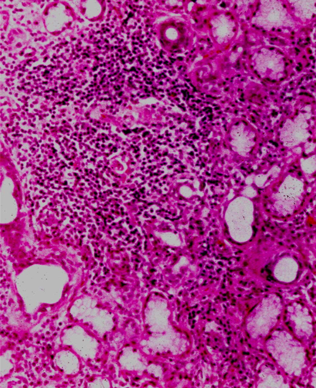
Lip minor salivary gland biopsy showing lymphocytic infiltration in a patient with Sjögren's syndrome. Image courtesy of Dr Peter H Schur.
Evaluation for Sjögren's syndrome as the cause of neuropathy
All (admittedly small) case series suggest that non-serological ancillary testing is more sensitive than serum autoantibodies in Sjögren's syndrome-associated neuropathy. These tests should therefore be pursued in patients with neuropathies of unclear aetiology in whom there is a high suspicion for Sjögren's syndrome but who have negative serum autoantibodies.9 ,25 We recommend sending anti-SSA, anti-SSB and antinuclear antibodies as part of the initial evaluation of all patients with sensory ganglionopathy, small fibre neuropathy, or other neuropathies if the initial evaluation is unrevealing, especially if there are autonomic features. If serologies are negative, we recommend lip minor salivary gland biopsy.9 ,25
Peripheral neuropathy in a patient with known Sjögren's syndrome should not necessarily be attributed to this underlying disease without evaluating for other potential causes. A general initial evaluation for neuropathy typically includes screening for diabetes mellitus, vitamin B12 deficiency, and/or a monoclonal gammopathy.26 In the appropriate clinical scenarios, clinicians should consider chronic alcohol use, HIV, amyloidosis, hereditary diseases, and/or paraneoplastic syndromes.
Treatment
While isolated sicca symptoms of Sjögren's syndrome may be managed symptomatically, extraglandular involvement generally requires immunosuppressive therapy—though there are only limited data to support this27 ,28—including corticosteroids, azathioprine, cyclophosphamide, intravenous immunoglobulin (IVIg), plasma exchange, infliximab and rituximab.4 ,9 ,27–31 Data from small series suggest that different subtypes of neuropathy in Sjögren's syndrome may respond better to particular immunosuppressive therapies.4 ,30 ,31 Patients with sensorimotor neuropathy, non-ataxic sensory neuropathy, and painful small fibre neuropathy may respond better to IVIg than patients with ganglionopathy,4 ,30 while patients with multiple mononeuropathies or cranial neuropathies may benefit more from corticosteroids.4 Unfortunately, the disease often progresses despite these therapies.4 The presence of cryoglobulinaemia and/or vasculitis in patients with Sjögren's syndrome-associated neuropathy may predict a response to rituximab.31 Decisions about the appropriate immunosuppressive regimen for a patient with neurologic manifestations of Sjögren's syndrome should be made in collaboration with the patient's rheumatologist.
The symptom management of Sjögren's syndrome-associated painful small fibre neuropathy includes antiepileptics (eg, gabapentin, pregabalin), tricyclic antidepressants (eg, nortriptyline), and serotonin-norepinephrine reuptake inhibitors (SNRIs) (eg, venlafaxine, duloxetine), as in the treatment of neuropathic pain from other aetiologies.9 However, neurologists should be cautious using tricyclic antidepressants in patients with Sjögren's syndrome, as they may worsen dry mouth and autonomic symptoms, if present.9
We generally treat the first presentation of ganglionopathy in a patient with Sjögren's syndrome with IVIg (400 mg/kg/day for 5 days for a total of 2 g). If there is minimal or no improvement, or if symptoms initially improve but recur, we initiate monthly IVIg infusions, with the goal of spacing these to longer intervals as tolerated if there is sustained improvement. We treat small fibre neuropathy symptomatically first, reserving IVIg for severe and/or refractory cases.
Central nervous system manifestations
Sjögren's syndrome may affect the spinal cord, brainstem, optic nerves, cerebellum, and cerebral hemispheres.1 ,2 ,8 ,32 In a cohort of 1010 Sjögren's cases, 2% had central nervous system (CNS) disease.11 CNS involvement is much less common than peripheral nervous system involvement, yielding less data on Sjögren's syndrome-associated CNS disease. CNS symptoms precede the diagnosis of Sjögren's syndrome by up to 2 years in 80% of patients.8 ,33 CNS disease in Sjögren's syndrome more frequently leads to severe disability (defined as Modified Oxford Handicap Scale ≥2) than does peripheral nervous system disease (78% of those with CNS disease vs 26% of those with peripheral disorders).8 Neurologists must therefore recognise the typical presentations of CNS disease that Sjögren's syndrome can cause. These include transverse myelitis, neuromyelitis optica (NMO), and brain lesions with morphology and locations similar to those seen in NMO.
Transverse myelitis and NMO
Transverse myelitis is an inflammatory disorder of the spinal cord that presents acutely or subacutely with weakness, sensory loss, and/or bowel and bladder involvement. It may be monophasic, or may recur in conditions such as multiple sclerosis, NMO, and autoimmune disease (for review, see Sá, 2009).34 When transverse myelitis accompanies Sjögren's syndrome, it often spans more than three levels of the spinal cord—a longitudinally extensive transverse myelitis.
NMO or Devic's disease is a CNS demyelinating disease characterised by relapsing longitudinally extensive transverse myelitis and optic neuritis, with a characteristic autoantibody to aquaporin-4 (NMO-IgG).35 By contrast, the myelitis of multiple sclerosis tends to affect multiple spinal levels with discrete (fewer than three spinal levels in length) and radially oriented lesions (figure 2).35 While NMO may cause brain lesions, they are less frequently symptomatic than in multiple sclerosis, and have a predilection for the hypothalamus, brainstem, and periventricular regions (figure 3).35 ,36 The morphology and locations of brainstem and periventricular white matter lesions in the two diseases are also generally distinctive.36 ,37 We review the emerging relationship between NMO and Sjögren's syndrome below; the relationship between multiple sclerosis and Sjögren's syndrome is controversial.38–41
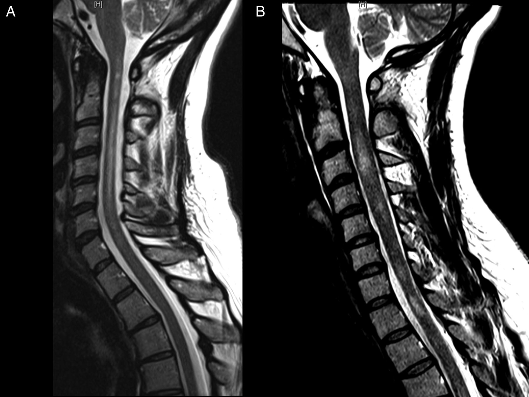
(A) Longitudinally extensive myelitis in a patient with neuromyelitis optica as seen on axial T2-weighted MRI. Image courtesy of Dr James Stankiewicz. (B) Discrete spinal cord lesions in a patient with multiple sclerosis as seen on T2-weighted MRI.

Fluid attenuated inversion recovery (FLAIR) MRI scan of the brain in a patient with neuromyelitis optica, showing lesions adjacent to the cerebral aqueduct (A) and fourth ventricle (B), as well as throughout the midbrain and pons (C–F).
Sjögren's syndrome and transverse myelitis
Between 1% and 5% of cases of acute myelitis may be associated with Sjögren's syndrome.34 ,42 In a series of 153 patients with NMO, 2% had Sjögren's syndrome.43 The incidence of myelitis in patients with Sjögren's syndrome is unknown, but estimated at less than 1%.44
Clinical features
In a systematic review of previously reported patients with Sjögren's syndrome and longitudinally extensive transverse myelitis, 78% (33 of 41 patients) had sicca symptoms, 60% (24 of 40 patients) had multiple relapses, and 54% had optic neuritis (28 of 52 patients)42; 89% of these patients (17 of 19 patients) had positive NMO-IgG antibody.
When comparing eight patients with Sjögren's syndrome-associated myelitis with a group of eight patients with NMO without Sjögren's syndrome, the two groups were similar in the number of patients with relapses (eight in both groups), their average number of relapses (seven in both groups over an average follow-up of 5 years), the presence of cerebrospinal fluid (CSF) oligoclonal bands (one in the Sjögren's syndrome group, zero in the NMO group), the number and location of MRI brain abnormalities, mean length of spinal cord lesions, likelihood of enhancing lesions on MRI (seven of eight in both groups), and NMO-IgG (aquaporin-4 antibody) positivity (four of five tested in the Sjögren's syndrome group, five of six tested in the NMO group).45 The only significant differences between groups were in age of onset (46.9±12.3 years for Sjögren's syndrome vs 34.4±6.3 years for NMO) and CSF total protein (0.92 g/L±0.11 (normal 0.15–0.45) for Sjögren's syndrome versus 0.34±0.22 for NMO).
Sjögren's syndrome-associated myelitis
Spinal cord lesions
In a review of 34 previously reported cases of Sjögren's syndrome-associated myelitis, 15% of patients had cervical cord lesions only, 21% had thoracic cord lesions only, 59% had lesions spanning the cervical and thoracic cord, and 6% had lesions spanning the entire spinal cord.42 Longitudinally extensive transverse myelitis that spans the cervical and/or thoracic spinal cord is the most common pattern in Sjögren's syndrome.
Brain lesions
In a retrospective study of 12 patients with Sjögren's syndrome and recurrent CNS lesions (brain and/or spinal cord), all had MRI evidence of brain lesions, and all of these were characteristic of NMO (located in the brainstem, hypothalamus, or adjacent to the third and/or fourth ventricles).33 Sixty-seven per cent had myelitis, 50% had optic neuritis, and 42% had both. Forty-two per cent had brain involvement initially, whereas 25% presented with spinal cord involvement. Seventy-five per cent of those tested for NMO-IgG had positive results (six of eight), one of whom had only brain involvement, while five had longitudinally extensive transverse myelitis. The authors concluded that patients with Sjögren's syndrome whose CNS lesions suggest NMO should be evaluated for NMO (by serum NMO-IgG), even if there is no evidence of longitudinally extensive transverse myelitis or optic neuritis.
NMO and Sjögren's syndrome
NMO and Sjögren's syndrome-associated myelitis clearly share many features. Are they associated or is their co-occurrence merely coincidental? Among 109 patients with connective tissue disease (Sjögren's syndrome, SLE, and/or vasculitis), 78% of patients with NMO-spectrum disorders (NMO, longitudinally extensive transverse myelitis, relapsing optic neuritis) were positive for aquaporin-4 antibodies, whereas none of the patients with other types of neurological disease were positive for aquaporin-4 antibodies.46 Although 16% of 153 patients with NMO had positive anti-SSA and/or anti-SSB antibodies, and 43% had positive antinuclear antibodies,43 only five of these patients (3%) fulfilled criteria for SLE and/or Sjögren's syndrome (two with SLE, two with Sjögren's syndrome, one with both). Notably, three out of these five patients had symptoms of NMO before the diagnosis of autoimmune disease (two patients had symptoms 1 year before and one had symptoms 8 years before). These data suggest that clinical features of NMO in rheumatological diseases such as Sjögren's syndrome and SLE more likely represent coexistent NMO rather than neurological complications of Sjögren's syndrome or SLE.35 ,43 ,46 By contrast, non-specific autoantibodies in patients with NMO (ie, antinuclear antibodies, anti-SSA/SSB) do not necessarily signify concurrent rheumatologic disease.35 ,43 ,47
We recommend evaluating for Sjögren's syndrome in all patients with longitudinally extensive myelitis or demyelinating brain lesions in a distribution atypical for multiple sclerosis. We also test for NMO-IgG in these patients, as well as in patients with known Sjögren's syndrome who present with clinical and/or radiological features of NMO.
Ancillary testing for Sjögren's syndrome in CNS disease
SS-A and SS-B in Sjögren's syndrome-associated myelitis
In a review of previously reported cases of Sjögren's syndrome with longitudinal myelitis, 85% (51 of 60) were positive for serum anti-SSA and/or anti-SSB.42 In one small study, although 77% of patients (10 of 13) with recurrent transverse myelitis had anti-SSA compared to only 33% (4 of 12) with monophasic transverse myelitis,48 the clinical significance of this finding is unclear as the authors did not report the number of patients meeting criteria for Sjögren's syndrome.49
Oligoclonal bands
CSF oligoclonal bands occur in 85% of patients with multiple sclerosis, but only 15–30% of patients with NMO.35 Patients with Sjögren's syndrome-associated CNS disease generally have fewer oligoclonal bands (1–2 bands) than patients with multiple sclerosis (2–10 bands).38 In one series, 30% of 54 patients with Sjögren's syndrome-associated CNS disease had oligoclonal bands, but the authors did not report the type of CNS disease in relationship to the presence and number of bands.8 In a series of eight patients with Sjögren's syndrome-associated myelitis, only one had oligoclonal bands (the patient had one band).45 Two or more oligoclonal bands in the setting of brain, spinal cord, or optic nerve inflammatory disease should therefore suggest multiple sclerosis, while fewer than two oligoclonal bands in this setting raises the possibility of NMO or underlying rheumatological disease, such as Sjögren's syndrome or SLE.
Salivary gland biopsy
In a review of 63 previously reported cases of Sjögren's syndrome-associated myelitis, salivary gland biopsy showed evidence of Sjögren's syndrome in 100% of 36 patients tested.42 However, this likely reflects the fact that the inclusion criteria for these studies required salivary gland biopsy positivity for diagnosis of Sjögren's syndrome.42 In a cohort of 16 patients with NMO (10 of whom were positive for NMO-IgG) and 9 patients with longitudinally extensive transverse myelitis (3 of whom were positive for NMO-IgG), 80% of patients tested (16 of 20) had a positive salivary gland biopsy, whereas only 30% (7 of 23) had seropositivity for anti-SSA.50 Given that only four patients in this study fulfilled criteria for Sjögren's syndrome, the clinical significance of these findings remains uncertain.
These data clearly support the practice of screening for NMO in patients with Sjögren's syndrome-associated myelitis, optic neuritis, and/or brain lesions. However, the significance of markers of systemic autoimmunity in NMO remains unclear if there are no other clinical features of rheumatological diseases, such as Sjögren's syndrome or SLE. Since Sjögren's syndrome-associated and SLE-associated neurological disease often precedes the systemic features of Sjögren's syndrome and SLE, we recommend sending autoimmune serologies to evaluate for underlying rheumatological disease in patients with clinical and/or radiological features of NMO, especially if NMO-IgG is negative. Serum NMO-IgG has a sensitivity of 48.3–76.7% and specificity of 97.7–100%.51
Treatment
Intravenous corticosteroids are first-line therapy for patients with Sjögren's syndrome-associated myelitis, and cyclophosphamide can be used in patients who worsen or do not improve with corticosteroids, though data come only from case reports and small series.27 ,52 There are limited data on alternatives, including azathioprine, chlorambucil, ciclosporin, methotrexate, or plasma exchange for refractory cases or in patients who cannot tolerate cyclophosphamide.27 ,52
If a patient with Sjögren's syndrome is ultimately found to have NMO, long-term immunosuppressive therapy is generally indicated to prevent subsequent relapses. According to the European Federation of Neurological Sciences guidelines, the first-line therapy for NMO is either rituximab or the combination of azathioprine and prednisolone, while cyclophosphamide, mitoxantrone, mycophenolate mofetil, intravenous immunoglobulin, methotrexate, and plasma exchange represent options for second-line therapy.53
We treat patients with Sjögren's syndrome-associated myelitis first with 1 g of intravenous methylprednisolone for 3–5 days, and consider intravenous immunoglobulin, plasma exchange, or cyclophosphamide in patients with minimal or no response to corticosteroids. We check the serum NMO-IgG in these patients, and if positive or if the patient has a history of prior demyelinating events (eg, optic neuritis or myelitis), we consider treatment with rituximab to reduce the risk of further attacks of demyelination.
Conclusion
Sjögren's syndrome can affect any component of the peripheral or central nervous system. We recommend evaluating for Sjögren's syndrome in all patients presenting with sensory ganglionopathy, small fibre neuropathy, or other neuropathies with unrevealing initial evaluation, as well as in patients with longitudinally extensive transverse myelitis and/or demyelinating-appearing brain lesions in patterns atypical for multiple sclerosis. All patients with these neurological disorders should be screened for sicca symptoms and other symptoms and signs of rheumatologic disease (eg, arthralgias, rash, Raynaud's phenomenon). Initial laboratory testing should include antinuclear antibodies, anti-SSA, and anti-SSB, but these may be insensitive, and one should proceed to lip minor salivary gland biopsy if serology is non-diagnostic. Patients with known Sjögren's syndrome who develop CNS disease should undergo testing for NMO-IgG. Patients with neurological manifestations of Sjögren's syndrome are ideally evaluated and cared for through collaborations between rheumatologists and neurologists to provide definitive diagnosis, as well as to optimise disease-specific and symptomatic treatment.
Key points
-
Consider Sjögren's syndrome in patients with sensory ganglionopathy or small fibre neuropathy, as well as other neuropathies (eg, sensorimotor, autonomic, trigeminal) if the initial diagnostic evaluation is unrevealing.
-
Clues to Sjögren's syndrome-associated neuropathies include concurrent autonomic features (eg, pupillary abnormalities, orthostasis) and non-length-dependent presentations of small fibre neuropathy.
-
Serum autoantibodies have low sensitivity in Sjögren's syndrome-associated neuropathy. A diagnostic evaluation for Sjögren's syndrome therefore requires a salivary gland biopsy, Schirmer's test, or a Rose Bengal test if serum autoantibodies are unrevealing.
-
Consider Sjögren's syndrome in transverse myelitis, especially if it is longitudinally extensive, in a patient with previous optic neuritis, is associated with two or fewer oligoclonal bands in the CSF, and/or is accompanied by brain lesions characteristic of NMO (hypothalamic, brainstem, around the third and/or fourth ventricle, and/or in patterns atypical for multiple sclerosis).
-
Patients with Sjögren's syndrome or SLE who develop longitudinally extensive transverse myelitis, optic neuritis and/or brain lesions typical for NMO (and/or atypical for multiple sclerosis) should be screened for NMO with serum NMO-IgG (aquaporin-4 antibodies), as NMO may coexist with rheumatologic disease.
-
There are limited data on the treatment of Sjögren's syndrome-associated neurological disease. Immunosuppression is generally necessary, and the appropriate regimen should be determined in collaboration with a rheumatologist, based on the neurological syndrome and the other systemic features of the disease.
References
Footnotes
-
Contributors Both authors contributed to the conception, drafting, and revision of the manuscript, and both authors approved the final version for submission for publication.
-
Competing interests ALB receives royalties from Clinical Pathophysiology Made Ridiculously Simple (Medmaster, Inc.) and The Improvising Mind (Oxford University Press). MAS was a partial owner and director of medical education for Lighthouse Learning, a CME content company that ceased doing business at the end of 2012.
-
Provenance and peer review Commissioned; externally peer reviewed. Reviewed by Jackie Palace, Oxford, UK.
Linked Articles
- Editors' choice
Other content recommended for you
- Myelin oligodendrocyte glycoprotein (MOG) antibody-associated longitudinally extensive transverse myelitis (LETM) and primary Sjogren syndrome: a rare association
- Neuromyelitis optica spectrum disorder presenting in an octogenarian
- The borderland of neuromyelitis optica
- Neuromyelitis optica: an overview
- Early factors associated with later conversion to multiple sclerosis in patients presenting with isolated myelitis
- SLE presenting as demyelinative autoimmune visual loss
- Multiple autoimmune disorders in a patient with neuromyelitis optica spectrum disorder presenting with rhabdomyolysis
- The dorsal root ganglion under attack: the acquired sensory ganglionopathies
- Overlapping CNS inflammatory diseases: differentiating features of NMO and MS
- Demyelination in rheumatic diseases