Article Text

Abstract
Objectives Autoantibodies reactive with Ro52 are often found in sera of patients with Sjögren's syndrome (SS). This study was undertaken to investigate the role of Ro52-induced immune responses in pathogenesis of SS.
Methods New Zealand Mixed (NZM) 2758 mice were immunised with Ro52 in alum adjuvant. Control mice were immunised either with maltose-binding protein or injected with alum alone. Mice were monitored for anti-Ro52 antibody, sialoadenitis and pilocarpine-induced salivation. Antibody binding to salivary gland (SG) cells was analysed in vivo and in vitro by immunofluorescence. Sera from immunised mice were passively transferred into untreated or alum injected NZM2758 mice.
Results By day 30 post-immunisation, Ro52 immunised mice generated immunoprecipitating anti-Ro52 antibodies and they had the maximum drop in saliva production. Both Ro52 immunised and control mice showed evidence of mild sialoadenitis. However, only Ro52 immunised mice had antibody deposition in their SG. Passive transfer of Ro52-immune sera induced SG dysfunction in recipient mice, only if the recipients were primed with alum. In vitro, antibodies from Ro52-immune sera were internalised by a SG cell line and this uptake was inhibited by cytochalasin D treatment.
Conclusions Our data show for the first time that antibodies induced by Ro52 are capable of inducing SG dysfunction, and that this phenomenon is dependent on the activation of innate immunity. The mouse model described in this study implies that autoantibody deposition in the SG might be an important step in the induction of xerostomia and pathogenesis of SS.
- Autoantibodies
- Autoimmunity
- Sjögren's Syndrome
Statistics from Altmetric.com
Introduction
Autoantibodies reactive with Ro52 are found in almost 70% of patients with Sjögren's syndrome (SS), a chronic autoimmune disorder mainly affecting the salivary and lacrimal glands.1 ,2 Ro52, also known as TRIM21, is an E3 ubiquitin ligase that either positively or negatively regulates type I interferon (IFN) responses.3–7 The presence of antibodies against Ro52 and Ro60 is used in the classification criteria of SS.8 Earlier reports have collectively termed these antibodies as anti-SSA (Sjögren's syndrome antigen A), without clearly distinguishing their specificity. However, recent analysis of sera from patients with SS clearly shows that the frequency of anti-Ro52 autoantibodies is higher than that of anti-Ro60 autoantibodies.2 ,9 ,10 The presence of anti-Ro52 autoantibodies is linked to higher lymphocytic focus scores within the salivary gland (SG).11 Furthermore, B cells reactive with Ro52 have been detected within the lymphocytic foci in SG of patients with SS.12 Despite this extensive knowledge and epidemiological data on anti-Ro52 antibody, it is still not clear whether this autoantibody specificity exerts pathogenic effects in SS.
To specifically address the role of Ro52-induced immune responses in SS, we developed an immunisation model using the New Zealand Mixed (NZM) 2758 mouse strain. The NZM mouse strains have been derived by breeding and backcrossing the New Zealand Black (NZB) and New Zealand White (NZW) strains of mice.13 ,14 The NZM2758 mice do not spontaneously develop anti-Ro52 antibodies and SS. However, this mouse strain has susceptibility genes for development of SS. We have recently reported that the treatment of NZM2758 mice with the aluminium containing adjuvant alum, induces a SS-like disorder.15 In this study, we investigated the effects of Ro52-induced immune responses on the development of SS-like disorder in NZM2758 mice.
Methods
Proteins
The full-length mRo52 cDNA (NM_009277.2) was cloned into the pMAL-c5E vector (New England BioLabs, Ipswich, Massachusetts, USA) to generate a maltose-binding protein (MBP)-Ro52 fusion protein. Empty vector was used for the production of MBP. Both proteins were purified under native conditions following manufacturer's instructions.
Mice
All experiments were approved by the Institutional Animal Care and Use Committees. The NZM2758 strains of mice were bred and maintained under specific pathogen-free conditions in the University of Virginia and The Oklahoma Medical Research Foundation vivarium. Outbred CD1 mice were purchased from National Cancer Institute, Frederick, Maryland, and used for generation of hyperimmune sera. Female mice were immunised either with 50 μg of MBP-mRo52 or MBP adsorbed onto adjuvant alum (Thermo Scientific, Rockford, Illinois, USA). An additional group of mice were injected with only alum as described previously.15 Mice were bled at different time points to obtain serum for antibody analyses. Pilocarpine-induced saliva was measured as described previously.16
Quantitative immunoprecipitation (IP) assay
Reactivity to native Ro52 was analysed by a quantitative immunoprecipitation assay as described previously.17 In vitro transcribed and translated 35S-Met labelled mRo52 was used as substrate.
Histology
Submandibular SG (SMG) harvested from mice were fixed in 10% buffered formalin and sections were stained with H&E. The slides were evaluated for sialoadenitis as reported previously.18
Direct immunofluorescence staining
SG were fixed in paraformaldehyde–lysine–periodate, transferred to 30% sucrose in PBS and were embedded in Optimal Cutting Temperature Compound (OCT) for cryostat sectioning. Five micron sections were used for the detection of IgG antibody deposits as described previously.19
Internalisation of antibodies
SCA9-15 cells were seeded onto glass coverslips and grown overnight at 37°C in 5% CO2. Cells were incubated with different mouse sera for 1 h and then fixed with Cytofix/CytoPerm Kit (BD Biosciences, San Jose, California, USA) for 30 min at room temperature. Bound antibody was detected by incubation with fluorescein isothiocyanate (FITC)-conjugated goat anti-mouse IgG (SouthernBiotech, Birmingham, Alabama, USA) and mounted in Prolong Gold with 4',6-diamidino-2-phenylindole (DAPI) (Life Technologies, Grand Island, New York, USA). In some experiments, cells were pretreated with 1.5 μg/mL cytochalasin D (Enzo Life Sciences, Farmingdale, New York, USA) for 15 min at 37°C followed by incubation with different mouse sera. To visualise actin filaments, cells were co-stained with Phalloidin Alexa Fluor 568 conjugate (Life Technologies). Images were captured by optical sectioning on a Zeiss LSM-510 META laser scanning confocal microscope.
Passive transfer of serum
Recipient female NZM2758 mice were either untreated or injected with alum as described previously.15 Pooled sera from either Ro52 or MBP or alum immunised mice were transferred into recipient mice (100 μL/mouse) on day 30 by intraperitoneal route. Saliva production was measured after 24 h.
Patients
The primary SS (pSS) classification was based on the American European Consensus Group (AECG) criteria.8 Clinical data from the Oklahoma Sjögren's Syndrome Center of Research Translation (OSSCORT) pSS cohort were analysed for autoantibody reactivity, biopsy scores and whole unstimulated salivary flow.20 The patient cohort and data collection have been described in detail previously.20 All procedures were approved by the Oklahoma Medical Research Foundation Institutional Review Board (IRB).
Statistical methods
To compare differences in saliva production between different groups of mice, non-parametric Kruskal–Wallis test was used. Mann–Whitney test was used to compare anti-Ro52 antibody levels. Spearman's test was used to determine the correlation between anti-Ro52 antibody levels and saliva volumes. Graph Pad Prism software was used to perform all statistical tests.
Results
Immunisation of NZM2758 mice with Ro52 induces anti-Ro52 antibodies and SG dysfunction
Day 30 post-immunisation sera were analysed for reactivity to native mRo52. Figure 1A shows that seven out of eight mice generated IgG antibodies capable of immunoprecipitating Ro52. None of the five MBP immunised mice had these antibodies. Similar results were obtained in an additional cohort of mice (five per group).

Salivary gland dysfunction is most prominent in Ro52 immunised mice and it correlates with levels of anti-Ro52 antibody. (A) Immunoprecipitating anti-mRo52 antibodies are generated in seven out of eight New Zealand Mixed 2758 mice immunised with Ro52. Data are represented as ΔCPM, which is CPM with serum sample minus CPM without serum. None of the maltose-binding protein (MBP) immunised mice have antibodies over the cut-off, which is mean ΔCPM+2SD for MBP immunised mice. Mann–Whitney test was used to determine the statistical significance. (B) In comparison with untreated mice, Ro52 immunised mice show a 65% drop and MBP immunised mice show a 28% drop in mean saliva volumes. Kruskal–Wallis test was used to determine the statistical significance and ****indicates p<0.0001. The number of mice used provides the experiment >99% power at a significance level (alpha) of 0.05 (two-tailed) (C). Inverse correlation is seen between saliva volume and level of anti-Ro52 antibody (p=0.0016; r=−0.802) as determined by Spearman's correlation test.
To determine the effects of Ro52 immunisation on SG function, pilocarpine-induced saliva was measured on day 35 (figure 1B). In comparison with untreated mice, both MBP and Ro52 immunised mice showed a significant drop in the mean saliva volume. However, maximum drop (65%, p<0.001) was observed in the Ro52 immunised group. The 28% drop in the mean saliva volume in MBP immunised mice is similar to that observed in alum injected NZM2758 mice previously.15 Thus, the loss of function in MBP immunised mice can be attributed to the effects of innate immune activation by alum. The anti-Ro52 antibody levels showed an inverse correlation (r=0.802, p=0.0016) with the amount of saliva produced (figure 1C). Collectively, the results demonstrate that SG dysfunction could be exacerbated by Ro52 immunisation.
Mild sialoadenitis and immunoglobulin deposition in SMG of immunised mice
Our previous studies have demonstrated that 4 months after alum treatment, NZM2758 mice show evidence for sialoadenitis.15 In the Ro52 immunised mice, since a significant drop in saliva production occurred just 1 month post-immunisation, SMG were obtained at this time point and H&E sections were analysed for sialoadenitis. Only one out of nine Ro52 immunised mice and four out of eight MBP immunised mice had small inflammatory foci in their SMG (figure 2A, B). CD4+ T cells were readily detected in these foci (see online supplementary figure S1). However, there was no statistically significant difference in the incidence (Fisher's exact test, p=0.132) or the severity of sialoadenitis between the MBP immunised and Ro52 immunised groups.

Mild sialoadenitis was seen in New Zealand Mixed 2758 mice injected either with Ro52 or maltose-binding protein (MBP), but IgG deposition was only seen in Ro52 immunised mice. (A and B) H&E stained sections of submandibular salivary gland (SMG) obtained on day 35 from MBP immunised mouse (A) and Ro52 immunised mouse (B) showing mild sialoadenitis (arrows). (C and D) Representative pictures of direct immunofluorescence staining of SMG from mice injected either with MBP (C) or with Ro52 (D). IgG deposition was detected by direct immunofluorescence using FITC-conjugated goat anti-mouse IgG. Scale bar=50 μm.
To determine the possibility that direct deposition of antibodies within the SG might influence glandular dysfunction, the presence of immunoglobulin within the SMG was analysed by direct immunofluorescence. IgG deposits were readily detected along the base of acinar and ductal cells and the interacinar regions in SG from Ro52 immunised mice, but not in the MBP immunised mice (figure 2C, D, see online supplementary figure S2).
Antibody-mediated SG dysfunction
To further investigate the role of antibodies in glandular dysfunction, we performed passive transfer of sera from immunised mice into either naïve untreated mice or alum injected mice. As shown in figure 3 (left panel), passive transfer of sera from Ro52 immunised mice did not affect saliva production in untreated NZM2758 recipients. However, injection of alum 1 month prior to Ro52-immune serum transfer induced a significant drop in saliva production compared with control mice injected with alum alone (figure 3, right panel). Transfer of sera from alum or MBP immunised mice did not significantly affect saliva production in alum injected recipient mice. Immunoglobulins eluted from SMG of Ro52-immune sera recipient mice reacted with Ro52 (see online supplementary figure S3), suggesting that anti-Ro52 antibodies are deposited in SMG.

Passive transfer of immune sera from Ro52 immunised mice rapidly induces salivary gland dysfunction only in recipients primed with alum. New Zealand Mixed 2758 mice were either untreated (left panel) or injected with alum (right panel). On day 30, sera obtained from mice immunised either with alum alone or maltose-binding protein (MBP) or Ro52 were used for passive transfer and saliva production was measured after 24 h. There was no drop in saliva production in untreated mice receiving Ro52-immune sera. Compared with untreated mice, all groups of alum injected mice showed a drop in the mean saliva volume. Of the mice preinjected with alum, only transfer of Ro52-immune sera led to a significant reduction in saliva production (p=0.0075, Kruskal–Wallis test, p<0.05 (*), p<0.01 (**)).
Internalisation of antibodies by SG cell line SCA9-15
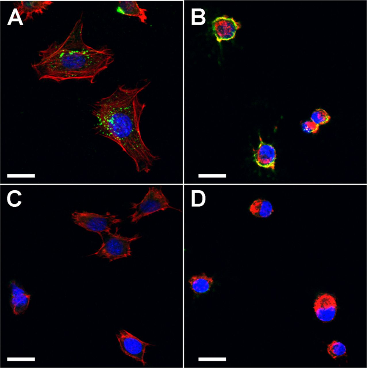
Since antibodies from Ro52-immune sera deposited in the SG, we analysed the reactivity of these sera with a mouse SG ductal cell line, SCA9-15. Antibodies from Ro52-immune sera entered live cells and mainly stained the cytoplasm as dots (figure 4). This staining pattern was not seen in sera obtained from mice immunised with MBP. Cytochalasin D treatment of cells prevented the entry of antibodies inside the cell and antibodies were retained on the surface, indicating that the internalisation was an active process of uptake (figure 5). In addition, preincubation of immune sera with Ro52 significantly reduced the internalisation and intracellular staining (see online supplementary figure S4).

Antibodies from Ro52-immune sera are actively internalised by salivary gland cell line. SCA9-15 cells were grown on coverslips and incubated with immune sera at 1:250 dilution. Bound antibodies were detected with FITC-conjugated goat anti-mouse IgG (A and D, green) and nuclei were stained with DAPI (B and E, blue). Antibodies from Ro52-immune sera show strong cytoplasmic staining in comparison with maltose-binding protein-immune sera (C and F, overlay). Scale bar=20 μm.
Cytochalasin D treatment prevents internalisation of antibodies by SCA9-15 cells. Confocal images show that cells incubated with Ro52-immune sera have cytoplasmic IgG (green) staining (A), which is not seen in cells incubated with control sera (C). Cytochalasin D treatment (B and D) localises Ro52-antibody reactivity to the surface (B), which is not seen in cells incubated with control sera (D). Cells treated with cytochalasin D become rounded due to collapse of actin filaments (red) stained with Phalloidin Alexa Fluor 568. Nuclei (blue) were stained with DAPI. Similar results were obtained in three additional experiments. Scale bar=10 μm.
Ro52 immunised NZM2758 mice mimic a subset of patients with SS
At day 30 post-immunisation time point, Ro52 immunised mice did not show evidence for sialoadenitis. However, they had circulating anti-Ro52 antibodies and SG dysfunction. Interestingly, a small subset of patients with SS in the Oklahoma SS cohort show similar features (table 1). Of the 321 patients, 73 patients (∼23%) were biopsy negative by AECG criteria (focus score <1). Among these, 49 were anti-Ro positive and 24 were anti-Ro negative. Among the 49 anti-Ro positive patients, 10 patients also had anti-La antibodies.
Summary of anti-Ro reactivity and objective dry mouth in patients with pSS in Oklahoma cohort with negative histopathology of minor salivary gland biopsy
Discussion
In this study, we demonstrate for the first time that antibodies induced by Ro52 immunisation can cause SG dysfunction. Apart from our study, the direct pathogenic role of anti-Ro52 antibodies has only been demonstrated in the experimental rat models of congenital heart block.22 ,23 Considered together, these studies clearly establish the pathogenic potential of anti-Ro52 antibodies.
Although multiple mouse models have been described for SS, each model recapitulates only certain features of this complex disorder.24 The experimental model system described in this study resembles clinical observations reported in a subset of patients with pSS who meet the AECG classification criteria (table 1). These patients do not have sialoadenitis, are anti-Ro or/and La positive and have xerostomia. However, only longitudinal studies in both patients and our model system would clarify whether this represents an early stage of the disease, which is followed by the development of severe sialoadenitis. In this regard, it should be noted that in the Ro60-peptide immunisation model, only 45% of mice developed sialoadenitis by 8 months post-immunisation.25 However, the severity of sialoadenitis did not correlate with the extent of SG dysfunction. The Ro52-model system clearly demonstrates the role of autoantibodies in SG dysfunction and suggests that this might represent an early feature of SS in some patients.
Passive transfer experiments show that the ability of antibodies to induce SG dysfunction was dependent on prior activation of innate immunity through alum. Alum used in this study has been reported to activate innate immune responses through the inflammasome pathway, leading to the production of proinflammatory cytokines.26 ,27 Inflammatory conditions in SG of patients with pSS have been shown to induce ductal expression of Ro52.28 In addition, expression and location of Ro52 in different cellular compartments have been modulated by activation of innate immunity through toll-like receptor 3 (TLR3), interferon-alpha (IFN-α) and nitric oxide.29–31 In NZM2758 mice, we observed that alum treatment caused significant upregulation of circulating KC, IL-1α, MIG, MIP2 and PDGF-β (see online supplementary figure S5). At present it is unclear as to how these cytokines influence antibody-mediated SG dysfunction. It is possible that systemic upregulation of proinflammatory cytokines can make the SG conducive for antibody-mediated injury, such as changes in the expression and localisation of proteins targeted by the Ro52-immune sera. Anti-Ro/La antibody responses in patients with SS have been associated with a positive type I IFN signature.32 Thus, whether an elevated type I IFN response occurs in Ro52 immunised mice and contributes towards glandular dysfunction needs to be evaluated.
In both active immunisation and passive transfer studies, immunoglobulin deposition was detected in the mouse SG. These data suggest that antibody deposition within the SG might be an important factor for the induction of glandular dysfunction, particularly in cases lacking lymphocytic foci. IgG antibody deposition in the SG ducts has been previously demonstrated in a limited study of patients with SS.33 In addition, circulating antibodies reacting with SG ductal antigens have been previously reported.34 Clearly additional studies with patient samples are warranted to further confirm the association between ductal antigen recognition, immunoglobulin deposition and xerostomia.
Since immunoglobulin deposition was observed in SG of Ro52 immunised mice, we analysed the reactivity of immune sera with a mouse SG cell line SCA9-15. Live SCA9-15 cells internalised antibodies, specifically from the Ro52-immune sera. The antibody uptake was prevented by cytochalasin D treatment, and the antibodies showed cell surface binding. Although preincubation of immune sera with Ro52 considerably inhibited antibody binding to SCA9-15 cells, the precise target of these antibodies on cell surface is not known. Previous studies have demonstrated that apoptotic human ductal cells cause subcellular redistribution and surface exposure of Ro52, Ro60 and La.35 However, in Ro52 immunised NZM2758 mice we did not observe epitope spreading and generation of anti-Ro60 and anti-La antibodies (see online supplementary figure S6). Thus, it is possible that antibodies in Ro52-immune sera bind either Ro52 or a cross-reactive protein(s) expressed on the surface of SG cells. Indeed, anti-Ro52 antibodies have been shown to cross-react with serotoninergic 5-HT4 receptor on cell surface.36 Whether such cross-reactive antibodies are generated in Ro52 immunised NZM2758 mice will be investigated in future. Alternatively, the antibody uptake might be occurring through the Fcγ receptors. A previous report has demonstrated that human SG cell line A-253 internalises anti-Ro and anti-La antibodies from patients with SS through Fcγ receptor binding.37 Although plausible, this mechanism cannot explain the lack of uptake of antibodies from MBP immunised mice.
The internalised antibodies in SCA9-15 cells were localised in the cytoplasm as dots (figure 4). Previous studies have demonstrated that Ro52 is present in cytoplasmic bodies that cannot be classified as endosomes, lysosomes, mitochondria, caveolae or proteasome-enriched structures.38 A characteristic feature of Ro52 cytoplasmic bodies is their transport along the microtubule network. It would be of interest to determine whether such structures are seen in SG epithelial cells and whether anti-Ro52 antibodies localise to these structures in vivo.
In summary, this study demonstrates the critical role played by Ro52-induced autoantibodies in pathogenesis of SS, particularly in the induction of SG dysfunction. Considering that this phenomenon was also dependent on the activation of innate immunity, novel immunotherapies targeting both innate immunity and adaptive immunity might prove to be highly beneficial for treating SS.
Acknowledgments
Technical assistance by the OMRF imaging core facility is acknowledged.
References
Supplementary materials
Supplementary Data
This web only file has been produced by the BMJ Publishing Group from an electronic file supplied by the author(s) and has not been edited for content.
Files in this Data Supplement:
- Data supplement 1 - Online supplement
- Data supplement 2 - Online figures
Footnotes
Handling editor Tore K Kvien
BMS and PK contributed equally.
Contributors Data acquisition, analysis, interpretation: BMS, PK, NW, AP, PDR, PD, HB and USD. Clinical data acquisition, analysis, interpretation: AR, KG, KSH, DUS, SY, DML, LR, RHS and KLS. Study design: PK, BMS, HB and USD. Manuscript preparation and review: All.
Funding This study has been funded by research grants from the National Institute of Dental and Craniofacial Research, DE019883 (USD), DE022977 (USD), National Institute of Allergy and Infectious Diseases, AI079621 (USD) and National Institute of Arthritis and Musculoskeletal and Skin Diseases, P50AR060804 (KLS). Financial support was also provided by the Oklahoma Medical Research Foundation (USD and HB).
Competing interests None.
Ethics approval Oklahoma Medical Research Foundation, IRB.
Provenance and peer review Not commissioned; externally peer reviewed.