


Asymptomatic bilateral pulmonary embolism in Churg–Strauss syndrome
- A. Maria*,
- P. Guilpain*⇓,
- A. Forestier*,
- E. Delhom#,
- A. Schiffman*,
- S. Rivière*,
- A.K. Van Kien¶,
- H. Leray-Moragues+,
- I. Serre§,
- T. Vincentf,
- J-F Eliaouf and
- A. Le Quellec*
- *Service de Médecine Interne A – Maladies Multi-Organiques, Centre de Compétence Maladies Auto-Immunes, #Service d'Imagerie Médicale, ¶Service de Médecine Interne et Maladies Vasculaires, fDépartement d'Immunologie, Université Montpellier 1, Hôpital Saint-Eloi, CHRU de Montpellier, +Service de Néphrologie, and §Service d'Anatomopathologie, Université Montpellier 1, Hôpital Lapeyronie, CHRU de Montpellier, Montpellier, France.
- P. Guilpain, Service de Médecine Interne A, Hôpital Saint-Eloi, CHRU de Montpellier, 80 avenue Augustin Fliche, 34295 Montpellier, Cedex 5, France. E-mail: p-guilpain{at}chu-montpellier.fr
To the Editor:
Churg–Strauss syndrome (CSS) is a systemic small-sized vessel vasculitis, characterised by severe asthma, transient pulmonary infiltrates, and blood and tissue eosinophilia [1]. CSS can affect several organs, including the lungs, heart, kidneys and peripheral nervous system. Anti-neutrophil cytoplasm autoantibodies (ANCA) mainly directed against myeloperoxidase (MPO) are detected in ∼40% of patients and are associated with renal involvement. Several studies have focused on venous thromboembolic events (VTE), which are an emerging clinical condition in ANCA-associated vasculitides (AAV) [2, 3]. Herein, we report the case of a patient with newly diagnosed CSS and totally asymptomatic pulmonary embolism, and discuss the features, pathogenesis and management of VTE in CSS.
In February 2011, a 61-yr-old male was referred for recent asthenia, diffuse arthromyalgia and blood eosinophilia. His past medical history included high blood pressure treated with amlodipine. There were no other risk factors for VTE other than age and inflammatory state. His recent history included late-onset asthma, which started 1 yr earlier and required various combined inhaled corticosteroids, bronchodilators and several short courses of oral corticosteroids.
In early December 2010, the patient complained of general weakness, weight loss and diffuse myalgia. In late December, another asthma flare-up was successfully treated with a course of oral prednisolone (1 mg·kg−1 per day for 5 days), which was effective for both myalgia and weakness. Unfortunately, muscle and joint pain immediately relapsed as soon as corticosteroids were discontinued, while respiratory symptoms had improved. In February 2011, a thoracic computed tomography (CT) scan was performed, focusing on the parenchyma not vessels. No lesions were reported. At the same time, distal paresthesia appeared and subsequently concerned the lower left and upper right limbs. Following this, the patient was referred to our department (Internal Medicine A, CHU, Montpellier, France).
On admission, the patient did not complain about dyspnoea, cough or chest pain. His blood pressure was 130/80 mmHg and cardiac frequency was 90 beats per min, his temperature rose to 39°C and his oxygen saturation was 98%. Pulmonary auscultation did not disclose wheezes or ronchi. Cardiac auscultation and vascular examination were normal and there was no sign of phlebitis. Neurological examination detected dysesthesia and hypoesthesia in the right cubital and left common peroneal nerve areas but no objective weakness was noted and all reflexes were present. There was no joint swelling or cutaneous eruption. Abdominal examination was normal. No hepatosplenomegaly and no enlarged lymph node were observed.
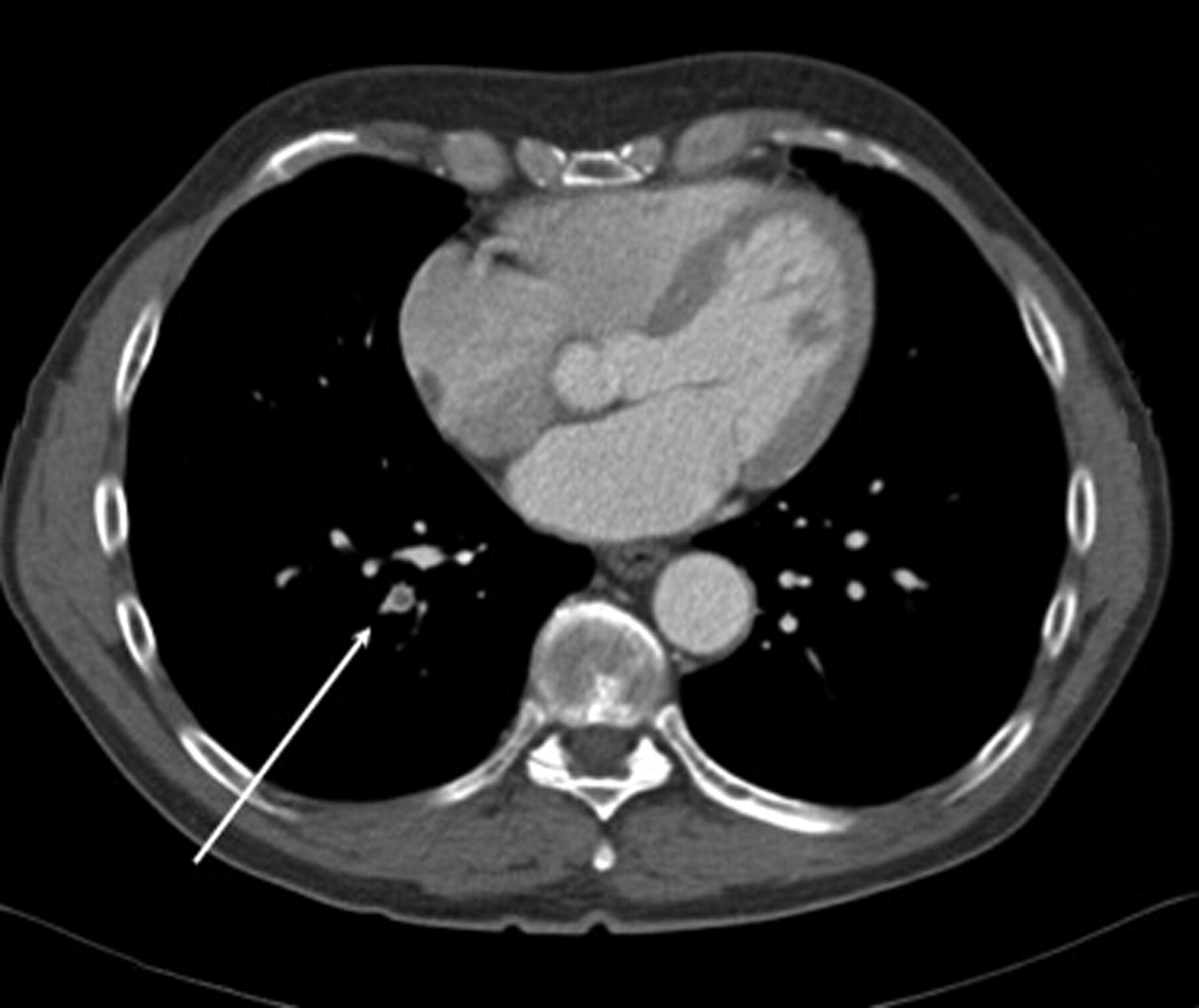
Blood tests showed a white blood cell count of 14.6×10⁹ cells per L with 5.4×10⁹ eosinophils per L (37% of white blood cells), C-reactive protein (CRP) was 94 mg·L−1, fibrinogen was 7.3 g·L−1 and serum creatinine was 123 μmol·L−1 (clearance 60 mL·min−1 Modification of Diet in Renal Disease score). Testing for ANCA was highly positive (1/1,600 perinuclear) and anti-MPO antibodies (69 UI·mL−1) were detected by Luminex® (Luminex Corporation, Austin, TX, USA). Urine sediment analysis showed 2.0×10³ leukocytes per L with eosinophiluria, and 4.0×10⁵ red blood cells per L. Proteinuria level was 0.55 g per 24 h. Blood and urine cultures remained sterile. An echocardiogram was normal and systolic pulmonary arterial pressure was 25 mmHg. The thoracic CT scan, which had been performed 2 weeks earlier, was then re-read. It did not include specific angiosequences, but eventually disclosed proximal bilateral pulmonary embolism (fig. 1). Ultrasonography of the lower limb veins did not demonstrate any venous thrombosis. Electromyography confirmed mononeuritis multiplex involving the right cubital and left common peroneal nerves. Percutaneous renal biopsy demonstrated pauci-immune extra-capillary crescentic glomerulonephritis, with associated tubulitis and no eosinophilic infiltration within the interstitium.

Thoracic computed tomography scan showing occlusion of the right posterior basal segmental artery.
CSS with neurological and renal involvement was diagnosed and oral corticosteroids were started (1 mg·kg−1 per day prednisone). Because of renal involvement, the patient also received three intravenous pulses of methylprednisolone (15 mg·kg−1 per day for 3 days) and pulses of intravenous cyclophosphamide (0.6 g·m−2). Because bilateral pulmonary embolism was also diagnosed, an anticoagulant treatment with low-molecular-weight heparin was immediately started and rapidly switched to oral anticoagulants. Under this regimen, the patient quickly improved and experienced a good outcome of CSS and pulmonary embolism with 8-month follow-up.
Thrombotic factors that could have been implicated in the development of pulmonary embolism were also investigated. Thoracic and abdominal CT scans did not show any tumour process, and no coagulation abnormality could be detected: there was no mutation for pro-thrombin and Factor V Leiden, no anti-thrombin III, protein C or protein S deficiency, no lupus anticoagulant, no anti-cardiolipin antibodies, no anti-β2GP1 antibodies and no hyperhomocysteinaemia.
The prevalence of VTE in AAV seems to be higher than previously expected. A specific survey for VTE in CSS yielded a prevalence of 6.5% in a cohort of 1,130 patients with AAV and of 8.1% in the 232 CSS patients of the study by Allenbach et al. [2]. The prevalence of VTE in CSS was not different to that observed in granulomatosis with polyangiitis (Wegener's granulomatosis) and microscopic polyangiitis. In this cohort of AAV patients, the first VTE was deep venous thrombosis in 62.2% of cases, pulmonary embolism in 21.6% of cases, or both in 16.2% of cases. VTE is not constantly observed at diagnosis of vasculitis. In the cohort of AAV patients reported by Allenbach et al. [2], the interval between the diagnosis of AAV and the first VTE vary to an extended degree (median (range) 5.8 (-2.4–156.0) months). A similar observation is reported in CSS, since VTE can also occur during flare-ups [4] and remission [3].
Risk factors for VTE in AAV have not yet been identified. In the cohort of Allenbach et al. [2], multivariate and univariate analysis found VTE to be associated with older age at diagnosis, male sex, previous VTE and stroke with motor deficit. Nephrotic range proteinuria was a risk factor in univariate analysis. There was no association with CRP level, ANCA positivity and disease activity assessed by the Birmingham Vasculitis Activity Score.
Similarly, the mechanisms leading to VTE in CSS remain unclear and speculative [5]. No association has been reported with innate or acquired thrombophilia but, to date, such conditions have not been well investigated. As mentioned previously, no link was established with ANCA positivity. Similarly to other inflammatory diseases, increased amounts of von Willebrand Factor and interleukin-6 released by endothelial cells, hepatic synthesis of CRP and fibrinogen may promote platelet aggregation and plasma hypercoagulability. The role of eosinophils is not established in the development of VTE and, in the study by Allenbach et al [2], the similar prevalence of VTE in other AAV classically devoid of eosinophilia (granulomatosis with polyangiitis and microscopic polyangiitis) do not argue for a specific role of these cells. Nevertheless, VTE can occur in the course of other hypereosinophilic states, such as hypereosinophilic syndromes, which has been attributed to the pro-thrombotic properties of eosinophil granule proteins. Indeed, eosinophil cationic proteins can bind to Hageman Factor (XII), activate the intrinsic pathway of coagulation and also interfere with anticoagulant activity of heparin and of endogenous heparan sulfate in vitro [4]. The main oxidative product of eosinophils, hypothiocyanous acid, may also lead to a pro-thrombotic state, through the induction of tissue factor activity in human umbilical vein endothelial cells [6].
Our case illustrates the risk of VTE in CSS, which may be promoted by inflammatory state and hypereosinophilia. In our patient, pulmonary embolism was totally asymptomatic and was unexpectedly diagnosed on a thoracic CT scan. A delayed or missed diagnosis could have led to respiratory failure and death in the present case. Thus, pulmonary embolism may be considered as a pitfall in CSS since respiratory signs may be falsely attributed to the vasculitis. Therefore, our observation raises the question of systematic screening for VTE in CSS.
We do not reasonably think that angiography CT scans should be performed systematically in this condition. CSS glomerulonephritis and renal insufficiency must definitely be taken into consideration because of the risk of worsening following the administration of a contrast agent. Furthermore, incidental pulmonary embolism is not so uncommon in the general population (∼2.6% of all patients according to a recent meta-analysis) [7] and in the clinical conditions with increased risk of VTE, such as malignant diseases. In these cases, systematic screening for VTE is not recommended but this screening is frequently addressed by the necessity for radiological monitoring of cancer.
In vasculitides, there is no recommendation for VTE to date and the length of curative anticoagulation remains to be established. However, the continuation of anticoagulation could be proposed until remission, which is usually obtained within 6 months.
Footnotes
Provenance
Submitted article, peer reviewed.
Statement of Interest
None declared.
- ©ERS 2012