
Main menu

  • Home
  • Current issue
  • Past issues
  • Authors/reviewers
    • Instructions for authors
    • Submit a manuscript
    • COVID-19 submission information
    • Institutional open access agreements
    • Peer reviewer login
  • Alerts
  • Subscriptions
  • ERS Publications
    • European Respiratory Journal
    • ERJ Open Research
    • European Respiratory Review
    • Breathe
    • ERS Books
    • ERS publications home

User menu

  • Log in
  • Subscribe
  • Contact Us
  • My Cart

Search

  • Advanced search
  • ERS Publications
    • European Respiratory Journal
    • ERJ Open Research
    • European Respiratory Review
    • Breathe
    • ERS Books
    • ERS publications home

Login

European Respiratory Society

Advanced Search

  • Home
  • Current issue
  • Past issues
  • Authors/reviewers
    • Instructions for authors
    • Submit a manuscript
    • COVID-19 submission information
    • Institutional open access agreements
    • Peer reviewer login
  • Alerts
  • Subscriptions

Clinical significance and applications of oscillometry

David A. Kaminsky, Shannon J. Simpson, Kenneth I. Berger, Peter Calverley, Pedro L. de Melo, Ronald Dandurand, Raffaele L. Dellacà, Claude S. Farah, Ramon Farré, Graham L. Hall, Iulia Ioan, Charles G. Irvin, David W. Kaczka, Gregory G. King, Hajime Kurosawa, Enrico Lombardi, Geoffrey N. Maksym, François Marchal, Ellie Oostveen, Beno W. Oppenheimer, Paul D. Robinson, Maarten van den Berge, Cindy Thamrin
European Respiratory Review 2022 31: 210208; DOI: 10.1183/16000617.0208-2021
David A. Kaminsky
1Dept of Medicine, Pulmonary and Critical Care Medicine, University of Vermont, Larner College of Medicine, Burlington, VT, USA
23These authors have contributed equally to this manuscript
  • Find this author on Google Scholar
  • Find this author on PubMed
  • Search for this author on this site
Shannon J. Simpson
2Children's Lung Health, Telethon Kids Institute, School of Allied Health, Curtin University, Perth, Australia
23These authors have contributed equally to this manuscript
  • Find this author on Google Scholar
  • Find this author on PubMed
  • Search for this author on this site
Kenneth I. Berger
3Division of Pulmonary, Critical Care, and Sleep Medicine, NYU School of Medicine and André Cournand Pulmonary Physiology Laboratory, Belleuve Hospital, New York, NY, USA
  • Find this author on Google Scholar
  • Find this author on PubMed
  • Search for this author on this site
Peter Calverley
4Institute of Ageing and Chronic Disease, University of Liverpool, Liverpool, UK
  • Find this author on Google Scholar
  • Find this author on PubMed
  • Search for this author on this site
Pedro L. de Melo
5Dept of Physiology, Biomedical Instrumentation Laboratory, Institute of Biology and Faculty of Engineering, State University of Rio de Janeiro, Rio de Janeiro, Brazil
  • Find this author on Google Scholar
  • Find this author on PubMed
  • Search for this author on this site
  • ORCID record for Pedro L. de Melo
Ronald Dandurand
6Lakeshore General Hospital, Pointe-Claire, QC, Canada
7Montreal Chest Institute, Meakins-Christie Labs, Oscillometry Unit of the Centre for Innovative Medicine, McGill University Health Centre and Research Institute, and McGill University, Montreal, QC, Canada
  • Find this author on Google Scholar
  • Find this author on PubMed
  • Search for this author on this site
Raffaele L. Dellacà
8Dipartimento di Elettronica, Informazione e Bioingegneria – DEIB, Politecnico di Milano University, Milan, Italy
  • Find this author on Google Scholar
  • Find this author on PubMed
  • Search for this author on this site
  • ORCID record for Raffaele L. Dellacà
Claude S. Farah
9Dept of Respiratory Medicine, Concord Repatriation General Hospital, Sydney, Australia
  • Find this author on Google Scholar
  • Find this author on PubMed
  • Search for this author on this site
  • ORCID record for Claude S. Farah
Ramon Farré
10Unitat de Biofísica i Bioenginyeria, Facultat de Medicina, Universitat de Barcelona-IDIBAPS, Barcelona, Spain
11CIBER de Enfermedades Respiratorias, Madrid, Spain
  • Find this author on Google Scholar
  • Find this author on PubMed
  • Search for this author on this site
Graham L. Hall
2Children's Lung Health, Telethon Kids Institute, School of Allied Health, Curtin University, Perth, Australia
  • Find this author on Google Scholar
  • Find this author on PubMed
  • Search for this author on this site
Iulia Ioan
12Dept of Paediatric Lung Function Testing, Children's Hospital, Vandoeuvre-lès-Nancy, France
13EA 3450 DevAH – Laboratory of Physiology, Faculty of Medicine, University of Lorraine, Vandoeuvre-lès-Nancy, France
  • Find this author on Google Scholar
  • Find this author on PubMed
  • Search for this author on this site
  • ORCID record for Iulia Ioan
Charles G. Irvin
1Dept of Medicine, Pulmonary and Critical Care Medicine, University of Vermont, Larner College of Medicine, Burlington, VT, USA
  • Find this author on Google Scholar
  • Find this author on PubMed
  • Search for this author on this site
David W. Kaczka
14Depts of Anaesthesia, Biomedical Engineering and Radiology, University of Iowa, Iowa City, IA, USA
  • Find this author on Google Scholar
  • Find this author on PubMed
  • Search for this author on this site
  • ORCID record for David W. Kaczka
  • For correspondence: david.kaminsky@uvm.edu
Gregory G. King
15Dept of Respiratory Medicine and Airway Physiology and Imaging Group, Royal North Shore Hospital, St Leonards, Australia
16Woolcock Institute of Medical Research, The University of Sydney, Sydney, Australia
  • Find this author on Google Scholar
  • Find this author on PubMed
  • Search for this author on this site
Hajime Kurosawa
17Dept of Occupational Health, Tohoku University School of Medicine, Sendai, Japan
  • Find this author on Google Scholar
  • Find this author on PubMed
  • Search for this author on this site
Enrico Lombardi
18Paediatric Pulmonary Unit, Meyer Paediatric University Hospital, Florence, Italy
  • Find this author on Google Scholar
  • Find this author on PubMed
  • Search for this author on this site
Geoffrey N. Maksym
19School of Biomedical Engineering, Dalhousie University, Halifax, NS, Canada
  • Find this author on Google Scholar
  • Find this author on PubMed
  • Search for this author on this site
François Marchal
12Dept of Paediatric Lung Function Testing, Children's Hospital, Vandoeuvre-lès-Nancy, France
13EA 3450 DevAH – Laboratory of Physiology, Faculty of Medicine, University of Lorraine, Vandoeuvre-lès-Nancy, France
  • Find this author on Google Scholar
  • Find this author on PubMed
  • Search for this author on this site
Ellie Oostveen
20Dept of Respiratory Medicine, Antwerp University Hospital and University of Antwerp, Belgium
  • Find this author on Google Scholar
  • Find this author on PubMed
  • Search for this author on this site
Beno W. Oppenheimer
3Division of Pulmonary, Critical Care, and Sleep Medicine, NYU School of Medicine and André Cournand Pulmonary Physiology Laboratory, Belleuve Hospital, New York, NY, USA
  • Find this author on Google Scholar
  • Find this author on PubMed
  • Search for this author on this site
Paul D. Robinson
21Woolcock Institute of Medical Research, Children's Hospital at Westmead, Sydney, Australia
  • Find this author on Google Scholar
  • Find this author on PubMed
  • Search for this author on this site
Maarten van den Berge
22Dept of Pulmonary Diseases, University of Groningen, University Medical Centre Groningen, Groningen, The Netherlands
  • Find this author on Google Scholar
  • Find this author on PubMed
  • Search for this author on this site
Cindy Thamrin
16Woolcock Institute of Medical Research, The University of Sydney, Sydney, Australia
  • Find this author on Google Scholar
  • Find this author on PubMed
  • Search for this author on this site
  • ORCID record for Cindy Thamrin
  • Article
  • Figures & Data
  • Info & Metrics
  • PDF
Loading

Abstract

Recently, “Technical standards for respiratory oscillometry” was published, which reviewed the physiological basis of oscillometric measures and detailed the technical factors related to equipment and test performance, quality assurance and reporting of results. Here we present a review of the clinical significance and applications of oscillometry. We briefly review the physiological principles of oscillometry and the basics of oscillometry interpretation, and then describe what is currently known about oscillometry in its role as a sensitive measure of airway resistance, bronchodilator responsiveness and bronchial challenge testing, and response to medical therapy, particularly in asthma and COPD. The technique may have unique advantages in situations where spirometry and other lung function tests are not suitable, such as in infants, neuromuscular disease, sleep apnoea and critical care. Other potential applications include detection of bronchiolitis obliterans, vocal cord dysfunction and the effects of environmental exposures. However, despite great promise as a useful clinical tool, we identify a number of areas in which more evidence of clinical utility is needed before oscillometry becomes routinely used for diagnosing or monitoring respiratory disease.

Abstract

This paper provides a current review of the interpretation, clinical significance and application of oscillometry in respiratory medicine, with special emphasis on limitations of evidence and suggestions for future research. https://bit.ly/3GQPViA

Introduction

The mechanical properties of the lungs are disrupted in many disease states and exposures and contribute to major respiratory symptoms such as dyspnoea. Both the clinical management and our understanding of respiratory disease have benefitted from widely available tests like spirometry and the measurement of absolute lung volumes and gas transfer. However, these measurements require significant patient co-operation and a willingness to undertake maximal respiratory efforts. Oscillometry is a noninvasive method for measuring the mechanical properties of the respiratory system, which can enhance our understanding and management of lung disease [1]. Recently, the European Respiratory Journal published “Technical standards for respiratory oscillometry” [2], which reviewed the physiological basis of oscillometric measures and detailed the technical factors related to standardisation of equipment and test performance, quality assurance and reporting of results. Here we provide a review of the clinical significance and applications of oscillometry. We briefly review the physiological principles of oscillometric measurements and the basics of oscillometry interpretation, highlight potential clinical applications, and identify future work required for oscillometry to become routinely used in clinical practice.

The physiology and performance of oscillometry

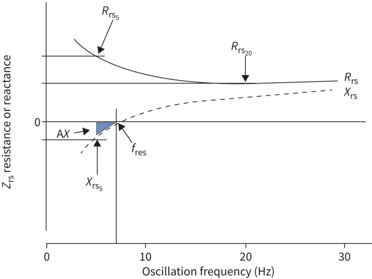
Oscillometry measures the mechanical impedance of the respiratory system (Zrs), representing the resistive and reactive forces that must be overcome to drive an oscillating flow signal into the respiratory system. The forces arise in the respiratory system from 1) the resistance of the airways and tissues to flow (Rrs), 2) the elastance (stiffness) of the lung parenchyma and chest wall in response to changes in volume (encompassed in reactance, Xrs), and 3) the inertance of accelerating gas in the airways (Irs). Zrs has generally been reported from a single frequency or over the frequency range of 5–40 Hz as an average across the whole breathing cycle (i.e. both inspiration and expiration). It has also been reported separately during the inspiratory and expiratory phases. These concepts and how they are measured by oscillometry are described in detail in the technical document. We provide a brief summary of these details in table 1, figure 1 and the supplementary material.

View this table:
  • View inline
  • View popup
TABLE 1

Summary of oscillometric parameters with a focus on the medium frequency range likely to be used in clinical practice (5–40 Hz)

FIGURE 1
  • Download figure
  • Open in new tab
  • Download powerpoint
FIGURE 1

Impedance spectrum. Respiratory system impedance (Zrs) is plotted against frequency. Zrs is composed of a real component seen by resistance (Rrs) and an imaginary component expressed as reactance (Xrs). Rrs and Xrs at specific frequencies are noted by the frequency at which they are measured (e.g. Rrs5=Rrs at 5 Hz, Rrs20=Rrs at 20 Hz). The point at which Xrs crosses zero is the resonant frequency (fres). Below fres, Xrs is dominated by elastance, and above fres, Xrs is dominated by inertance. An integrated measure of low frequency Xrs is the area inscribed by Xrs and Zrs=0 starting at the lowest frequency up to fres, known as the area of the Xrs curve, AX. The lowest frequency defining AX is shown at 5 Hz, but it may be estimated starting from any frequency. Note that the Zrs spectrum shown is characteristic for a healthy adult. In healthy young children, the values and frequency dependence of Rrs would be relatively increased, Xrs would be more negative and fres would be shifted markedly to the right (resulting in increased AX).

Much of the evidence for clinical utility of oscillometry, including the studies cited in the current document, have been collected using a wide range of equipment, protocols and reporting formats. Although there is increasing availability of commercial oscillometry devices, comparability across devices remains a challenge, especially with regards to differences in the frequency dependence of resistance and reactance at higher-than-normal impedance [3, 4]. As such, the applicability of existing reference equations (supplementary table S1) remains uncertain for abnormal disease states and across different devices and manufacturers. Until this is resolved, comparisons between healthy and diseased cohorts should ideally be made using the same device, and differences in results based on variability in equipment, protocols and reference values should be considered when interpreting data presented in this review. Efforts are currently underway by the Global Lung Function Initiative to gather data from around the world to develop more universal, robust reference equations that take into account specific equipment and protocols.

Additionally, while oscillometry is a simple test to perform in clinic, its interpretation remains a challenge for many. Further aspects need to be better established before oscillometry gains an evidence-based position in clinical practice, as summarised in table 2. Many of these aspects are highlighted in the following sections, but in particular it is important to note that establishing minimal important clinical differences to assess change in lung function over time is a crucial area for future research.

View this table:
  • View inline
  • View popup
TABLE 2

Gaps in knowledge and future needs in defining the clinical applications of oscillometry

Oscillometry during infancy

Lung function measurements during infancy have largely been confined to a few specialist centres due to technical complexity, lack of commercially available equipment, and potential risks associated with the measurement (e.g. requirement for sedation). Additionally, the use of a face mask in infants complicates the interpretation of impedance data, since much of the measured Rrs may be attributed to nasal resistance [5]. To date, there is no evidence that impedance measurements in infants contribute to clinical decision-making. However, oscillometric studies have contributed to the pathophysiological understanding of respiratory disease in infants, particularly those with wheezing disorders, as described in the supplementary material.

Preterm birth and bronchopulmonary dysplasia beyond the neonatal intensive care unit

Emerging evidence suggests that oscillometry is a clinically useful measure of lung function in survivors of very preterm birth. Compared to preschoolers delivered at term, those born prematurely have increased Rrs, more negative Xrs and increased area of reactance (AX) and resonant frequency (fres) [6]. Deficits are even greater in premature babies with bronchopulmonary dysplasia (BPD) [7–9]. Xrs measured in the first week of life has been shown to improve prognostication of respiratory outcome in very preterm infants on noninvasive respiratory support. Recently, Evans et al. [10] showed that while all oscillometric outcomes were abnormal in those born very prematurely, Xrs was particularly sensitive at distinguishing term versus preterm preschoolers, suggesting a role for oscillometry in monitoring preterm lung disease. Deficits in oscillometric outcomes persist up to at least adolescence and correlate with respiratory symptoms [11, 12].

To date, only one study has measured oscillometry longitudinally in this population [13]. Interestingly, Xrs and AX deteriorated over time in those with BPD in parallel with declines in spirometry measures, and were found to decrease faster in those children exposed to environmental tobacco smoke and those with respiratory symptoms. Deterioration in these oscillometry outcomes was not associated with structural lung damage on chest computed tomography (CT) scans. The pathophysiology underpinning increased lung stiffness in this population remains unclear, but may reflect failed alveolarisation, structural damage to the parenchyma or inflammatory changes to the lungs.

Asthma

Oscillometric measurements can distinguish adult asthmatics from healthy controls [14–16], asthmatics with different degrees of airway obstruction [17], and between groups of adults with asthma compared to COPD [15, 18–21]. Oscillometry may be especially helpful in diagnosing asthma in patients with preserved spirometry as a more sensitive indicator of abnormal airway physiology [22]. However, while helpful in adults, the delineation between healthy controls and asthmatics using oscillometry is less clear in preschool-aged children, with several studies showing no difference between groups [23–25]. These conflicting findings may be due to differing diagnostic criteria, disease severity or degree of control in the paediatric populations studied who may have smaller differences between health and disease than adults.

Oscillometric outcomes other than resistance and reactance (such as AX and Rrs5–20) may also be useful in supporting a diagnosis of asthma, predicting future loss of asthma control or monitoring clinical treatment changes. In particular, the term “small airway dysfunction” (SAD) has gained traction. The large ATLANTIS cohort revealed that Rrs5–20, AX and Xrs5 were all strong contributors to SAD, with high prevalence in asthma compared to other physiological measures thought to reflect small airways [26]. However, a limitation of the study is that the definition of SAD relied on a statistical, data-driven approach, rather than an independent measure. It also is important to realise that no measures are specific for SAD, including Rrs5–20, which is sensitive to heterogeneouis peripheral airway narrowing, but can also be affected by heterogeneity in the central airways and upper airway shunt (table 1). Furthermore, these relative contributions are likely dependent on disease. We urge caution in the strict interpretation of Rrs5–20 as relating solely to small airways disease, especially when measured using devices that are known to be associated with enhanced frequency dependence of resistance [3, 27–29]. The clinical significance of Rrs5–20 continues to be evaluated in terms of its mechanism [30, 31], prevalence [32] and correlations with measures of severity and control in both children and adults [26, 33, 34].

In addition, oscillometry has provided novel insights into the pathophysiology of asthma via the effects of lung volume on oscillatory mechanics, as well as short- and long-term variations in mechanics over time. These topics are further addressed in the supplementary material. These variations may be a marker of instability, and potentially useful to detect exacerbations or loss of control, particularly in a home telemonitoring setting [35]. Intrabreath changes in oscillometry parameters may also provide additional information beyond conventional parameters: in preschoolers it improved detection of acute obstruction and recurrent wheezers from healthy controls [36], while in adults with severe asthma it distinguished those with poor control from those with good control [37]. While oscillometry measures are generally altered in asthmatics at the population level, oscillometry is arguably of most benefit in clinical practice for the diagnosis of asthma (via bronchodilation or bronchoprovocation) and monitoring response to intervention, as discussed in the following sections.

Bronchodilator response testing

As discussed in the recent technical standards [2], cut-offs indicating a significant bronchodilator response (BDR) using oscillometric indices are reported in healthy adults and children (i.e. 40% decrease in Rrs at 5 Hz, 50% increase in Xrs at 5 Hz and 80% decrease in AX relative to baseline) (supplementary table S2) [2]. Recent work has shown results for BDR in adults [38, 39] that are similar to published data using different instruments [4], suggesting that BDR in oscillometry appears to be robust to different devices and populations. Nevertheless, given the diversity of age ranges, equipment and protocols, further work is still needed to best define a BDR, its significance relative to important patient outcomes and consistency of response over time.

Several studies have shown that a BDR based on oscillometric parameters is better than one based on forced expiratory volume in 1 s (FEV1) at differentiating asthmatic from healthy children [40–42]. However, data relating to BDR in children with “early life” wheeze (i.e. no formal diagnosis of asthma) are less clear, with some studies reporting no differences in BDR between wheezy and non-wheezy preschoolers [43]. In adults, bronchodilation was identified more often using oscillometry than spirometry, and in one study changes in Xrs and AX (but not Rrs) correlated with spirometric responses and identified more subjects with poor asthma control compared to spirometry [44]. Other studies have shown that BDR assessed with oscillometry correlates with poor asthma control [14, 45].

Bronchial challenge testing

Oscillometry can also be used as an alternative to spirometry for conducting bronchial challenges in adults [46–58] and children [59–65]. A summary of studies to date attempting to define airway hyperresponsiveness (AHR) based on oscillometry is shown in supplementary table S3. Results vary widely, ranging from a 20 to 50% increase in Rrs5 and a 20–80% decrease in Xrs5. Provocation studies in children are feasible in patients as young as 3 years old using oscillometry [59, 61]. However, conclusive data indicating which outcome measures and threshold values best reflect a positive bronchial challenge test are currently lacking in this age group. The main advantage of oscillometry for bronchial challenge testing is increased sensitivity of detecting bronchoconstriction, which might shorten the test and reduce the cumulative dose of the agent. However, there may be underestimation of the response occurring in the lungs in children during a challenge due to loss of the oscillatory flow into the upper and large airways, known as upper airway shunt [66]. More information about upper airway shunt is found in the supplementary material.

Deep breaths (i.e. inflation to total lung capacity (TLC)) during spirometry testing, or with some inhalation agents, potentially affect diagnosis, given both the bronchoprotective and bronchodilator effect of deep inhalation in health and in asthma [67–69]. Consequently, oscillometry may be more sensitive than spirometry for detecting AHR in mild asthmatic patients, given their maintained, albeit reduced, response to a deep breath, resulting in a lower provocative dose of methacholine [47, 70] and therefore a shorter testing protocol. However, since avoiding deep breaths may result in AHR even in healthy individuals [71], oscillometry may be less sensitive than spirometry in distinguishing healthy from asthmatic individuals. Of note, patients may prfer oscillometry over spirometry during bronchial challenge testing because they do not have to repeatedly take the deep breaths needed to perform spirometry.

AHR detected by oscillometry is repeatable [47, 49, 61, 72] and correlates with responsiveness based on FEV1 [73], but the correlations of spirometry and oscillometry are inconsistent. There is wide variability across studies, which may be explained by differences in methodology and study populations. However, it appears that oscillometric indices may provide additional information to spirometry. For example, some subjects report symptoms during bronchial challenge without accompanying changes in FEV1, which may be related to the effects of deep inhalation mitigating changes in FEV1; however, concomitant changes in Rrs and AX suggest there is narrowing and/or closure of small airways [74, 75] not detected by spirometry. In summary, oscillometry is a useful tool to detect airway narrowing before spirometry during bronchial challenge testing, but the correlations and thresholds of response are variable, and further studies are needed to determine if oscillometry indices are more clinically relevant than spirometry.

Treatment responses in asthma

In addition to treatment with bronchodilators, differences in Rrs and Xrs in response to different inhaled corticosteroids (ICS) [76, 77], montelukast [78], ICS/long acting β2-agonist (LABA) formulations [79], and ICS versus ICS/LABA [80] have been demonstrated. In general, Rrs, Xrs and AX are more sensitive than spirometry [81]. Recent work has shown that oscillometry and other parameters related to peripheral airway function can be correlated with an improvement in symptoms for patients with poorly controlled asthma receiving ICS/LABA therapy [82]. One study has attempted to determine whether oscillometry could distinguish any benefit of small versus large particle size ICS therapy based on peripheral versus central airway obstruction, respectively; however, this study only looked at the differential treatment effect on symptoms, not oscillometry, and used an arbitrary oscillometric definition of peripheral airway function [83]. Oscillometric indices are also sensitive to improvements in asthma in response to mepolizumab therapy [84]. These findings suggest an important role of oscillometry in identifying and monitoring treatment responses in asthma, complementary to traditional spirometry.

Thus, in asthma, individual oscillometry studies have demonstrated physiological correlations of specific indices with symptoms, imaging, spirometry, changes in response to bronchodilators, bronchial challenges and treatment, and prediction of treatment response. What needs to follow are larger scale trials beyond proof of concept that demonstrate actual clinical utility in terms of improving patient care in asthma, as well as what constitutes a minimal clinically significant difference in oscillometry parameters.

Cystic fibrosis

A number of studies in children with cystic fibrosis (CF) reveal normal oscillometric indices [85–87], with only one study of preschool children with CF reporting abnormal values [88]. Associations between oscillometric indices and other clinical parameters in children with CF are variable [25, 86, 88–92]. There are limited data on the use of oscillometry in adults with CF; however, by one report, resistance and reactance are abnormal in adults with CF and correlate with other measures of lung function [93]. Therefore, the clinical utility of oscillometry in CF remains uncertain, particularly when other clinical tests have shown superior ability to detect early CF lung disease and successfully monitor improvement in lung function with intervention.

COPD

Oscillometry may play an important role in the early detection of the adverse effects of smoking before COPD is diagnosed [94]. Several studies have found a high prevalence of abnormal Zrs in smokers with normal spirometry, mainly in Rrs and Xrs near 5 Hz [95–98], with up to 60% of smokers with normal spirometry (FEV1/forced vital capacity (FVC)>0.70) having some abnormality on oscillometry. Smokers also have greater prevalence of significant BDR compared to nonsmokers [38]. The clinical significance of these findings still needs to be determined by pathologic correlations and prospective clinical studies to establish relevance and utility.

Patients with COPD have significantly higher Rrs and more negative Xrs values than healthy people [99], changes that are proportional to the degree of airway obstruction [100]. Empirical studies demonstrate that Xrs is related to the degree of gas trapping and hyperinflation in the lungs and reflects the amount of communicating lung volume [101]. Xrs and fres relate better to FEV1, and measures of hyperinflation, i.e. inspiratory capacity/TLC and residual volume/TLC ratios, than do resistance measurements [102, 103]. Magnetic resonance imaging-derived measurements of abnormal gas mixing correlate best with Rrs5–19 and AX in COPD patients [104]. Recent work with parametric response mapping by CT has demonstrated strong correlations with oscillometry-derived Rrs5–19 in patients with COPD [105], which is presumed to reflect small airways disease (but may also reflect large airway heterogeneity). A similar finding has been shown using endobronchial optical coherency tomography, which demonstrated that Rrs5–20 correlated with small airways pathology in heavy smokers and patients with COPD [106]. Oscillometric parameters likely related to gas trapping (i.e. Xrs ) in patients with COPD correlated with changes in exercise capacity following completion of pulmonary rehabilitation [107]. Oscillometry may also help in the categorisation of COPD severity [108].

As in asthma, oscillometry parameters are sensitive to treatment in patients with COPD. For example, Xrs near 5 Hz rather than Rrs appears to be more sensitive to bronchodilators [109–113] or ICS/LABA combination treatment [114], or recovery from exacerbations [102, 115–117]. The magnitude of the BDR depends on the disease stage. In the earlier stages of COPD, improvements in oscillometric parameters are greater compared to healthy subjects and are related mainly to an increased bronchodilation of the central airways, improvements in ventilation homogeneity based on the slope of the resistive component of Zrs, and total mechanical load [118].

Examination of intrabreath oscillometry has been especially significant in COPD, where it has been used to demonstrate evidence of tidal expiratory flow limitation (EFLT). Reactance and resistance are higher during expiration compared to inspiration in patients with COPD [119–121], reflecting dynamic airway compression and expiratory airflow limitation [122]. The underlying mechanisms of airway collapse during tidal breathing are uncertain. An empirically defined threshold of the difference between inspiratory and expiratory Xrs has been shown to be a sensitive and specific method for detecting EFLT [123, 124]. The inspiratory–expiratory difference in Xrs and its variability over time is also associated with worse dyspnoea [125]. This index is also associated with more rapid deterioration in exercise capacity over time and an increased likelihood of exacerbations irrespective of the degree of spirometric impairment [126, 127]. EFLT during tidal breathing is an important determinant of dynamic hyperinflation and its identification using oscillometry during noninvasive ventilation allows determining the lowest positive end-expiratory pressure (PEEP) able to abolish it. Recent preliminary studies showed that oscillometry can be incorporated in home noninvasive ventilators for abolishing EFLT by continuously tailoring PEEP to varying lung mechanics [128] leading to reduced hypercapnia and ineffective efforts in stable COPD during nocturnal noninvasive ventilation [129]. Currently, some major limitations to the use of intrabreath monitoring of oscillometry are the lack of normative data and standardisation of analysis and reporting of data.

Oscillometry is often thought to be a better reflection of breathing conditions of everyday life than spirometry, as it directly assesses mechanical impediment to airflow during normal breathing, rather than following deep inhalation; this arguably makes it more useful in COPD, where dyspnoea often occurs even at rest, or where severely obstructed patients can have difficulties with forced manoeuvres. It also makes it amenable to home telemonitoring in COPD, in which feasibility and potential clinical utility has been demonstrated [130] (see supplementary material). Furthermore, the deep inspiration required in spirometry has variable effects on airway calibre in COPD, which may affect clinical correlations [131, 132]. Oscillometry has also revealed greater variations in lung function over time [133, 134] and greater bronchodilator responses in COPD than expected from spirometry [38], again shedding light on disease pathogenesis that goes beyond fixed airway obstruction and reversibility in the larger airways.

In summary, oscillometry may aide in earlier detection of smoking-related effects on the lung and adds insight into the pathophysiology of COPD; however, we still need more data to assess how oscillometry relates to clinical phenotypes of COPD (e.g. predominance of emphysema versus bronchitis) and how it will affect clinical management of patients with COPD.

Bronchiolitis obliterans

Because of its sensitivity to small airway disease, oscillometry has been used to study patients with or at risk of bronchiolitis obliterans. In children, post-infectious bronchiolitis obliterans can be detected by greater changes in Xrs compared to spirometry [135]. In adult lung and bone marrow transplant recipients, bronchiolitis obliterans syndrome may be detected earlier by oscillometry than by conventional spirometry [136–138]. The lack of a need for deep inspiration with oscillometry is also beneficial in patients in the acute postoperative lung transplant period.

Obesity

Impedance measured by oscillometry has been well studied in obesity [139–145]. The effects of obesity are most apparent in Xrs, which suggests that there is an increase in heterogeneous airway narrowing and in airway closure in the lung periphery, although increased stiffness of the chest wall can also contribute [146]. By contrast, there are minimal changes in spirometry and typically no changes in FEV1/FVC ratio in obesity, with the exception of very severe obesity (>40 kg·m−2) [147].

In obesity, Rrs is increased, possibly due to reduced operating lung volume [148–150]. However, this is not the whole reason for this increase [140, 144, 151] as pulmonary circulatory congestion and airway oedema occur in obesity [152, 153] and are correlated with the magnitude of abnormality in both Rrs and Xrs [143]. These changes in airway function make interpretation of impedance in obesity difficult when assessing a patient with potential coexisting airway disease. Obese individuals with clinically and physiologically confirmed asthma have a more negative Xrs than obese, non-asthmatic individuals, probably because of enhanced peripheral airway closure [142, 146]. Changes in Rrs and Xrs with obesity and following bariatric surgery are more pronounced in the supine position with oscillometry being more sensitive to weight loss compared with spirometry [142, 146, 154]. The responses to methacholine bronchial challenge in obese subjects are different compared to non-obese subjects, particularly with regard to bronchoprotection [155] and exaggerated decreases in Xrs [139, 141, 155]. These nuanced differences cannot be easily determined with spirometry. Therefore, obesity is associated with a greater response to bronchoconstriction in terms of changes in the respiratory system elastic properties, but the clinical and pathophysiologic significance of these observations is uncertain.

In children, obesity has been found to be associated with a pattern different from that of obese adults (reduced FEV1/FVC with normal or even increased FEV1 and FVC), called “airway dysanapsis” [156]. Oscillometry may help distinguish the effects of obesity or asthma on the cause of a low FEV1/FVC in children due to dysanapsis or airway narrowing [157]. Increased Rrs5–20 and AX were also found in a cross-sectional study on overweight and obese adolescents [158]. Further research is needed to better define the effects of obesity on oscillometric parameters in overweight and obese children. In both adults and children, it remains unclear how oscillometry data in obesity will contribute to clinical diagnosis and management.

Restrictive diseases

The clinical application of oscillometry in restrictive lung disease is less established in comparison to obstructive disease. Most oscillometric studies of restrictive lung diseases have investigated interstitial lung disease (ILD). Although ILD and obstructive lung diseases are clinically distinct, their corresponding Zrs spectra demonstrate similar patterns of abnormalities, perhaps due to the higher airway resistance found at lower lung volume in ILD. For example, a patient with ILD may demonstrate elevated Rrs, enhanced frequency dependence of Rrs, and more negative Xrs at low frequencies consistent with increased elastance [159–162]. Such findings correlate with the severity of the restriction assessed by either TLC or vital capacity and with the severity of radiographic abnormalities [162–165]. Recent work has shown that while oscillometric parameters in both patients with ILD and COPD correlate well with ventilation unevenness and Rrs, correlations were poor with alterations in static compliance due to emphysema or pulmonary fibrosis [166]. The longitudinal change of Zrs measured by oscillometry in ILD has not been studied.

Oscillometry has also been used to study lung function in pulmonary restriction caused by neuromuscular disease. Conventional physiological studies document diminished vital capacity due to both reduced lung compliance and reduced outward pull of the chest wall in children and adults with neuromuscular disease [167, 168]. While it has been suggested that oscillometry may be useful for the evaluation of neuromuscular disease [169], the data are limited. Technical difficulties in establishing an adequate mouth seal in patients with bulbar weakness may limit applicability in this group. However, oscillometry would appear to have a major advantage over spirometry in detecting lung disease in patients with neuromuscular weakness in general because it does not require muscle force to generate the deep inspiration involved in spirometry.

Vocal cord dysfunction

Paradoxical inspiratory adduction of the vocal cords or reduction in subglottal cross-sectional area induces acute symptoms that is often mistaken for (or associated with) asthma [170]. Detection of inspiratory flow limitation is not widely assessed in clinic and is poorly tolerated, particularly in children. Model analysis has indicated the potential of oscillometry for assessing vocal cord dysfunction (VCD) [171, 172]. It is recommended to carefully examine intratidal changes in Zrs for large positive swings during inspiration [173], especially when tidal inspiratory flow limitation may be suspected [174], and to identify markedly positive differences between inspiratory and expiratory Rrs [175]. Owing to expiratory glottal narrowing, these differences are usually negative in control subjects or stable asthmatics and may be enhanced further in acute bronchoconstriction [175–177]. Large case-control studies are necessary to establish the sensitivity and specificity in VCD.

Sleep apnoea

Obstructive sleep apnoea (OSA) is a very prevalent disease in adults, characterised by recurrent upper airway collapse and obstruction, resulting in nocturnal apnoea and hypopnoea. Given that increase in upper airway resistance is a landmark of OSA, oscillometry is particularly suited for detecting such airway obstruction [178] and hence for application in this disease [179, 180]. Initially, oscillometry was applied to help diagnose OSA in awake patients, since increased susceptibility to airway collapse can be detected by measuring changes in Rrs during continuous negative airway pressure [181]. The effect of posture on oscillometric measures is also enhanced in supine patients with OSA compared to supine patients without OSA, increasing the sensitivity of oscillometry in OSA [182]. Although initial data showed that oscillometry was able to distinguish OSA patients from healthy adults [181], its application for simplified diagnosis has not yet been implemented in routine clinical practice. However, oscillometry is useful for monitoring upper airway collapse during sleep [183–185]. Oscillometry also provides insight into how airway resistance is normalised by application of continuous positive airway pressure (CPAP) [186], and how sleep apnoea and asthma may interact to worsen airway obstruction [187]. Further information on the use of oscillometry in the setting of CPAP is found in the supplementary material.

Environmental and occupational exposures

Oscillometry has been used in monitoring and detection of lung function among people exposed to various environmental or occupational irritants or hazards. Firefighters and those exposed to asbestos have been shown to have abnormalities in oscillometry parameters even with normal spirometric indices [188]. Similar findings are seen in symptomatic people exposed to World Trade Center dust during the 9/11 attacks and suggest the detection of distal airway dysfunction even when spirometry is normal [189, 190]. Exposures to specific inhaled contaminants have been shown to result in changes in Zrs that often but not invariably correlate with changes in spirometry; hence, the two measures may be complementary [191]. Oscillometry has the potential to detect changes years after an environmental exposure. For example, infants exposed to poor air quality from a coal mine fire for a 6-week episode when less than 2 years old had worse AX 3 years after the fire [192]. Furthermore, a study in adolescents showed that exposure to air pollution from local traffic during infancy was associated with abnormal oscillometry parameters suggesting distal airways disease, especially in those with asthma [193]. These studies all suggest that oscillometry may be useful for monitoring lung function in occupational settings and in detecting early changes in lung function in association with environmental exposures.

Of unique concern at the time of this writing is the prevention of spreading contaminated aerosols during lung function testing during the current coronavirus disease 2019 pandemic. Although no data are available, oscillometry is thought to be safer than spirometry in this regard, given that deep inhalation and forced exhalation are not required [194].

Critical care

The measurement of Zrs in critically ill patients receiving ventilatory support poses many unique challenges to the clinician, especially regarding technique and safety [195–200], including managing the interface between the oscillometric generator and the patient's airway [201–203], and the influence of the ventilator circuit on the excitation waveform [204–215]. Further details related to the technical issues of applying oscillometry through the ventilatory circuit are discussed in the supplementary material. Despite technical challenges, the use of oscillometry in ventilated and critically ill patients has yielded tremendous information on the mechanical derangements and pathophysiological processes associated with various respiratory diseases [198, 200, 216–220], such as lung derecruitment [221], parenchymal overdistention [222] and EFLT [124, 223, 224]. In the paediatric patients, oscillometry may be useful for monitoring the effects of positional changes and adjustment of PEEP [220, 225–228] and for improving prediction of respiratory outcomes in extremely preterm newborns receiving invasive ventilation [229]. A recent American Thoracic Society/European Respiratory Society workshop report on the evaluation of respiratory function, including oscillometry in the neonatal and paediatric intensive care units, has recently been published [230]. Oscillometry thus has the potential not only to optimise ventilator settings [226, 231–235], but also to enhance our understanding of the immediate impact of various surgical interventions on lung function [204, 205, 216, 236].

Summary and future research

In summary, oscillometry has shown to be of value for the diagnosing lung disease in our youngest patients and throughout life, and monitoring disease progression, acute exacerbations and treatment effects. While many gaps in our understanding are closing, many still need to be filled in order to ensure a smooth transition of oscillometry from a research tool to a reliable, robust, clinical tool (table 2). In undertaking this review of clinical literature using oscillometry, we have identified several areas where more evidence is required before oscillometry may be used routinely in clinical practice. Looking to the future, we envision many opportunities to develop oscillometry for a wide range of clinical applications [1].

Supplementary material

Supplementary Material

Please note: supplementary material is not edited by the Editorial Office, and is uploaded as it has been supplied by the author.

Supplementary material ERR-0208-2021.SUPPLEMENT

Footnotes

  • Provenance: Submitted article, peer reviewed.

  • This article has supplementary material available from err.ersjournals.com

  • Conflict of interest: D.A. Kaminsky reports personal payments made as faculty speaker for Cardiorespiratory Diagnostics Seminar from MGC Diagnostics, Inc. outside the submitted work. Past Chair of ATS Proficiency Standards for Pulmonary Function Laboratories Committee, unpaid.

  • Conflict of interest: S.J. Simpson has nothing to disclose.

  • Conflict of interest: K.I. Berger has nothing to disclose.

  • Conflict of interest: P. Calverley reports receiving consulting fees paid by Phillips Respironics for advisory work on a novel COPD ventilator. Payment or honoraria for lectures, presentations, speakers bureaus, manuscript writing or educational events received from Phillips Respironics, outside the submitted work.

  • Conflict of interest: P.L. de Melo reports patent 28727 issued.

  • Conflict of interest: R.J. Dandurand reports grants or contracts paid to the institution from AstraZeneca, Boehringer-Ingelheim, Covis Pharma, Grifols, MGC Diagnostics, Teva Pharma, Thorasys, and Vyaire, outside the submitted work. Speaking payment from Novartis for L'oscillométrie en clinique: qu'ajoute-t-elle aux évaluations pulmonaires?, 18 September 2019, Boehringer-Ingelheim for L'oscillométrie: vieille physiologie avec un avenir brilliant, 17 November 2020, and Latin American Respiratory Physiology Society for Oscillometry in Asthma and COPD: Interpretation Strategies, 14 November 2020, outside the submitted work. Chairman, Oscillometry Harmonisation Study Group, an international committee academic and industry experts working to standardise oscillometry devices, and Chairman, Respiratory Effectiveness Group Technologies Working Group, Cambridge, U.K. (https://www.regresearchnetwork.org).

  • Conflict of interest: R.L. Dellacà reports royalties or licenses from Restech, Philips and Vyaire. Payment or honoraria for lectures, presentations, speakers bureaus, manuscript writing or educational events received from Restech and Philips, outside the submitted work. Support for attending meetings from Philips and Vyaire. Patent issued, owned and licensed from Politecnico di Milano University. Member of the Board of Directors for Restech. Stocks owned for Restech. Free loan of equipment for studies received from Vyaire and Restech.

  • Conflict of interest: C.S. Farah has nothing to disclose.

  • Conflict of interest: R. Farré has nothing to disclose.

  • Conflict of interest: G.L. Hall has nothing to disclose.

  • Conflict of interest: I. Ioan has nothing to disclose.

  • Conflict of interest: C.G. Irvin received consulting fees from Medical Graphics Corporation, outside the submitted work.

  • Conflict of interest: D.W. Kaczka reports support for the present manuscript from University of Iowa. Grants or contracts from Dept of Defence and NIH, outside the submitted work. Consulting fees received from ZOLL Medical, Inc. Payment or honoraria for lectures, presentations, speakers bureaus, manuscript writing or educational events received from CHEST, ASME, Medical Society of New Zealand, and Johns Hopkins University, outside the submitted work. US patent 10,675,423 B2 (patent on MFOV technique, inventor) and PCT patent pending, patent on MFOV technique pending. Stock or stock options held for OscillaVent, Inc. Loan of ventilator for other projects from ZOLL Medical Inc.

  • Conflict of interest: G.G. King reports grants or contracts from Restech Italy, NHMRC, Boehringer Ingelheim, CycloPharm, GlaxoSmithKline, Menarini, MundiPharma, Philanthropic individuals and societies, outside the submitted work. Payment or honoraria for lectures, presentations, speakers bureaus, manuscript writing or educational events received from AstraZeneca, Boehringer Ingelheim, Chiesi, Cipla, CycloPharm, GlaxoSmithKline, Menarini, MundiPharma and Novartis, outside the submitted work. Leadership or fiduciary role in other board, society, committee or advocacy group for ERS Technical Standards for Respiratory Oscillometry.

  • Conflict of interest: H. Kurosawa reports receiving a grant from CHEST Co. Ltd. Royalties or licence for CHEST Co. Ltd. Payment or honoraria for lectures received from CHEST Co. Ltd. Nippon, Boehringer Ingelheim, Novartis, and Teijin Pharma, outside the submitted work.

  • Conflict of interest: E. Lombardi reports grants or contracts from Restech and Sanofi, outside the submitted work. Payment or honoraria for lectures, presentations, speakers bureaus, manuscript writing or educational events received from Angelini, Chiesi, GSK, Novartis and Sanofi, outside the submitted work. Participation on a Data Safety Monitoring Board or Advisory Board for GSK and Novartis.

  • Conflict of interest: G.N. Maksym reports grants or contracts from National Research Council of Canada, Cyclomedica Inc. Australia, and Lung Association of Nova Scotia, outside the submitted work. Accommodation Expenses received from Thorasys, Thoracic Medical Systems Inc. for attending European Society Meeting 2019. Patents planned, issued or pending for Method and system to acquire oscillometry measurements, owned by Thorasys, Thoracic Medical Systems Inc. Stock or stock options held for Thorasys, Thoracic Medical Systems Inc.

  • Conflict of interest: F. Marchal has nothing to disclose.

  • Conflict of interest: E. Oostveen has nothing to disclose.

  • Conflict of interest: B.W. Oppenheimer has nothing to disclose.

  • Conflict of interest: P.D. Robinson has nothing to disclose.

  • Conflict of interest: M. van den Berge reports grants or contracts from GlaxoSmithKline, Novartis, Astra Zeneca, Roche, and Genentech, outside the submitted work.

  • Conflict of interest: C. Thamrin report grants or contracts from Restech SRL and THORASYS Thoracic. Equipment on loan for research studies from Restech SRL and THORASYS Thoracic, outside the submitted work.

  • Received September 11, 2021.
  • Accepted October 29, 2021.
  • Copyright ©The authors 2022
http://creativecommons.org/licenses/by-nc/4.0/

This version is distributed under the terms of the Creative Commons Attribution Non-Commercial Licence 4.0. For commercial reproduction rights and permissions contact permissions{at}ersnet.org

References

  1. ↵
    1. Calverley PMA,
    2. Farré R
    . Oscillometry: old physiology with a bright future. Eur Respir J 2020; 56: 2001815. doi:10.1183/13993003.01815-2020
    OpenUrlAbstract/FREE Full Text
  2. ↵
    1. King GG,
    2. Bates J,
    3. Berger KI, et al.
    Technical standards for respiratory oscillometry. Eur Respir J 2020; 55: 1900753. doi:10.1183/13993003.00753-2019
    OpenUrlAbstract/FREE Full Text
  3. ↵
    1. Dandurand RJ,
    2. Lavoie JP,
    3. Lands LC, et al.
    Comparison of oscillometry devices using active mechanical test loads. ERJ Open Res 2019; 5: 00160-2019. doi:10.1183/23120541.00160-2019
    OpenUrlAbstract/FREE Full Text
  4. ↵
    1. Oostveen E,
    2. Boda K,
    3. van der Grinten CP, et al.
    Respiratory impedance in healthy subjects: baseline values and bronchodilator response. Eur Respir J 2013; 42: 1513–1523. doi:10.1183/09031936.00126212
    OpenUrlAbstract/FREE Full Text
  5. ↵
    1. Hall GL,
    2. Hantos Z,
    3. Wildhaber JH, et al.
    Contribution of nasal pathways to low frequency respiratory impedance in infants. Thorax 2002; 57: 396–399. doi:10.1136/thorax.57.5.396
    OpenUrlAbstract/FREE Full Text
  6. ↵
    1. Zannin E,
    2. Neumann RP,
    3. Dellaca R, et al.
    Forced oscillation measurements in the first week of life and pulmonary outcome in very preterm infants on noninvasive respiratory support. Pediatr Res 2019; 86: 382–388. doi:10.1038/s41390-019-0432-6
    OpenUrl
  7. ↵
    1. Udomittipong K,
    2. Sly PD,
    3. Patterson HJ, et al.
    Forced oscillations in the clinical setting in young children with neonatal lung disease. Eur Respir J 2008; 31: 1292–1299. doi:10.1183/09031936.00058107
    OpenUrlAbstract/FREE Full Text
    1. Verheggen M,
    2. Wilson AC,
    3. Pillow JJ, et al.
    Respiratory function and symptoms in young preterm children in the contemporary era. Pediatr Pulmonol 2016; 51: 1347–1355. doi:10.1002/ppul.23487
    OpenUrl
  8. ↵
    1. Vrijlandt EJ,
    2. Boezen HM,
    3. Gerritsen J, et al.
    Respiratory health in prematurely born preschool children with and without bronchopulmonary dysplasia. J Pediatr 2007; 150: 256–261. doi:10.1016/j.jpeds.2006.12.007
    OpenUrlCrossRefPubMed
  9. ↵
    1. Evans DJ,
    2. Schultz A,
    3. Verheggen M, et al.
    Identifying pediatric lung disease: a comparison of forced oscillation technique outcomes. Pediatr Pulmonol 2019; 54: 751–758. doi:10.1002/ppul.24286
    OpenUrlPubMed
  10. ↵
    1. Thunqvist P,
    2. Gustafsson PM,
    3. Schultz ES, et al.
    Lung function at 8 and 16 years after moderate-to-late preterm birth: a prospective cohort study. Pediatrics 2016; 137: e20152056. doi:10.1542/peds.2015-2056
    OpenUrlAbstract/FREE Full Text
  11. ↵
    1. Simpson SJ,
    2. Logie KM,
    3. O'Dea CA, et al.
    Altered lung structure and function in mid-childhood survivors of very preterm birth. Thorax 2017; 72: 702–711. doi:10.1136/thoraxjnl-2016-208985
    OpenUrlAbstract/FREE Full Text
  12. ↵
    1. Simpson SJ,
    2. Turkovic L,
    3. Wilson AC, et al.
    Lung function trajectories throughout childhood in survivors of very preterm birth: a longitudinal cohort study. Lancet Child Adolesc Health 2018; 2: 350–359. doi:10.1016/S2352-4642(18)30064-6
    OpenUrl
  13. ↵
    1. Nair A,
    2. Ward J,
    3. Lipworth BJ
    . Comparison of bronchodilator response in patients with asthma and healthy subjects using spirometry and oscillometry. Ann Allergy Asthma Immunol 2011; 107: 317–322. doi:10.1016/j.anai.2011.07.011
    OpenUrlCrossRefPubMed
  14. ↵
    1. Clement J,
    2. Landser FJ,
    3. Van de Woestijne KP
    . Total resistance and reactance in patients with respiratory complaints with and without airways obstruction. Chest 1983; 83: 215–220. doi:10.1378/chest.83.2.215
    OpenUrlCrossRefPubMed
  15. ↵
    1. Tsuburai T,
    2. Suzuki S,
    3. Tsurikisawa N, et al.
    Use of forced oscillation technique to detect airflow limitations in adult Japanese asthmatics. Arerugi 2012; 61: 184–193.
    OpenUrlPubMed
  16. ↵
    1. Cavalcanti JV,
    2. Lopes AJ,
    3. Jansen JM, et al.
    Detection of changes in respiratory mechanics due to increasing degrees of airway obstruction in asthma by the forced oscillation technique. Respir Med 2006; 100: 2207–2219. doi:10.1016/j.rmed.2006.03.009
    OpenUrlCrossRefPubMed
  17. ↵
    1. Mori K,
    2. Shirai T,
    3. Mikamo M, et al.
    Colored 3-dimensional analyses of respiratory resistance and reactance in COPD and asthma. COPD 2011; 8: 456–463. doi:10.3109/15412555.2011.626818
    OpenUrlPubMed
    1. Paredi P,
    2. Goldman M,
    3. Alamen A, et al.
    Comparison of inspiratory and expiratory resistance and reactance in patients with asthma and chronic obstructive pulmonary disease. Thorax 2010; 65: 263–267. doi:10.1136/thx.2009.120790
    OpenUrlAbstract/FREE Full Text
    1. van Noord JA,
    2. Clement J,
    3. van de Woestijne KP, et al.
    Total respiratory resistance and reactance in patients with asthma, chronic bronchitis, and emphysema. Am Rev Respir Dis 1991; 143: 922–927. doi:10.1164/ajrccm/143.5_Pt_1.922
    OpenUrlCrossRefPubMed
  18. ↵
    1. Yang SC,
    2. Lin BY
    . Comparison of airway hyperreactivity in chronic obstructive pulmonary disease and asthma. Chang Gung Med J 2010; 33: 515–523.
    OpenUrlPubMed
  19. ↵
    1. Kim SR,
    2. Park KH,
    3. Son NH, et al.
    Application of impulse oscillometry in adult asthmatic patients with preserved lung function. Allergy Asthma Immunol Res 2020; 12: 832–843. doi:10.4168/aair.2020.12.5.832
    OpenUrl
  20. ↵
    1. Hellinckx J,
    2. De Boeck K,
    3. Bande-Knops J, et al.
    Bronchodilator response in 3–6.5 years old healthy and stable asthmatic children. Eur Respir J 1998; 12: 438–443. doi:10.1183/09031936.98.12020438
    OpenUrlAbstract
    1. Malmberg LP,
    2. Pelkonen AS,
    3. Haahtela T, et al.
    Exhaled nitric oxide rather than lung function distinguishes preschool children with probable asthma. Thorax 2003; 58: 494–499. doi:10.1136/thorax.58.6.494
    OpenUrlAbstract/FREE Full Text
  21. ↵
    1. Thamrin C,
    2. Gangell CL,
    3. Udomittipong K, et al.
    Assessment of bronchodilator responsiveness in preschool children using forced oscillations. Thorax 2007; 62: 814–819. doi:10.1136/thx.2006.071290
    OpenUrlAbstract/FREE Full Text
  22. ↵
    1. Postma DS,
    2. Brightling C,
    3. Baldi S, et al.
    Exploring the relevance and extent of small airways dysfunction in asthma (ATLANTIS): baseline data from a prospective cohort study. Lancet Respir Med 2019; 7: 402–416. doi:10.1016/S2213-2600(19)30049-9
    OpenUrl
  23. ↵
    1. Kuo CR,
    2. Jabbal S,
    3. Lipworth B
    . I say IOS you say AOS: comparative bias in respiratory impedance measurements. Lung 2019; 197: 473–481. doi:10.1007/s00408-019-00247-y
    OpenUrl
    1. Soares M,
    2. Richardson M,
    3. Thorpe J, et al.
    Comparison of forced and impulse oscillometry measurements: a clinical population and printed airway model study. Sci Rep 2019; 9: 2130. doi:10.1038/s41598-019-38513-x
    OpenUrl
  24. ↵
    1. Tanimura K,
    2. Hirai T,
    3. Sato S, et al.
    Comparison of two devices for respiratory impedance measurement using a forced oscillation technique: basic study using phantom models. J Physiol Sci 2014; 64: 377–382. doi:10.1007/s12576-014-0329-4
    OpenUrlPubMed
  25. ↵
    1. Bell AJ,
    2. Foy BH,
    3. Richardson M, et al.
    Functional CT imaging for identification of the spatial determinants of small-airways disease in adults with asthma. J Allergy Clin Immunol 2019; 144: 83–93. doi:10.1016/j.jaci.2019.01.014
    OpenUrl
  26. ↵
    1. Foy BH,
    2. Soares M,
    3. Bordas R, et al.
    Lung computational models and the role of the small airways in asthma. Am J Respir Crit Care Med 2019; 200: 982–991. doi:10.1164/rccm.201812-2322OC
    OpenUrl
  27. ↵
    1. Anderson WJ,
    2. Zajda E,
    3. Lipworth BJ
    . Are we overlooking persistent small airways dysfunction in community-managed asthma? Ann Allergy Asthma Immunol 2012; 109: 185–189.e2. doi:10.1016/j.anai.2012.06.022
    OpenUrlCrossRefPubMed
  28. ↵
    1. Manoharan A,
    2. Anderson WJ,
    3. Lipworth J, et al.
    Small airway dysfunction is associated with poorer asthma control. Eur Respir J 2014; 44: 1353–1355. doi:10.1183/09031936.00082314
    OpenUrlFREE Full Text
  29. ↵
    1. Shi Y,
    2. Aledia AS,
    3. Galant SP, et al.
    Peripheral airway impairment measured by oscillometry predicts loss of asthma control in children. Clin Immunol 2013; 131: 718–723.
    OpenUrl
  30. ↵
    1. Gobbi A,
    2. Dellacà R,
    3. King GG, et al.
    Towards predicting individual risk in asthma using daily home monitoring of resistance. Am J Respir Crit Care Med 2017; 195: 265–267. doi:10.1164/rccm.201603-0537LE
    OpenUrlCrossRefPubMed
  31. ↵
    1. Czövek D,
    2. Shackleton C,
    3. Hantos Z, et al.
    Tidal changes in respiratory resistance are sensitive indicators of airway obstruction in children. Thorax 2016; 71: 907–915. doi:10.1136/thoraxjnl-2015-208182
    OpenUrlAbstract/FREE Full Text
  32. ↵
    1. Chiabai J,
    2. Friedrich FO,
    3. Fernandes MTC, et al.
    Intrabreath oscillometry is a sensitive test for assessing disease control in adults with severe asthma. Ann Allergy Asthma Immunol 2021; 127: 372–377. doi:10.1016/j.anai.2021.06.005
    OpenUrl
  33. ↵
    1. Jetmalani K,
    2. Brown N,
    3. Boustany C, et al.
    Normal limits for oscillometric bronchodilator responses and relationships with clinical factors. ERJ Open Res 2021; 7: 00439-2021. doi:10.1183/23120541.00439-2021
    OpenUrlAbstract/FREE Full Text
  34. ↵
    1. Johansson H,
    2. Wollmer P,
    3. Sundström J, et al.
    Bronchodilator response in FOT parameters in middle-aged adults from SCAPIS: normal values and relationship to asthma and wheezing. Eur Respir J 2021; 58: 2100229. doi:10.1183/13993003.00229-2021
    OpenUrlAbstract/FREE Full Text
  35. ↵
    1. Marotta A,
    2. Klinnert MD,
    3. Price MR, et al.
    Impulse oscillometry provides an effective measure of lung dysfunction in 4-year-old children at risk for persistent asthma. J Allergy Clin Immunol 2003; 112: 317–322. doi:10.1067/mai.2003.1627
    OpenUrlCrossRefPubMed
    1. Song TW,
    2. Kim KW,
    3. Kim ES, et al.
    Utility of impulse oscillometry in young children with asthma. Pediatr Allergy Immunol 2008; 19: 763–768. doi:10.1111/j.1399-3038.2008.00734.x
    OpenUrlCrossRefPubMed
  36. ↵
    1. Oostveen E,
    2. Dom S,
    3. Desager K, et al.
    Lung function and bronchodilator response in 4-year-old children with different wheezing phenotypes. Eur Respir J 2010; 35: 865–872. doi:10.1183/09031936.00023409
    OpenUrlAbstract/FREE Full Text
  37. ↵
    1. Harrison J,
    2. Gibson AM,
    3. Johnson K, et al.
    Lung function in preschool children with a history of wheezing measured by forced oscillation and plethysmographic specific airway resistance. Pediatr Pulmonol 2010; 45: 1049–1056. doi:10.1002/ppul.21223
    OpenUrlCrossRefPubMed
  38. ↵
    1. Cottee AM,
    2. Seccombe LM,
    3. Thamrin C, et al.
    Bronchodilator response assessed by the forced oscillation technique identifies poor asthma control with greater sensitivity than spirometry. Chest 2020; 157: 1435–1441. doi:10.1016/j.chest.2019.12.035
    OpenUrl
  39. ↵
    1. Kuo CR,
    2. Chan R,
    3. Lipworth B
    . Impulse oscillometry bronchodilator response and asthma control. J Allergy Clin Immunol Pract 2020; 8: 3610–3612. doi:10.1016/j.jaip.2020.07.031
    OpenUrl
  40. ↵
    1. Bohadana AB,
    2. Peslin R,
    3. Megherbi SE, et al.
    Dose-response slope of forced oscillation and forced expiratory parameters in bronchial challenge testing. Eur Respir J 1999; 13: 295–300. doi:10.1034/j.1399-3003.1999.13b13.x
    OpenUrlAbstract
  41. ↵
    1. Broeders ME,
    2. Molema J,
    3. Hop WC, et al.
    Bronchial challenge, assessed with forced expiratory manoeuvres and airway impedance. Respir Med 2005; 99: 1046–1052. doi:10.1016/j.rmed.2005.01.006
    OpenUrlCrossRefPubMed
    1. Imahashi Y,
    2. Kanazawa H,
    3. Ijiri N, et al.
    Analysis of the contributing factors to airway hyperresponsiveness by a forced oscillation technique in patients with asthma. Osaka City Med J 2014; 60: 53–62.
    OpenUrl
  42. ↵
    1. McClean MA,
    2. Htun C,
    3. King GG, et al.
    Cut-points for response to mannitol challenges using the forced oscillation technique. Respir Med 2011; 105: 533–540. doi:10.1016/j.rmed.2010.10.013
    OpenUrlCrossRefPubMed
    1. Naji N,
    2. Keung E,
    3. Kane J, et al.
    Comparison of changes in lung function measured by plethymography and IOS after bronchoprovocation. Respir Med 2013; 107: 503–510. doi:10.1016/j.rmed.2012.12.022
    OpenUrl
    1. Rozen D,
    2. Bracamonte M,
    3. Sergysels R
    . Comparison between plethysmographic and forced oscillation techniques in the assessment of airflow obstruction. Respiration 1983; 44: 197–203. doi:10.1159/000194549
    OpenUrlPubMed
    1. Schmekel B,
    2. Smith HJ
    . The diagnostic capacity of forced oscillation and forced expiration techniques in identifying asthma by isocapnic hyperpnoea of cold air. Eur Respir J 1997; 10: 2243–2249. doi:10.1183/09031936.97.10102243
    OpenUrlAbstract
    1. Suzuki S,
    2. Chonan T,
    3. Sasaki H, et al.
    Time-course of response in exercise-induced bronchoconstriction. Ann Allergy 1984; 53: 341–346.
    OpenUrlPubMed
    1. van Noord JA,
    2. Clement J,
    3. van de Woestijne KP, et al.
    Total respiratory resistance and reactance as a measurement of response to bronchial challenge with histamine. Am Rev Respir Dis 1989; 139: 921–926. doi:10.1164/ajrccm/139.4.921
    OpenUrlPubMed
    1. Wesseling GJ,
    2. Vanderhoven-Augustin IM,
    3. Wouters EF
    . Forced oscillation technique and spirometry in cold air provocation tests. Thorax 1993; 48: 254–259. doi:10.1136/thx.48.3.254
    OpenUrlAbstract/FREE Full Text
    1. Wouters EF,
    2. Polko AH,
    3. Schouten HJ, et al.
    Contribution of impedance measurement of the respiratory system to bronchial challenge tests. J Asthma 1988; 25: 259–267. doi:10.3109/02770908809073211
    OpenUrlCrossRefPubMed
    1. Mansur AH,
    2. Manney S,
    3. Ayres JG
    . Methacholine-induced asthma symptoms correlate with impulse oscillometry but not spirometry. Respir Med 2008; 102: 42–49. doi:10.1016/j.rmed.2007.08.007
    OpenUrlCrossRefPubMed
  43. ↵
    1. Tsurikisawa N,
    2. Oshikata C,
    3. Tsuburai T, et al.
    Physiologic airway responses to inhaled histamine and acetylcholine in patients with mild asthma as analyzed by forced oscillation. Arerugi 2015; 64: 952–970.
    OpenUrl
  44. ↵
    1. Alblooshi AS,
    2. Simpson SJ,
    3. Stick SM, et al.
    The safety and feasibility of the inhaled mannitol challenge test in young children. Eur Respir J 2013; 42: 1420–1423. doi:10.1183/09031936.00041713
    OpenUrlFREE Full Text
    1. Bisgaard H,
    2. Klug B
    . Lung function measurement in awake young children. Eur Respir J 1995; 8: 2067–2075. doi:10.1183/09031936.95.08122067
    OpenUrlAbstract/FREE Full Text
  45. ↵
    1. Hall GL,
    2. Gangell C,
    3. Fukushima T, et al.
    Application of a shortened inhaled adenosine-5′-monophosphate challenge in young children using the forced oscillation technique. Chest 2009; 136: 184–189. doi:10.1378/chest.08-2848
    OpenUrlCrossRefPubMed
    1. Kalliola S,
    2. Malmberg LP,
    3. Kajosaari M, et al.
    Assessing direct and indirect airway hyperresponsiveness in children using impulse oscillometry. Ann Allergy Asthma Immunol 2014; 113: 166–172. doi:10.1016/j.anai.2014.04.026
    OpenUrl
    1. Malmberg LP,
    2. Makela MJ,
    3. Mattila PS, et al.
    Exercise-induced changes in respiratory impedance in young wheezy children and nonatopic controls. Pediatr Pulmonol 2008; 43: 538–544. doi:10.1002/ppul.20805
    OpenUrlCrossRefPubMed
    1. Nielsen KG,
    2. Bisgaard H
    . The effect of inhaled budesonide on symptoms, lung function and cold air and methacholine responsivenesss in 2- to 5-year-old asthmatic children. Am J Respir Crit Care Med 2000; 162: 1500–1506. doi:10.1164/ajrccm.162.4.2002019
    OpenUrlCrossRefPubMed
  46. ↵
    1. Schulze J,
    2. Smith HJ,
    3. Fuchs J, et al.
    Methacholine challenge in young children as evaluated by spirometry and impulse oscillometry. Respir Med 2012; 106: 627–634. doi:10.1016/j.rmed.2012.01.007
    OpenUrlCrossRefPubMed
  47. ↵
    1. Marchal F,
    2. Mazurek H,
    3. Habib M, et al.
    Input respiratory impedance to estimate airway hyperreactivity in children: standard method versus head generator. Eur Respir J 1994; 7: 601–607. doi:10.1183/09031936.94.07030601
    OpenUrlAbstract
  48. ↵
    1. Pellegrino R,
    2. Sterk PJ,
    3. Sont JK, et al.
    Assessing the effect of deep inhalation on airway calibre: a novel approach to lung function in bronchial asthma and COPD. Eur Respir J 1998; 12: 1219–1227. doi:10.1183/0903.1936.98.12051219
    OpenUrlAbstract
    1. Skloot G,
    2. Togias A
    . Bronchodilation and bronchoprotection by deep inspiration and their relationship to bronchial hyperresponsiveness. Clin Rev Allergy Immunol 2003; 24: 55–72. doi:10.1385/CRIAI:24:1:55
    OpenUrlCrossRefPubMed
  49. ↵
    1. Lutchen KR,
    2. Jensen A,
    3. Atileh H, et al.
    Airway constriction pattern is a central component of asthma severity. The role of deep inspirations. Am J Respir Crit Care Med 2001; 164: 207–215. doi:10.1164/ajrccm.164.2.2008119
    OpenUrlCrossRefPubMed
  50. ↵
    1. Weersink EJ,
    2. van den Elshout FJ,
    3. van Herwaarden CV, et al.
    Bronchial responsiveness to histamine and methacholine measured with forced expirations and with the forced oscillation technique. Respir Med 1995; 89: 351–356. doi:10.1016/0954-6111(95)90007-1
    OpenUrlCrossRefPubMed
  51. ↵
    1. Skloot G,
    2. Permutt S,
    3. Togias A
    . Airway hyperresponsiveness in asthma: a problem of limited smooth muscle relaxation with inspiration. J Clin Invest 1995; 96: 2393–2403. doi:10.1172/JCI118296
    OpenUrlCrossRefPubMed
  52. ↵
    1. Choi SH,
    2. Sheen YH,
    3. Kim MA, et al.
    Clinical implications of oscillatory lung function during methacholine bronchoprovocation testing of preschool children. Biomed Res Int 2017; 2017: 9460190. doi: 10.1155/2017/9460190
    OpenUrl
  53. ↵
    1. Seccombe LM,
    2. Peters MJ,
    3. Buddle L, et al.
    Exercise-induced bronchoconstriction identified using the forced oscillation technique. Front Physiol 2019; 10: 1411. doi:10.3389/fphys.2019.01411
    OpenUrl
  54. ↵
    1. Berger KI,
    2. Kalish S,
    3. Shao Y, et al.
    Isolated small airway reactivity during bronchoprovocation as a mechanism for respiratory symptoms in WTC dust-exposed community members. Am J Ind Med 2016; 59: 767–776. doi:10.1002/ajim.22639
    OpenUrl
  55. ↵
    1. Segal LN,
    2. Goldring RM,
    3. Oppenheimer BW, et al.
    Disparity between proximal and distal airway reactivity during methacholine challenge. COPD 2011; 8: 145–152. doi:10.3109/15412555.2011.560127
    OpenUrlCrossRef
  56. ↵
    1. Hoshino M
    . Comparison of effectiveness in ciclesonide and fluticasone propionate on small airway function in mild asthma. Allergol Int 2010; 59: 59–66. doi:10.2332/allergolint.09-OA-0122
    OpenUrlCrossRefPubMed
  57. ↵
    1. Yamaguchi M,
    2. Niimi A,
    3. Ueda T, et al.
    Effect of inhaled corticosteroids on small airways in asthma: investigation using impulse oscillometry. Pulm Pharmacol Ther 2009; 22: 326–332. doi:10.1016/j.pupt.2009.01.005
    OpenUrlCrossRefPubMed
  58. ↵
    1. Moeller A,
    2. Lehmann A,
    3. Knauer N, et al.
    Effects of montelukast on subjective and objective outcome measures in preschool asthmatic children. Pediatr Pulmonol 2008; 43: 179–186. doi:10.1002/ppul.20753
    OpenUrlCrossRefPubMed
  59. ↵
    1. Hozawa S,
    2. Terada M,
    3. Hozawa M
    . Comparison of budesonide/formoterol Turbuhaler with fluticasone/salmeterol Diskus for treatment effects on small airway impairment and airway inflammation in patients with asthma. Pulm Pharmacol Ther 2011; 24: 571–576. doi:10.1016/j.pupt.2011.05.004
    OpenUrlPubMed
  60. ↵
    1. Houghton CM,
    2. Lawson N,
    3. Borrill ZL, et al.
    Comparison of the effects of salmeterol/fluticasone propionate with fluticasone propionate on airway physiology in adults with mild persistent asthma. Respir Res 2007; 8: 52. doi:10.1186/1465-9921-8-52
    OpenUrlPubMed
  61. ↵
    1. Larsen G,
    2. Morgan W,
    3. Heldt G, et al.
    Impulse oscillometry versus spirometry in a long-term study of controller therapy for pediatric asthma. J Allergy Clin Immunol 2009; 123: 861–867. doi:10.1016/j.jaci.2008.10.036
    OpenUrlCrossRefPubMed
  62. ↵
    1. Tang FSM,
    2. Rutting S,
    3. Farrow CE, et al.
    Ventilation heterogeneity and oscillometry predict asthma control improvement following step-up inhaled therapy in uncontrolled asthma. Respirology 2020; 25: 827–835. doi:10.1111/resp.13772
    OpenUrl
  63. ↵
    1. Sugawara H,
    2. Saito A,
    3. Yokoyama S, et al.
    A retrospective analysis of usefulness of impulse oscillometry system in the treatment of asthma. Respir Res 2020; 21: 226. doi:10.1186/s12931-020-01494-x
    OpenUrl
  64. ↵
    1. Antonicelli L,
    2. Tontini C,
    3. Marchionni A, et al.
    Forced oscillation technique as method to document and monitor the efficacy of mepolizumab in treating severe eosinophilic asthma. Allergy 2020; 75: 433–436. doi:10.1111/all.13938
    OpenUrl
  65. ↵
    1. Kerby GS,
    2. Rosenfeld M,
    3. Ren CL, et al.
    Lung function distinguishes preschool children with CF from healthy controls in a multi-center setting. Pediatr Pulmonol 2012; 47: 597–605. doi:10.1002/ppul.21589
    OpenUrlCrossRefPubMed
  66. ↵
    1. Nielsen KG,
    2. Pressler T,
    3. Klug B, et al.
    Serial lung function and responsiveness in cystic fibrosis during early childhood. Am J Respir Crit Care Med 2004; 169: 1209–1216. doi:10.1164/rccm.200303-347OC
    OpenUrlCrossRefPubMed
  67. ↵
    1. Solymar L,
    2. Aronsson P,
    3. Sixt R
    . The forced oscillation technique in children with respiratory disease. Pediatr Pulmonol 1985; 1: 256–261. doi:10.1002/ppul.1950010507
    OpenUrlCrossRefPubMed
  68. ↵
    1. Gangell CL,
    2. Horak F,
    3. Patterson HJ, et al.
    Respiratory impedance in children with cystic fibrosis using forced oscillations in clinic. Eur Respir J 2007; 30: 892–897. doi:10.1183/09031936.00003407
    OpenUrlAbstract/FREE Full Text
    1. Moreau L,
    2. Crenesse D,
    3. Berthier F, et al.
    Relationship between impulse oscillometry and spirometric indices in cystic fibrosis children. Acta Paediatr 2009; 98: 1019–1023. doi:10.1111/j.1651-2227.2009.01246.x
    OpenUrlCrossRefPubMed
    1. Ramsey KA,
    2. Ranganathan SC,
    3. Gangell CL, et al.
    Impact of lung disease on respiratory impedance in young children with cystic fibrosis. Eur Respir J 2015; 46: 1672–1679. doi:10.1183/13993003.00156-2015
    OpenUrlAbstract/FREE Full Text
    1. Ren CL,
    2. Rosenfeld M,
    3. Mayer OH, et al.
    Analysis of the associations between lung function and clinical features in preschool children with cystic fibrosis. Pediatr Pulmonol 2012; 47: 574–581. doi:10.1002/ppul.21590
    OpenUrlCrossRefPubMed
  69. ↵
    1. Zannin E,
    2. Nyilas S,
    3. Ramsey KA, et al.
    Within-breath changes in respiratory system impedance in children with cystic fibrosis. Pediatr Pulmonol 2019; 54: 737–742. doi:10.1002/ppul.24281
    OpenUrl
  70. ↵
    1. Lima AN,
    2. Faria AC,
    3. Lopes AJ, et al.
    Forced oscillations and respiratory system modeling in adults with cystic fibrosis. Biomed Eng 2015; 14: 11. doi:10.1186/s12938-015-0007-7
    OpenUrl
  71. ↵
    1. Ribeiro CO,
    2. Faria ACD,
    3. Lopes AJ, et al.
    Forced oscillation technique for early detection of the effects of smoking and COPD: contribution of fractional-order modeling. Int J Chron Obstruct Pulmon Dis 2018; 13: 3281–3295. doi:10.2147/COPD.S173686
    OpenUrl
  72. ↵
    1. Faria AC,
    2. Costa AA,
    3. Lopes AJ, et al.
    Forced oscillation technique in the detection of smoking-induced respiratory alterations: diagnostic accuracy and comparison with spirometry. Clinics 2010; 65: 1295–1304. doi:10.1590/S1807-59322010001200012
    OpenUrlCrossRefPubMed
    1. Faria AC,
    2. Lopes AJ,
    3. Jansen JM, et al.
    Evaluating the forced oscillation technique in the detection of early smoking-induced respiratory changes. Biomed Eng 2009; 8: 22. doi:10.1186/1475-925X-8-22
    OpenUrl
    1. Jetmalani K,
    2. Thamrin C,
    3. Farah CS, et al.
    Peripheral airway dysfunction and relationship with symptoms in smokers with preserved spirometry. Respirology 2017; 23: 512–518. doi:10.1111/resp.13215
    OpenUrl
  73. ↵
    1. Shinke H,
    2. Yamamoto M,
    3. Hazeki N, et al.
    Visualized changes in respiratory resistance and reactance along a time axis in smokers: a cross-sectional study. Respir Investig 2013; 51: 166–174. doi:10.1016/j.resinv.2013.02.006
    OpenUrl
  74. ↵
    1. Crim C,
    2. Celli B,
    3. Edwards LD, et al.
    Respiratory system impedance with impulse oscillometry in healthy and COPD subjects: ECLIPSE baseline results. Respir Med 2011; 105: 1069–1078. doi:10.1016/j.rmed.2011.01.010
    OpenUrlCrossRefPubMed
  75. ↵
    1. Di Mango AM,
    2. Lopes AJ,
    3. Jansen JM, et al.
    Changes in respiratory mechanics with increasing degrees of airway obstruction in COPD: detection by forced oscillation technique. Respir Med 2006; 100: 399–410. doi:10.1016/j.rmed.2005.07.005
    OpenUrlCrossRefPubMed
  76. ↵
    1. Milne S,
    2. Jetmalani K,
    3. Chapman DG, et al.
    Respiratory system reactance reflects communicating lung volume in chronic obstructive pulmonary disease. J Appl Physiol 2019; 126: 1223–1231. doi:10.1152/japplphysiol.00503.2018
    OpenUrlCrossRef
  77. ↵
    1. Milne S,
    2. Hammans C,
    3. Watson S, et al.
    Bronchodilator responses in respiratory impedance, hyperinflation and gas trapping in COPD. COPD 2018; 15: 341–349. doi:10.1080/15412555.2018.1458217
    OpenUrl
  78. ↵
    1. Tse HN,
    2. Tseng CZ,
    3. Wong KY, et al.
    Accuracy of forced oscillation technique to assess lung function in geriatric COPD population. Int J Chron Obstruct Pulmon Dis 2016; 11: 1105–1118. doi:10.2147/COPD.S102222
    OpenUrl
  79. ↵
    1. Eddy RL,
    2. Westcott A,
    3. Maksym GN, et al.
    Oscillometry and pulmonary magnetic resonance imaging in asthma and COPD. Physiol Rep 2019; 7: e13955. doi:10.14814/phy2.13955
    OpenUrl
  80. ↵
    1. Ostridge K,
    2. Gove K,
    3. Paas KHW, et al.
    Using novel computed tomography analysis to describe the contribution and distribution of emphysema and small airways disease in chronic obstructive pulmonary disease. Ann Am Thorac Soc 2019; 16: 990–997. doi:10.1513/AnnalsATS.201810-669OC
    OpenUrl
  81. ↵
    1. Su ZQ,
    2. Guan WJ,
    3. Li SY, et al.
    Significances of spirometry and impulse oscillometry for detecting small airway disorders assessed with endobronchial optical coherence tomography in COPD. Int J Chron Obstruct Pulmon Dis 2018; 13: 3031–3044. doi:10.2147/COPD.S172639
    OpenUrl
  82. ↵
    1. Zimmermann SC,
    2. Thamrin C,
    3. Chan AS, et al.
    Relationships between forced oscillatory impedance and 6-minute walk distance after pulmonary rehabilitation in COPD. Int J Chron Obstruct Pulmon Dis 2020; 15: 157–166. doi:10.2147/COPD.S225543
    OpenUrl
  83. ↵
    1. Amaral JL,
    2. Lopes AJ,
    3. Faria AC, et al.
    Machine learning algorithms and forced oscillation measurements to categorise the airway obstruction severity in chronic obstructive pulmonary disease. Comput Methods Programs Biomed 2015; 118: 186–197. doi:10.1016/j.cmpb.2014.11.002
    OpenUrlPubMed
  84. ↵
    1. Borrill ZL,
    2. Houghton CM,
    3. Tal-Singer R, et al.
    The use of plethysmography and oscillometry to compare long-acting bronchodilators in patients with COPD. Br J Clin Pharmacol 2008; 65: 244–252. doi:10.1111/j.1365-2125.2007.03013.x
    OpenUrlCrossRefPubMed
    1. Abe T,
    2. Setoguchi Y,
    3. Kono Y, et al.
    Effects of inhaled tiotropium plus transdermal tulobuterol versus tiotropium alone on impulse oscillation system (IOS)-assessed measures of peripheral airway resistance and reactance, lung function and quality of life in patients with COPD: a randomized crossover study. Pulm Pharmacol Ther 2011; 24: 617–624. doi:10.1016/j.pupt.2011.06.002
    OpenUrlCrossRefPubMed
    1. Molino A,
    2. Simioli F,
    3. Stanziola AA, et al.
    Effects of combination therapy indacaterol/glycopyrronium versus tiotropium on moderate to severe COPD: evaluation of impulse oscillometry and exacerbation rate. Multidiscip Respir Med 2017; 12: 25. doi:10.1186/s40248-017-0105-4
    OpenUrl
    1. Dellacà R,
    2. Pompilio PP,
    3. Walker PP, et al.
    Effect of bronchodilation on expiratory flow limitation and resting lung mechanics in COPD. Eur Respir J 2009; 33: 1329–1337. doi:10.1183/09031936.00139608
    OpenUrlAbstract/FREE Full Text
  85. ↵
    1. Diba C,
    2. King GG,
    3. Berend N, et al.
    Improved respiratory system conductance following bronchodilator predicts reduced exertional dyspnoea. Respir Med 2011; 105: 1345–1351. doi:10.1016/j.rmed.2011.03.013
    OpenUrlPubMed
  86. ↵
    1. Timmins SC,
    2. Diba C,
    3. Schoeffel RE, et al.
    Changes in oscillatory impedance and nitrogen washout with combination fluticasone/salmeterol therapy in COPD. Respir Med 2014; 108: 344–350. doi:10.1016/j.rmed.2013.10.004
    OpenUrlCrossRefPubMed
  87. ↵
    1. Jetmalani K,
    2. Timmins S,
    3. Brown NJ, et al.
    Expiratory flow limitation relates to symptoms during COPD exacerbations requiring hospital admission. Int Obstruct Pulmon Dis 2015; 10: 939–945. doi:10.2147/COPD.S78332
    OpenUrl
    1. Johnson MK,
    2. Birch M,
    3. Carter R, et al.
    Measurement of physiological recovery from exacerbation of chronic obstructive pulmonary disease using within-breath forced oscillometry. Thorax 2007; 62: 299–306. doi:10.1136/thx.2006.061044
    OpenUrlAbstract/FREE Full Text
  88. ↵
    1. Stevenson NJ,
    2. Walker PP,
    3. Costello RW, et al.
    Lung mechanics and dyspnea during exacerbations of chronic obstructive pulmonary disease. Am Crit Care Med 2005; 172: 1510–1516. doi:10.1164/rccm.200504-595OC
    OpenUrl
  89. ↵
    1. da Costa GM,
    2. Faria AC,
    3. Di Mango AM, et al.
    Respiratory impedance and response to salbutamol in healthy individuals and patients with COPD. Respiration 2014; 88: 101–111. doi:10.1159/000362691
    OpenUrl
  90. ↵
    1. Ohishi J,
    2. Kurosawa H,
    3. Ogawa H, et al.
    Application of impulse oscillometry for within-breath analysis in patients with chronic obstructive pulmonary disease: pilot study. BMJ Open 2011; 1: e000184. doi:10.1136/bmjopen-2011-000184
    OpenUrlAbstract/FREE Full Text
    1. Silva KK,
    2. Faria AC,
    3. Lopes AJ, et al.
    Within-breath respiratory impedance and airway obstruction in patients with chronic obstructive pulmonary disease. Clinics 2015; 70: 461–469. doi:10.6061/clinics/2015(07)01
    OpenUrl
  91. ↵
    1. Yamauchi Y,
    2. Kohyama T,
    3. Jo T, et al.
    Dynamic change in respiratory resistance during inspiratory and expiratory phases of tidal breathing in patients with chronic obstructive pulmonary disease. Int Obstruct Pulmon Dis 2012; 7: 259–269. doi:10.2147/COPD.S30399
    OpenUrl
  92. ↵
    1. Kurosawa H,
    2. Kohzuki M
    . Images in clinical medicine. Dynamic airway narrowing. N Engl J Med 2004; 350: 1036. doi:10.1056/NEJMicm030626
    OpenUrlCrossRefPubMed
  93. ↵
    1. Dellacà R,
    2. Santus P,
    3. Aliverti A, et al.
    Detection of expiratory flow limitation in COPD using the forced oscillation technique. Eur Respir J 2004; 23: 232–240. doi:10.1183/09031936.04.00046804
    OpenUrlAbstract/FREE Full Text
  94. ↵
    1. Dellaca RL,
    2. Duffy N,
    3. Pompilio PP, et al.
    Expiratory flow limitation detected by forced oscillation and negative expiratory pressure. Eur Respir J 2007; 29: 363–374. doi:10.1183/09031936.00038006
    OpenUrlAbstract/FREE Full Text
  95. ↵
    1. Aarli BB,
    2. Calverley PM,
    3. Jensen RL, et al.
    Variability of within-breath reactance in COPD patients and its association with dyspnoea. Eur Respir J 2015; 45: 625–634. doi:10.1183/09031936.00051214
    OpenUrlAbstract/FREE Full Text
  96. ↵
    1. Aarli BB,
    2. Calverley PM,
    3. Jensen RL, et al.
    The association of tidal EFL with exercise performance, exacerbations, and death in COPD. Int J Chron Obstruct Pulmon Dis 2017; 12: 2179–2188. doi:10.2147/COPD.S138720
    OpenUrl
  97. ↵
    1. Yamagami H,
    2. Tanaka A,
    3. Kishino Y, et al.
    Association between respiratory impedance measured by forced oscillation technique and exacerbations in patients with COPD. Int J Chron Obstruct Pulmon Dis 2018; 13: 79–89.
    OpenUrl
  98. ↵
    1. Milesi I,
    2. Porta R,
    3. Barbano L, et al.
    Automatic tailoring of the lowest PEEP to abolish tidal expiratory flow limitation in seated and supine COPD patients. Respir Med 2019; 155: 13–18. doi:10.1016/j.rmed.2019.06.022
    OpenUrl
  99. ↵
    1. Zannin E,
    2. Milesi I,
    3. Porta R, et al.
    Effect of nocturnal EPAP titration to abolish tidal expiratory flow limitation in COPD patients with chronic hypercapnia: a randomized, cross-over pilot study. Respir Res 2020; 21: 301. doi:10.1186/s12931-020-01567-x
    OpenUrl
  100. ↵
    1. Zimmermann SC,
    2. Huvanandana J,
    3. Nguyen CD, et al.
    Day-to-day variability of forced oscillatory mechanics for early detection of acute exacerbations in COPD. Eur Respir J 2020; 56: 1901739. doi:10.1183/13993003.01739-2019
    OpenUrlAbstract/FREE Full Text
  101. ↵
    1. Cairncross A,
    2. Jones R,
    3. James A, et al.
    Airway wall response to deep inspiration in COPD. Eur Respir J 2016; 48: Suppl. 60, OA3520. doi:10.1183/13993003.congress-2016.OA3520
    OpenUrl
  102. ↵
    1. Scichilone N,
    2. La Sala A,
    3. Bellia M, et al.
    The airway response to deep inspirations decreases with COPD severity and is associated with airway distensibility assessed by computed tomography. J Appl Physiol 2008; 105: 832–838. doi:10.1152/japplphysiol.01307.2007
    OpenUrlCrossRefPubMed
  103. ↵
    1. Rutting S,
    2. Badal T,
    3. Wallis R, et al.
    Long-term variability of oscillatory impedance in stable obstructive airways disease. Eur Respir J 2021; 58: 2004318. doi:10.1183/13993003.04318-2020
    OpenUrlAbstract/FREE Full Text
  104. ↵
    1. Timmins SC,
    2. Coatsworth N,
    3. Palnitkar G, et al.
    Day-to-day variability of oscillatory impedance and spirometry in asthma and COPD. Respir Physiol Neurobiol 2013; 185: 416–424. doi:10.1016/j.resp.2012.08.017
    OpenUrlCrossRefPubMed
  105. ↵
    1. Lee E,
    2. Yoon J,
    3. Cho HJ, et al.
    Respiratory reactance in children aged three to five years with postinfectious bronchiolitis obliterans is higher than in those with asthma. Acta Paediatr 2017; 106: 81–86. doi:10.1111/apa.13632
    OpenUrl
  106. ↵
    1. Cho E,
    2. Wu JKY,
    3. Birriel DC, et al.
    Airway oscillometry detects spirometric-silent episodes of acute cellular rejection. Am J Respir Crit Care Med 2020; 201: 1536–1544. doi:10.1164/rccm.201908-1539OC
    OpenUrl
    1. Hamakawa H,
    2. Sakai H,
    3. Takahashi A, et al.
    Forced oscillation technique as a non-invasive assessment for lung transplant recipients. Adv Exp Med Biol 2010; 662: 293–298. doi:10.1007/978-1-4419-1241-1_42
    OpenUrlCrossRefPubMed
  107. ↵
    1. Ochman M,
    2. Wojarski J,
    3. Wiorek A, et al.
    Usefulness of the impulse oscillometry system in graft function monitoring in lung transplant recipients. Transplant Proc 2018; 50: 2070–2074. doi:10.1016/j.transproceed.2017.12.060
    OpenUrl
  108. ↵
    1. Mahadev S,
    2. Farah CS,
    3. King GG, et al.
    Obesity, expiratory flow limitation and asthma symptoms. Pulm Pharmacol Ther 2013; 26: 438–443. doi:10.1016/j.pupt.2012.05.004
    OpenUrlCrossRefPubMed
  109. ↵
    1. Mahadev S,
    2. Salome CM,
    3. Berend N, et al.
    The effect of low lung volume on airway function in obesity. Respir Physiol Neurobiol 2013; 188: 192–199. doi:10.1016/j.resp.2013.05.021
    OpenUrlCrossRefPubMed
  110. ↵
    1. Salome CM,
    2. Munoz PA,
    3. Berend N, et al.
    Effect of obesity on breathlessness and airway responsiveness to methacholine in non-asthmatic subjects. Int J Obes 2008; 32: 502–509. doi:10.1038/sj.ijo.0803752
    OpenUrlCrossRef
  111. ↵
    1. Al-Alwan A,
    2. Bates JH,
    3. Chapman DG, et al.
    The nonallergic asthma of obesity. A matter of distal lung compliance. Am J Respir Crit Care Med 2014; 189: 1494–1502. doi:10.1164/rccm.201401-0178OC
    OpenUrlCrossRefPubMed
  112. ↵
    1. Oppenheimer BW,
    2. Berger KI,
    3. Ali S, et al.
    Pulmonary vascular congestion: a mechanism for distal lung unit dysfunction in obesity. PLoS ONE 2016; 11: e0152769. doi:10.1371/journal.pone.0152769
    OpenUrl
  113. ↵
    1. Oppenheimer BW,
    2. Berger KI,
    3. Segal LN, et al.
    Airway dysfunction in obesity: response to voluntary restoration of end expiratory lung volume. PLoS ONE 2014; 9: e88015. doi:10.1371/journal.pone.0088015
    OpenUrl
  114. ↵
    1. Oppenheimer BW,
    2. Macht R,
    3. Goldring RM, et al.
    Distal airway dysfunction in obese subjects corrects after bariatric surgery. Surg Obes Relat Dis 2012; 8: 582–589. doi:10.1016/j.soard.2011.08.004
    OpenUrlPubMed
  115. ↵
    1. Peters U,
    2. Dechman G,
    3. Hernandez P, et al.
    Improvement in upright and supine lung mechanics with bariatric surgery affects bronchodilator responsiveness and sleep quality. J Appl Physiol 2018; 125: 1305–1314. doi:10.1152/japplphysiol.00694.2017
    OpenUrl
  116. ↵
    1. Salome CM,
    2. King GG,
    3. Berend N
    . Physiology of obesity and effects on lung function. J Appl Physiol 2010; 108: 206–211. doi:10.1152/japplphysiol.00694.2009
    OpenUrlCrossRefPubMed
  117. ↵
    1. Dattani RS,
    2. Swerner CB,
    3. Stradling JR, et al.
    Exploratory study into the effect of abdominal mass loading on airways resistance and ventilatory failure. BMJ Open Respir Res 2016; 3: e000138. doi:10.1136/bmjresp-2016-000138
    OpenUrlAbstract/FREE Full Text
    1. Yap JC,
    2. Watson RA,
    3. Gilbey S, et al.
    Effects of posture on respiratory mechanics in obesity. J Appl Physiol 1995; 79: 1199–1205. doi:10.1152/jappl.1995.79.4.1199
    OpenUrlPubMed
  118. ↵
    1. Zerah F,
    2. Harf A,
    3. Perlemuter L, et al.
    Effects of obesity on respiratory resistance. Chest 1993; 103: 1470–1476. doi:10.1378/chest.103.5.1470
    OpenUrlCrossRefPubMed
  119. ↵
    1. Watson RA,
    2. Pride NB
    . Postural changes in lung volumes and respiratory resistance in subjects with obesity. J Appl Physiol 2005; 98: 512–517. doi:10.1152/japplphysiol.00430.2004
    OpenUrlCrossRefPubMed
  120. ↵
    1. Rochester DF,
    2. Enson Y
    . Current concepts in the pathogenesis of the obesity-hypoventilation syndrome. Mechanical and circulatory factors. Am J Med 1974; 57: 402–420. doi:10.1016/0002-9343(74)90135-1
    OpenUrlCrossRefPubMed
  121. ↵
    1. Kaltman AJ,
    2. Goldring RM
    . Role of circulatory congestion in the cardiorespiratory failure of obesity. Am J Med 1976; 60: 645–653. doi:10.1016/0002-9343(76)90499-X
    OpenUrlCrossRefPubMed
  122. ↵
    1. Peters U,
    2. Hernandez P,
    3. Dechman G, et al.
    Early detection of changes in lung mechanics with oscillometry following bariatric surgery in severe obesity. Appl Physiol Nutr Metab 2016; 41: 538–547. doi:10.1139/apnm-2015-0473
    OpenUrl
  123. ↵
    1. Skloot G,
    2. Schechter C,
    3. Desai A, et al.
    Impaired response to deep inspiration in obesity. J Appl Physiol 2011; 111: 726–734. doi:10.1152/japplphysiol.01155.2010
    OpenUrlCrossRefPubMed
  124. ↵
    1. Forno E,
    2. Weiner DJ,
    3. Mullen J, et al.
    Obesity and airway dysanapsis in children with and without asthma. Am J Respir Crit Care Med 2017; 195: 314–323. doi:10.1164/rccm.201605-1039OC
    OpenUrl
  125. ↵
    1. Jones MH,
    2. Roncada C,
    3. Fernandes MTC, et al.
    Asthma and obesity in children are independently associated with airway dysanapsis. Front Pediatr 2017; 5: 270. doi:10.3389/fped.2017.00270
    OpenUrl
  126. ↵
    1. Ekstrom S,
    2. Hallberg J,
    3. Kull I, et al.
    Body mass index status and peripheral airway obstruction in school-age children: a population-based cohort study. Thorax 2018; 73: 538–545. doi:10.1136/thoraxjnl-2017-210716
    OpenUrlAbstract/FREE Full Text
  127. ↵
    1. van Noord JA,
    2. Clement J,
    3. Cauberghs M, et al.
    Total respiratory resistance and reactance in patients with diffuse interstitial lung disease. Eur Respir J 1989; 2: 846–852.
    OpenUrlAbstract/FREE Full Text
    1. Sokai R,
    2. Ito S,
    3. Iwano S, et al.
    Respiratory mechanics measured by forced oscillation technique in rheumatoid arthritis-related pulmonary abnormalities: frequency-dependence, heterogeneity and effects of smoking. Springerplus 2016; 5: 335. doi:10.1186/s40064-016-1952-8
    OpenUrl
    1. de Mesquita Júnior JA,
    2. Lopes AJ,
    3. Jansen JM, et al.
    Using the forced oscillation technique to evaluate respiratory resistance in individuals with silicosis. J Bras Pneumol 2006; 32: 213–220. doi:10.1590/S1806-37132006000300007
    OpenUrlPubMed
  128. ↵
    1. Sugiyama A,
    2. Hattori N,
    3. Haruta Y, et al.
    Characteristics of inspiratory and expiratory reactance in interstitial lung disease. Respir Med 2013; 107: 875–882. doi:10.1016/j.rmed.2013.03.005
    OpenUrlPubMed
    1. Aronsson D,
    2. Hesselstrand R,
    3. Bozovic G, et al.
    Airway resistance and reactance are affected in systemic sclerosis. Eur Clin Respir J 2015; 2: 28667. doi:10.3402/ecrj.v2.28667
    OpenUrl
    1. Fujii M,
    2. Shirai T,
    3. Mori K, et al.
    Inspiratory resonant frequency of forced oscillation technique as a predictor of the composite physiologic index in interstitial lung disease. Respir Physiol Neurobiol 2015; 207: 22–27. doi:10.1016/j.resp.2014.12.009
    OpenUrlPubMed
  129. ↵
    1. Lopes AJ,
    2. Mogami R,
    3. Camilo GB, et al.
    Relationships between the pulmonary densitometry values obtained by CT and the forced oscillation technique parameters in patients with silicosis. Br J Radiol 2015; 88: 20150028. doi:10.1259/bjr.20150028
    OpenUrl
  130. ↵
    1. Takeichi N,
    2. Yamazaki H,
    3. Fujimoto K
    . Comparison of impedance measured by the forced oscillation technique and pulmonary functions, including static lung compliance, in obstructive and interstitial lung disease. Int J Chron Obstruct Pulmon Dis 2019; 14: 1109–1118. doi:10.2147/COPD.S198030
    OpenUrl
  131. ↵
    1. De Troyer A,
    2. Borenstein S,
    3. Cordier R
    . Analysis of lung volume restriction in patients with respiratory muscle weakness. Thorax 1980; 35: 603–610. doi:10.1136/thx.35.8.603
    OpenUrlAbstract/FREE Full Text
  132. ↵
    1. Gibson GJ,
    2. Pride NB,
    3. Davis JN, et al.
    Pulmonary mechanics in patients with respiratory muscle weakness. Am Rev Respir Dis 1977; 115: 389–395.
    OpenUrlPubMed
  133. ↵
    1. Allen J
    . Pulmonary complications of neuromuscular disease: a respiratory mechanics perspective. Paediatr Respir Rev 2010; 11: 18–23. doi:10.1016/j.prrv.2009.10.002
    OpenUrlCrossRefPubMed
  134. ↵
    1. Al-Alwan A,
    2. Kaminsky D
    . Vocal cord dysfunction in athletes: clinical presentation and review of the literature. Phys Sportsmed 2012; 40: 22–27. doi:10.3810/psm.2012.05.1961
    OpenUrlPubMed
  135. ↵
    1. Rigau J,
    2. Farre R,
    3. Trepat X, et al.
    Oscillometric assessment of airway obstruction in a mechanical model of vocal cord dysfunction. J Biomech 2004; 37: 37–43. doi:10.1016/S0021-9290(03)00256-2
    OpenUrlCrossRefPubMed
  136. ↵
    1. Farre R,
    2. Navajas D
    . Forced oscillation: a poorly exploited tool for simply assessing respiratory function in children. Respirology 2016; 21: 982–983. doi:10.1111/resp.12842
    OpenUrl
  137. ↵
    1. Komarow HD,
    2. Young M,
    3. Nelson C, et al.
    Vocal cord dysfunction as demonstrated by impulse oscillometry. J Allergy Clin Immunol Pract 2013; 1: 387–393. doi:10.1016/j.jaip.2013.05.005
    OpenUrl
  138. ↵
    1. Bey A,
    2. Botti S,
    3. Coutier-Marie L, et al.
    Bronchial or laryngeal obstruction induced by exercise? Front Pediatr 2017; 5: 150. doi:10.3389/fped.2017.00150
    OpenUrl
  139. ↵
    1. Ioan I,
    2. Marchal F,
    3. Coffinet L, et al.
    Breathing-related changes of respiratory resistance in vocal cord dysfunction. Respirology 2016; 21: 1134–1136. doi:10.1111/resp.12735
    OpenUrl
    1. Schweitzer C,
    2. Chone C,
    3. Marchal F
    . Influence of data filtering on reliability of respiratory impedance and derived parameters in children. Pediatr Pulmonol 2003; 36: 502–508. doi:10.1002/ppul.10359
    OpenUrlCrossRefPubMed
  140. ↵
    1. Higenbottam T
    . Narrowing of glottis opening in humans associated with experimentally induced bronchoconstriction. J Appl Physiol Respir Environ Exerc Physiol 1980; 49: 403–407.
    OpenUrlPubMed
  141. ↵
    1. Farré R,
    2. Peslin R,
    3. Rotger M, et al.
    Inspiratory dynamic obstruction detected by forced oscillation during CPAP. A model study. Am J Respir Crit Care Med 1997; 155: 952–956. doi:10.1164/ajrccm.155.3.9117031
    OpenUrlPubMed
  142. ↵
    1. Farré R,
    2. Montserrat JM,
    3. Navajas D
    . Noninvasive monitoring of respiratory mechanics during sleep. Eur Respir J 2004; 24: 1052–1060. doi:10.1183/09031936.04.00072304
    OpenUrlAbstract/FREE Full Text
  143. ↵
    1. Farré R,
    2. Montserrat JM,
    3. Navajas D
    . Assessment of upper airway mechanics during sleep. Respir Physiol Neurobiol 2008; 163: 74–81. doi:10.1016/j.resp.2008.06.017
    OpenUrlPubMed
  144. ↵
    1. Lorino AM,
    2. Lofaso F,
    3. Dahan E, et al.
    Respiratory impedance response to continuous negative airway pressure in awake controls and OSAS. Eur Respir J 2001; 17: 71–78. doi:10.1183/09031936.01.17100710
    OpenUrlPubMed
  145. ↵
    1. Cao J,
    2. Que C,
    3. Wang G, et al.
    Effect of posture on airway resistance in obstructive sleep apnea-hypopnea syndrome by means of impulse oscillation. Respiration 2009; 77: 38–43. doi:10.1159/000114146
    OpenUrlPubMed
  146. ↵
    1. Badia JR,
    2. Farré R,
    3. Montserrat JM, et al.
    Forced oscillation technique for the evaluation of severe sleep apnoea/hypopnoea syndrome: a pilot study. Eur Respir J 1998; 11: 1128–1134. doi:10.1183/09031936.98.11051128
    OpenUrlAbstract
    1. Badia JR,
    2. Farre R,
    3. Rigau J, et al.
    Forced oscillation measurements do not affect upper airway muscle tone or sleep in clinical studies. Eur Respir J 2001; 18: 335–339. doi:10.1183/09031936.01.00085001
    OpenUrlAbstract/FREE Full Text
  147. ↵
    1. Jobin V,
    2. Rigau J,
    3. Beauregard J, et al.
    Evaluation of upper airway patency during Cheyne–Stokes breathing in heart failure patients. Eur Respir J 2012; 40: 1523–1530. doi:10.1183/09031936.00060311
    OpenUrlAbstract/FREE Full Text
  148. ↵
    1. Navajas D,
    2. Farré R,
    3. Rotger M, et al.
    Assessment of airflow obstruction during CPAP by means of forced oscillation in patients with sleep apnea. Am J Respir Crit Care Med 1998; 157: 1526–1530. doi:10.1164/ajrccm.157.5.9710026
    OpenUrlCrossRefPubMed
  149. ↵
    1. Cao X,
    2. Bradley TD,
    3. Bhatawadekar SA, et al.
    Effect of simulated obstructive apnea on thoracic fluid volume and airway narrowing in asthma. Am J Respir Crit Care Med 2021; 203: 908–910. doi:10.1164/rccm.202012-4321LE
    OpenUrl
  150. ↵
    1. Schermer T,
    2. Malbon W,
    3. Newbury W, et al.
    Spirometry and impulse oscillometry (IOS) for detection of respiratory abnormalities in metropolitan firefighters. Respirology 2010; 15: 975–985. doi:10.1111/j.1440-1843.2010.01809.x
    OpenUrlCrossRefPubMed
  151. ↵
    1. Friedman SM,
    2. Maslow CB,
    3. Reibman J, et al.
    Case-control study of lung function in World Trade Center Health Registry area residents and workers. Am J Respir Crit Care Med 2011; 184: 582–589. doi:10.1164/rccm.201011-1909OC
    OpenUrlCrossRefPubMed
  152. ↵
    1. Oppenheimer BW,
    2. Goldring RM,
    3. Herberg ME, et al.
    Distal airway function in symptomatic subjects with normal spirometry following World Trade Center dust exposure. Chest 2007; 132: 1275–1282. doi:10.1378/chest.07-0913
    OpenUrlCrossRefPubMed
  153. ↵
    1. Keman S,
    2. Willemse B,
    3. Wesseling GJ, et al.
    A five year follow-up of lung function among chemical workers using flow-volume and impedance measurements. Eur Respir J 1996; 9: 2109–2115. doi:10.1183/09031936.96.09102109
    OpenUrlAbstract/FREE Full Text
  154. ↵
    1. Shao J,
    2. Zosky GR,
    3. Hall GL, et al.
    Early life exposure to coal mine fire smoke emissions and altered lung function in young children. Respirology 2020; 25: 198–205. doi:10.1111/resp.13617
    OpenUrlCrossRef
  155. ↵
    1. Schultz ES,
    2. Hallberg J,
    3. Gustafsson PM, et al.
    Early life exposure to traffic-related air pollution and lung function in adolescence assessed with impulse oscillometry. J Allergy Clin Immunol 2016; 138: 930–2.e5. doi:10.1016/j.jaci.2016.04.014
    OpenUrl
  156. ↵
    1. Gupta N,
    2. Sachdev A,
    3. Gupta D
    . Oscillometry–A reasonable option to monitor lung functions in the era of COVID-19 pandemic. Pediatr Pulmonol 2021; 56: 14–15. doi:10.1002/ppul.25121
    OpenUrl
  157. ↵
    1. van de Woestijne KP
    . The forced oscillation technique in intubated, mechanically-ventilated patients. Pediatr Pulmonol 1993; 6: 767–769.
    OpenUrl
    1. Gullo A
    1. Farre R
    . Monitoring respiratory mechanics by forced oscillation in ventilated patients. In: Gullo A, ed. Anaesthesia, Pain, Intensive Care and Emergency Medicine – APICE. Berlin Heidelberg, Springer-Verlag, 2001; pp. 195–204.
    1. Navajas D,
    2. Farre R
    . Forced oscillation assessment of respiratory mechanics in ventilated patients. Crit Care 2001; 5: 3–9. doi:10.1186/cc972
    OpenUrlPubMed
  158. ↵
    1. Beydon L,
    2. Malassine P,
    3. Lorino AM, et al.
    Respiratory resistance by end-inspiratory occlusion and forced oscillations in intubated patients. J Appl Physiol 1996; 80: 1105–1111. doi:10.1152/jappl.1996.80.4.1105
    OpenUrlPubMed
    1. Peslin R,
    2. Felico de Silva J,
    3. Chabot F, et al.
    Respiratory mechanics studied by multiple linear regression in unsedated ventilated patients. Eur Respir J 1992; 5: 871–878.
    OpenUrlAbstract/FREE Full Text
  159. ↵
    1. Peslin R,
    2. Felicio da Silva J,
    3. Duvivier C, et al.
    Respiratory mechanics studied by forced oscillations during artificial ventilation. Eur Respir J 1993; 6: 772–784.
    OpenUrlAbstract/FREE Full Text
  160. ↵
    1. Farre R,
    2. Ferrer M,
    3. Rotger M, et al.
    Servocontrolled generator to measure respiratory impedance from 0.25 to 26 Hz in ventilated patients at different PEEP levels. Eur Respir J 1995; 8: 1222–1227. doi:10.1183/09031936.95.08071222
    OpenUrlAbstract
    1. Kaczka DW,
    2. Ingenito EP,
    3. Lutchen KR
    . Technique to determine inspiratory impedance during mechanical ventilation: implications for flow-limited patients. Ann Biomed Eng 1999; 27: 340–355. doi:10.1114/1.146
    OpenUrlCrossRefPubMed
  161. ↵
    1. Navajas D,
    2. Farre R,
    3. Canet J, et al.
    Respiratory input impedance in anesthetized paralyzed patients. J Appl Physiol 1990; 69: 1372–1379. doi:10.1152/jappl.1990.69.4.1372
    OpenUrlPubMed
  162. ↵
    1. Albu G,
    2. Babik B,
    3. Kesmarky K, et al.
    Changes in airway and respiratory tissue mechanics after cardiac surgery. Ann Thorac Surg 2010; 89: 1218–1226. doi:10.1016/j.athoracsur.2009.12.062
    OpenUrlCrossRefPubMed
  163. ↵
    1. Babik B,
    2. Petak F,
    3. Asztalos T, et al.
    Components of respiratory resistance monitored in mechanically ventilated patients. Eur Respir J 2002; 20: 1538–1544. doi:10.1183/09031936.02.00000802
    OpenUrlAbstract/FREE Full Text
    1. Barker SJ,
    2. Tremper KK
    . Pressure losses of endotracheal tubes: preformed versus straight tubes. J Clin Eng 1986; 11: 371–376. doi:10.1097/00004669-198609000-00011
    OpenUrl
    1. Hentschel R,
    2. Buntzel J,
    3. Guttmann J, et al.
    Endotracheal tube resistance and inertance in a model of mechanical ventilation of newborns and small infants-the impact of ventilator settings on tracheal pressure swings. Physiol Meas 2011; 32: 1439–1451. doi:10.1088/0967-3334/32/9/007
    OpenUrlCrossRefPubMed
    1. Kaczka DW,
    2. Hager DN,
    3. Hawley ML, et al.
    Quantifying mechanical heterogeneity in canine acute lung injury: impact of mean airway pressure. Anesthesiology 2005; 103: 306–317. doi:10.1097/00000542-200508000-00014
    OpenUrlCrossRefPubMed
    1. Loring SH,
    2. Elliott EA,
    3. Drazen JM
    . Kinetic energy loss and convective acceleration in respiratory resistance measurements. Lung 1979; 156: 33–42. doi:10.1007/BF02713989
    OpenUrlPubMed
    1. Lorino AM,
    2. Beydon L,
    3. Mariette C, et al.
    A new correction technique for measuring respiratory impedance through an endotrachael tube. Eur Respir J 1996; 9: 1079–1086. doi:10.1183/09031936.96.09051079
    OpenUrlAbstract/FREE Full Text
    1. Navajas D,
    2. Farre R,
    3. Rotger M, et al.
    Recording pressure at the distal end of the endotracheal tube to measure respiratory impedance. Eur Respir J 1989; 2: 178–184.
    OpenUrlAbstract/FREE Full Text
    1. Navalesi P,
    2. Hernandez P,
    3. Laporta D, et al.
    Influence of site of tracheal pressure measurement on in situ estimation of endotracheal tube resistance. J Appl Physiol 1994; 77: 2899–2906. doi:10.1152/jappl.1994.77.6.2899
    OpenUrlPubMed
    1. Scholz AW,
    2. Weiler N,
    3. David M, et al.
    Respiratory mechanics measured by forced oscillations during mechanical ventilation through a tracheal tube. Physiol Meas 2011; 32: 571–583. doi:10.1088/0967-3334/32/5/006
    OpenUrlPubMed
    1. Schuessler TF,
    2. Bates JHT
    . A computer-controlled research ventilator for small animals: design and evaluation. IEEE Trans Biomed Eng 1995; 42: 860–866. doi:10.1109/10.412653
    OpenUrlCrossRefPubMed
  164. ↵
    1. Sullivan M,
    2. Paliotta J,
    3. Saklad M
    . Endotracheal tube as a factor in measurement of respiratory mechanics. J Appl Physiol 1976; 41: 590–592. doi:10.1152/jappl.1976.41.4.590
    OpenUrlPubMed
  165. ↵
    1. Kaczka DW,
    2. Ingenito EP,
    3. Body SC, et al.
    Inspiratory lung impedance in COPD: effects of PEEP and immediate impact of lung volume reduction surgery. J Appl Physiol 2001; 90: 1833–1841. doi:10.1152/jappl.2001.90.5.1833
    OpenUrlPubMed
    1. Lorx A,
    2. Szabó B,
    3. Hercsuth M, et al.
    Low-frequency assessment of airway and tissue mechanics in ventilated COPD patients. J Appl Physiol 2009; 107: 1884–1892. doi:10.1152/japplphysiol.00151.2009
    OpenUrlCrossRefPubMed
    1. Lorx A,
    2. Suki B,
    3. Hercsuth M, et al.
    Airway and tissue mechanics in ventilated patients with pneumonia. Respir Physiol Neurobiol 2010; 171: 101–109. doi:10.1016/j.resp.2010.03.004
    OpenUrlCrossRefPubMed
    1. Farre R,
    2. Ferrer M,
    3. Rotger M, et al.
    Respiratory mechanics in ventilated COPD patients: forced oscillation versus occlusion techniques. Eur Respir J 1998; 12: 170–176. doi:10.1183/09031936.98.12010170
    OpenUrlAbstract
  166. ↵
    1. Gauthier R,
    2. Beyaert C,
    3. Feillet F, et al.
    Respiratory oscillation mechanics in infants with bronchiolitis during mechanical ventilation. Pediatr Pulmonol 1998; 25: 18–31. doi:10.1002/(SICI)1099-0496(199801)25:1<18::AID-PPUL2>3.0.CO;2-K
    OpenUrlCrossRefPubMed
  167. ↵
    1. Dellaca RL,
    2. Andersson Olerud M,
    3. Zannin E, et al.
    Lung recruitment assessed by total respiratory system input reactance. Intensive Care Med 2009; 35: 2164–2172. doi:10.1007/s00134-009-1673-3
    OpenUrlCrossRefPubMed
  168. ↵
    1. Kaczka DW,
    2. Cao K,
    3. Christensen GE, et al.
    Analysis of regional mechanics in canine lung injury using forced oscillations and 3D image registration. Ann Biomed Eng 2011; 39: 1112–1124. doi:10.1007/s10439-010-0214-0
    OpenUrlCrossRefPubMed
  169. ↵
    1. Dellacà RL,
    2. Rotger M,
    3. Aliverti A, et al.
    Noninvasive detection of expiratory flow limitation in COPD patients during nasal CPAP. Eur Respir J 2006; 27: 983–991. doi:10.1183/09031936.06.00080005
    OpenUrlAbstract/FREE Full Text
  170. ↵
    1. Suh ES,
    2. Pompilio P,
    3. Mandal S, et al.
    Autotitrating external positive end-expiratory airway pressure to abolish expiratory flow limitation during tidal breathing in patients with severe COPD: a physiological study. Eur Respir J 2020; 56: 1902234. doi:10.1183/13993003.02234-2019
    OpenUrlAbstract/FREE Full Text
  171. ↵
    1. Dellaca RL,
    2. Veneroni C,
    3. Vendettuoli V, et al.
    Relationship between respiratory impedance and positive end-expiratory pressure in mechanically ventilated neonates. Intensive Care Med 2013; 39: 511–519. doi:10.1007/s00134-012-2795-6
    OpenUrl
  172. ↵
    1. Raffaeli G,
    2. Veneroni C,
    3. Ghirardello S, et al.
    Role of lung function monitoring by the forced oscillation technique for tailoring ventilation and weaning in neonatal ECMO: new insights from a case report. Front Pediatr 2018; 6: 332. doi:10.3389/fped.2018.00332
    OpenUrl
    1. Vendettuoli V,
    2. Veneroni C,
    3. Zannin E, et al.
    Positional effects on lung mechanics of ventilated preterm infants with acute and chronic lung disease. Pediatr Pulmonol 2015; 50: 798–804. doi:10.1002/ppul.23049
    OpenUrl
  173. ↵
    1. Wallström L,
    2. Veneroni C,
    3. Zannin E, et al.
    Forced oscillation technique for optimising PEEP in ventilated extremely preterm infants. Eur Respir J 2020; 55: 1901650. doi:10.1183/13993003.01650-2019
    OpenUrlAbstract/FREE Full Text
  174. ↵
    1. Veneroni C,
    2. Wallström L,
    3. Sindelar R, et al.
    Oscillatory respiratory mechanics on the first day of life improves prediction of respiratory outcomes in extremely preterm newborns. Pediatr Res 2019; 85: 312–317. doi:10.1038/s41390-018-0133-6
    OpenUrl
  175. ↵
    1. Peterson-Carmichael S,
    2. Seddon PC,
    3. Cheifetz IM, et al.
    An official American Thoracic Society/European Respiratory Society workshop report: evaluation of respiratory mechanics and function in the pediatric and neonatal intensive care units. Ann Am Thorac Soc 2016; 13 S1–11. doi:10.1513/AnnalsATS.201511-730ST
    OpenUrl
  176. ↵
    1. Dellacà RL,
    2. Zannin E,
    3. Kostic P, et al.
    Optimisation of positive end-expiratory pressure by forced oscillation technique in a lavage model of acute lung injury. Intensive Care Med 2011; 37: 1021–1030. doi:10.1007/s00134-011-2211-7
    OpenUrlCrossRefPubMed
    1. Sellares J,
    2. Acerbi I,
    3. Loureiro H, et al.
    Respiratory impedance during weaning from mechanical ventilation in a mixed population of critically ill patients. Br J Anaesth 2009; 103: 828–832. doi:10.1093/bja/aep301
    OpenUrlCrossRefPubMed
    1. Kostic P,
    2. Zannin E,
    3. Andersson Olerud M, et al.
    Positive end-expiratory pressure optimization with forced oscillation technique reduces ventilator induced lung injury: a controlled experimental study in pigs with saline lavage lung injury. Crit Care 2011; 15: R126. doi:10.1186/cc10236
    OpenUrlPubMed
    1. Zannin E,
    2. Dellaca RL,
    3. Kostic P, et al.
    Optimizing positive end-expiratory pressure by oscillatory mechanics minimizes tidal recruitment and distension: an experimental study in a lavage model of lung injury. Crit Care 2012; 16: R217. doi:10.1186/cc11858
    OpenUrlPubMed
  177. ↵
    1. Zannin E,
    2. Ventura ML,
    3. Dellaca RL, et al.
    Optimal mean airway pressure during high-frequency oscillatory ventilation determined by measurement of respiratory system reactance. Pediatr Res 2014; 75: 493–499. doi:10.1038/pr.2013.251
    OpenUrlCrossRefPubMed
  178. ↵
    1. Babik B,
    2. Asztalos T,
    3. Petak F, et al.
    Changes in respiratory mechanics during cardiac surgery. Anesth Analg 2003; 96: 1280–1287. doi:10.1213/01.ANE.0000055363.23715.40
    OpenUrlCrossRefPubMed
PreviousNext
Back to top
View this article with LENS
Vol 31 Issue 163 Table of Contents
European Respiratory Review: 31 (163)
  • Table of Contents
  • Index by author
Email

Thank you for your interest in spreading the word on European Respiratory Society .

NOTE: We only request your email address so that the person you are recommending the page to knows that you wanted them to see it, and that it is not junk mail. We do not capture any email address.

Enter multiple addresses on separate lines or separate them with commas.
Clinical significance and applications of oscillometry
(Your Name) has sent you a message from European Respiratory Society
(Your Name) thought you would like to see the European Respiratory Society web site.
CAPTCHA
This question is for testing whether or not you are a human visitor and to prevent automated spam submissions.
Print
Citation Tools
Clinical significance and applications of oscillometry
David A. Kaminsky, Shannon J. Simpson, Kenneth I. Berger, Peter Calverley, Pedro L. de Melo, Ronald Dandurand, Raffaele L. Dellacà, Claude S. Farah, Ramon Farré, Graham L. Hall, Iulia Ioan, Charles G. Irvin, David W. Kaczka, Gregory G. King, Hajime Kurosawa, Enrico Lombardi, Geoffrey N. Maksym, François Marchal, Ellie Oostveen, Beno W. Oppenheimer, Paul D. Robinson, Maarten van den Berge, Cindy Thamrin
European Respiratory Review Mar 2022, 31 (163) 210208; DOI: 10.1183/16000617.0208-2021

Citation Manager Formats

  • BibTeX
  • Bookends
  • EasyBib
  • EndNote (tagged)
  • EndNote 8 (xml)
  • Medlars
  • Mendeley
  • Papers
  • RefWorks Tagged
  • Ref Manager
  • RIS
  • Zotero

Share
Clinical significance and applications of oscillometry
David A. Kaminsky, Shannon J. Simpson, Kenneth I. Berger, Peter Calverley, Pedro L. de Melo, Ronald Dandurand, Raffaele L. Dellacà, Claude S. Farah, Ramon Farré, Graham L. Hall, Iulia Ioan, Charles G. Irvin, David W. Kaczka, Gregory G. King, Hajime Kurosawa, Enrico Lombardi, Geoffrey N. Maksym, François Marchal, Ellie Oostveen, Beno W. Oppenheimer, Paul D. Robinson, Maarten van den Berge, Cindy Thamrin
European Respiratory Review Mar 2022, 31 (163) 210208; DOI: 10.1183/16000617.0208-2021
Reddit logo Technorati logo Twitter logo Connotea logo Facebook logo Mendeley logo
Full Text (PDF)

Jump To

  • Article
    • Abstract
    • Abstract
    • Introduction
    • Oscillometry during infancy
    • Preterm birth and bronchopulmonary dysplasia beyond the neonatal intensive care unit
    • Asthma
    • Cystic fibrosis
    • COPD
    • Bronchiolitis obliterans
    • Obesity
    • Restrictive diseases
    • Vocal cord dysfunction
    • Sleep apnoea
    • Environmental and occupational exposures
    • Critical care
    • Summary and future research
    • Supplementary material
    • Footnotes
    • References
  • Figures & Data
  • Info & Metrics
  • PDF

Subjects

  • Respiratory clinical practice
  • Tweet Widget
  • Facebook Like
  • Google Plus One

More in this TOC Section

  • Forbearance with endobronchial stenting: cognisance before conviction
  • Prevalence, imaging patterns, and risk factors of ILD in CTD
  • Strength of association between comorbidities and asthma
Show more Reviews

Related Articles

Navigate

  • Home
  • Current issue
  • Archive

About the ERR

  • Journal information
  • Editorial board
  • Press
  • Permissions and reprints
  • Advertising
  • Sponsorship

The European Respiratory Society

  • Society home
  • myERS
  • Privacy policy
  • Accessibility

ERS publications

  • European Respiratory Journal
  • ERJ Open Research
  • European Respiratory Review
  • Breathe
  • ERS books online
  • ERS Bookshop

Help

  • Feedback

For authors

  • Instructions for authors
  • Publication ethics and malpractice
  • Submit a manuscript

For readers

  • Alerts
  • Subjects
  • RSS

Subscriptions

  • Accessing the ERS publications

Contact us

European Respiratory Society
442 Glossop Road
Sheffield S10 2PX
United Kingdom
Tel: +44 114 2672860
Email: journals@ersnet.org

ISSN

Print ISSN: 0905-9180
Online ISSN: 1600-0617

Copyright © 2023 by the European Respiratory Society