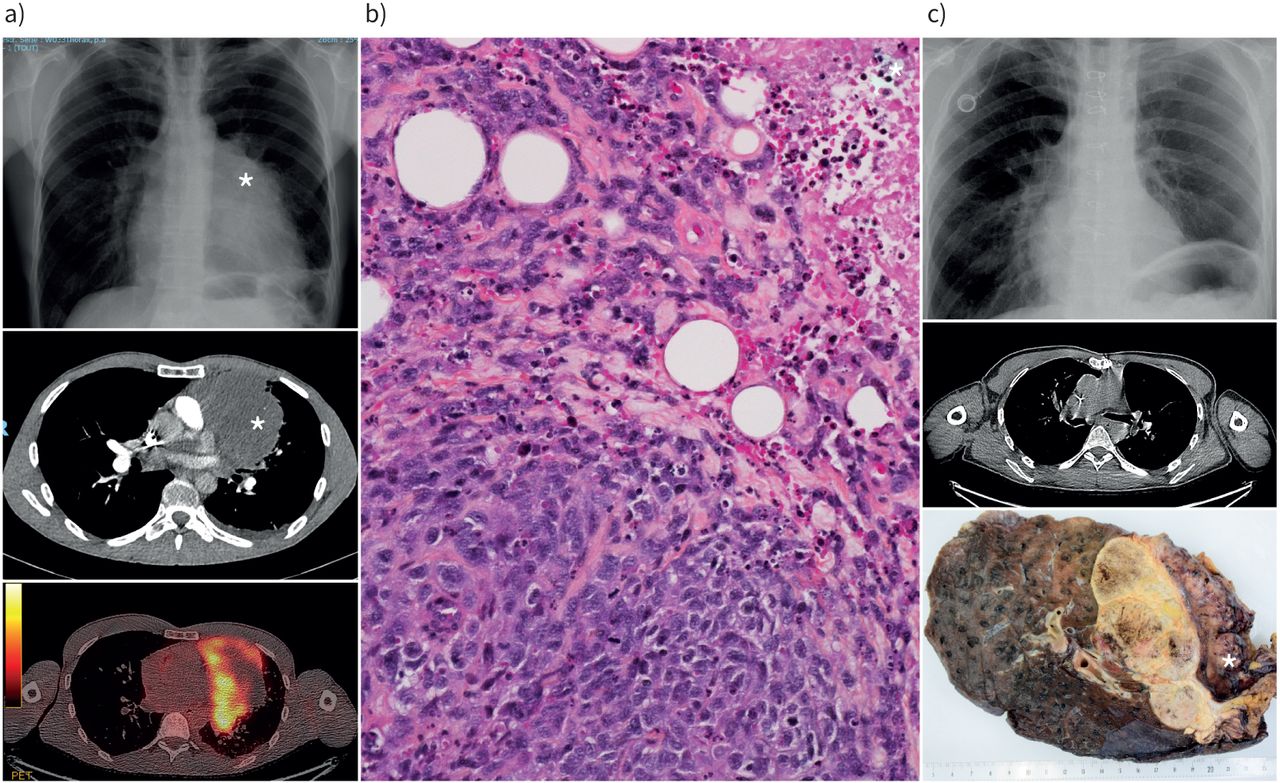
Main menu

  • Home
  • Current issue
  • Past issues
  • Authors/reviewers
    • Instructions for authors
    • Submit a manuscript
    • Institutional open access agreements
    • Peer reviewer login
  • Alerts
  • Subscriptions
  • ERS Publications
    • European Respiratory Journal
    • ERJ Open Research
    • European Respiratory Review
    • Breathe
    • ERS Books
    • ERS publications home

User menu

  • Log in
  • Subscribe
  • Contact Us
  • My Cart

Search

  • Advanced search
  • ERS Publications
    • European Respiratory Journal
    • ERJ Open Research
    • European Respiratory Review
    • Breathe
    • ERS Books
    • ERS publications home

Login

European Respiratory Society

Advanced Search

  • Home
  • Current issue
  • Past issues
  • Authors/reviewers
    • Instructions for authors
    • Submit a manuscript
    • Institutional open access agreements
    • Peer reviewer login
  • Alerts
  • Subscriptions

Mediastinal tumours and pseudo-tumours: a comprehensive review with emphasis on multidisciplinary approach

Maria-Rosa Ghigna, Vincent Thomas de Montpreville
European Respiratory Review 2021 30: 200309; DOI: 10.1183/16000617.0309-2020
Maria-Rosa Ghigna
Dept of Pathology, Marie Lannelongue Hospital, Le Plessis Robinson, France
  • Find this author on Google Scholar
  • Find this author on PubMed
  • Search for this author on this site
  • ORCID record for Maria-Rosa Ghigna
Vincent Thomas de Montpreville
Dept of Pathology, Marie Lannelongue Hospital, Le Plessis Robinson, France
  • Find this author on Google Scholar
  • Find this author on PubMed
  • Search for this author on this site
  • ORCID record for Vincent Thomas de Montpreville
  • For correspondence: v.thomasdemontpreville@hml.fr
  • Article
  • Figures & Data
  • Info & Metrics
  • PDF
Loading

Figures

  • Tables
  • FIGURE 1
    • Download figure
    • Open in new tab
    • Download powerpoint
    FIGURE 1

    Radiological and pathological appearance of a SMARCA4-deficient tumour. a) Initial chest radiograph and computed tomographic (CT) scan showing a mediastino-pulmonary tumour (*) with positivity on positron emission tomography; b) histology of the initial biopsy at medium magnification showing an undifferentiated tumour infiltrating fat, with mitoses and necrosis (*; top right); c) post-chemotherapy chest radiograph and CT scan showing tumour regression and macroscopic appearance of the surgical specimen showing a necrotic tumour infiltrating the left pulmonary upper lobe lung and pericardium (*).

  • FIGURE 2
    • Download figure
    • Open in new tab
    • Download powerpoint
    FIGURE 2

    Radiological and pathological typical features of a multilocular thymic cyst. a) Computed tomographic (CT) scan showing the lesion in the anterior mediastinum; b) positron emission tomography showing a moderate hypermetabolism (standardised uptake value 3.7); c) gross appearance of the multilocular cystic lesion; d) histological appearance at low magnification showing several cavities with thick fibroinflammatory walls.

  • FIGURE 3
    • Download figure
    • Open in new tab
    • Download powerpoint
    FIGURE 3

    Radiological and histological typical features of Müllerian cysts of the mediastinum. a, b) Magnetic resonance imaging showing a paravertebral cystic lesion; c) histological features at low magnification showing a cystic lesion resembling a fallopian tube; d) histological appearance of the ciliated epithelium of the cyst at high magnification; e) immunohistochemistry showing strong oestrogen receptor nuclear expression.

Tables

  • Figures
  • TABLE 1

    Frequencies of sex and age of 942 consecutive primary tumours and cysts of the mediastinum encountered in a 10-year period (2010–2019) at Marie Lannelongue Hospital (Le Plessis Robinson, France)

    CasesSexAge yearsTotal
    FMRangeMean
    Thymic lesions507 (53.8)
     Congenital cyst44 (8.55)202427–8164.10
     Multilocular cyst7 (1.39)4331–6229.61
     Cholesteroloma3 (0.59)0325–5335.07
     Myasthenia gravis138 (27.4)92468–6931.19
     Hyperplasia without myasthenia gravis36 (7.16)20168–7935.82
     Thymolipoma2 (0.4)1120–6140.86
     Thymomas239 (47.5)1211119–9058.43
     Thymic carcinomas27 (5.37)121514–8252.46
     Carcinoid tumours11 (2.19)2928–7253.33
    Nonthymic cysts61 (6.5)
     Bronchogenic20 (32.8)1287–6441.42
     Pleuro-pericardial38 (62.3)19199–7452.25
     Müllerian3 (9.84)3047–5752.09
    Lymphomas10411014–8441.20214 (22.7)
     Diffuse large B-cell81 (37.9)423915–8444.49
     Hodgkin lymphoma110 (51.4)555514–7736.72
     T-lymphoblastic10 (4.67)2818–6345.60
     Miscellaneous13 (6.07)5825–8358.06
    Germinal cell tumours16550–7430.7371 (7.5)
     Teratoma25 (35.2)14110–7428.16
     Malignant nonseminomatous37 (52.1)23515–6531.96
     Seminoma9 (12.7)0920–4832.80
    Benign connective tumours63 (6.7)
     Castleman disease4 (6.35)2230–7745.65
     Lipoma4 (6.35)2255–6159.50
     Angioma10 (15.9)370–7447.89
     Schwannoma25 (39.7)16910–7846.72
     Neurofibroma5 (7.94)2342–5644.37
     Ganglioneuroma6 (9.52)426–6030.03
     Paraganglioma9 (14.3)6315–7853.25
    Miscellaneous malignancies26 (2.8)
     Sarcomas21 (80.8)1296–7943.03
     Undifferentiated malignant tumours5 (19.2)3218–4841.30
    942 (100)

    Data are presented as n (%) or n, unless otherwise stated. F: female; M: male. Bold type represents significant features.

    • TABLE 2

      Frequency of myasthenia gravis and Masaoka–Koga stages according to histological types of thymomas in a series of 239 cases encountered in a 10-year period (2010–2019) at Marie Lannelongue Hospital (Le Plessis Robinson, France)

      CasesMyasthenia gravisMasaoka–Koga
      I/IIIII/IV
      A24 (10)1 (4.2)18 (75)6 (25)
      AB62 (25.9)7 (11)52 (83.9)10 (16.1)
      Micronodular5 (2.09)0 (0)4 (80)1 (20)
      B136 (15.1)15 (42)20 (58.8)14 (41.2)
      B269 (28.9)37 (54)40 (59.7)27 (40.3)
      B340 (16.7)22 (55)10 (25.6)29 (74.4)
      Unclassified3 (1.26)1 (33)1 (100)0 (0)
      Total239 (100)83 (35)145 (62.5)87 (37.5)

      Data are presented as n (%). Bold type represents significant features.

      PreviousNext
      Back to top
      View this article with LENS
      Vol 30 Issue 162 Table of Contents
      European Respiratory Review: 30 (162)
      • Table of Contents
      • Index by author
      Email

      Thank you for your interest in spreading the word on European Respiratory Society .

      NOTE: We only request your email address so that the person you are recommending the page to knows that you wanted them to see it, and that it is not junk mail. We do not capture any email address.

      Enter multiple addresses on separate lines or separate them with commas.
      Mediastinal tumours and pseudo-tumours: a comprehensive review with emphasis on multidisciplinary approach
      (Your Name) has sent you a message from European Respiratory Society
      (Your Name) thought you would like to see the European Respiratory Society web site.
      CAPTCHA
      This question is for testing whether or not you are a human visitor and to prevent automated spam submissions.
      Print
      Citation Tools
      Mediastinal tumours and pseudo-tumours: a comprehensive review with emphasis on multidisciplinary approach
      Maria-Rosa Ghigna, Vincent Thomas de Montpreville
      European Respiratory Review Dec 2021, 30 (162) 200309; DOI: 10.1183/16000617.0309-2020

      Citation Manager Formats

      • BibTeX
      • Bookends
      • EasyBib
      • EndNote (tagged)
      • EndNote 8 (xml)
      • Medlars
      • Mendeley
      • Papers
      • RefWorks Tagged
      • Ref Manager
      • RIS
      • Zotero

      Share
      Mediastinal tumours and pseudo-tumours: a comprehensive review with emphasis on multidisciplinary approach
      Maria-Rosa Ghigna, Vincent Thomas de Montpreville
      European Respiratory Review Dec 2021, 30 (162) 200309; DOI: 10.1183/16000617.0309-2020
      del.icio.us logo Digg logo Reddit logo Technorati logo Twitter logo CiteULike logo Connotea logo Facebook logo Google logo Mendeley logo
      Full Text (PDF)

      Jump To

      • Article
        • Abstract
        • Abstract
        • Introduction
        • Special considerations on biopsy sampling and histological analysis of mediastinal lesions
        • Anterior mediastinum (pre-vascular compartment)
        • Middle mediastinum
        • Posterior mediastinum
        • Conclusion
        • Footnotes
        • References
      • Figures & Data
      • Info & Metrics
      • PDF

      Subjects

      • Lung cancer
      • Tweet Widget
      • Facebook Like
      • Google Plus One

      More in this TOC Section

      Series

      • The oesophageal balloon for respiratory monitoring in ventilated patients
      • QoL in patients with chronic respiratory failure on HMV
      • Awake prone positioning in acute hypoxaemic respiratory failure
      Show more Series

      Thoracic oncology

      • Thymic tumours and their special features
      • Lung cancer is a hereditary disease
      • Immune checkpoint inhibitors in thoracic malignancies
      Show more Thoracic oncology

      Related Articles

      Navigate

      • Home
      • Current issue
      • Archive

      About the ERR

      • Journal information
      • Editorial board
      • Press
      • Permissions and reprints
      • Advertising
      • Sponsorship

      The European Respiratory Society

      • Society home
      • myERS
      • Privacy policy
      • Accessibility

      ERS publications

      • European Respiratory Journal
      • ERJ Open Research
      • European Respiratory Review
      • Breathe
      • ERS books online
      • ERS Bookshop

      Help

      • Feedback

      For authors

      • Instructions for authors
      • Publication ethics and malpractice
      • Submit a manuscript

      For readers

      • Alerts
      • Subjects
      • RSS

      Subscriptions

      • Accessing the ERS publications

      Contact us

      European Respiratory Society
      442 Glossop Road
      Sheffield S10 2PX
      United Kingdom
      Tel: +44 114 2672860
      Email: journals@ersnet.org

      ISSN

      Print ISSN: 0905-9180
      Online ISSN: 1600-0617

      Copyright © 2023 by the European Respiratory Society