
Main menu

  • Home
  • Current issue
  • Past issues
  • Authors/reviewers
    • Instructions for authors
    • Submit a manuscript
    • Institutional open access agreements
    • Peer reviewer login
    • WoS Reviewer Recognition Service
  • Alerts
  • Subscriptions
  • ERS Publications
    • European Respiratory Journal
    • ERJ Open Research
    • European Respiratory Review
    • Breathe
    • ERS Books
    • ERS publications home

User menu

  • Log in
  • Subscribe
  • Contact Us
  • My Cart

Search

  • Advanced search
  • ERS Publications
    • European Respiratory Journal
    • ERJ Open Research
    • European Respiratory Review
    • Breathe
    • ERS Books
    • ERS publications home

Login

European Respiratory Society

Advanced Search

  • Home
  • Current issue
  • Past issues
  • Authors/reviewers
    • Instructions for authors
    • Submit a manuscript
    • Institutional open access agreements
    • Peer reviewer login
    • WoS Reviewer Recognition Service
  • Alerts
  • Subscriptions

Pulmonary alveolar microlithiasis

Patrick Kosciuk, Cristopher Meyer, Kathryn A. Wikenheiser-Brokamp, Francis X. McCormack
European Respiratory Review 2020 29: 200024; DOI: 10.1183/16000617.0024-2020
Patrick Kosciuk
1Division of Pulmonary, Critical Care, and Sleep Medicine, University of Cincinnati, Cincinnati, OH, USA
  • Find this author on Google Scholar
  • Find this author on PubMed
  • Search for this author on this site
Cristopher Meyer
2Division of Radiology, University of Wisconsin, Madison, WI, USA
  • Find this author on Google Scholar
  • Find this author on PubMed
  • Search for this author on this site
Kathryn A. Wikenheiser-Brokamp
3Division of Pathology & Laboratory Medicine, Cincinnati Children's Hospital Medical Center, Cincinnati, OH, USA
4Dept of Pathology & Laboratory Medicine, University of Cincinnati, Cincinnati, OH, USA
  • Find this author on Google Scholar
  • Find this author on PubMed
  • Search for this author on this site
Francis X. McCormack
1Division of Pulmonary, Critical Care, and Sleep Medicine, University of Cincinnati, Cincinnati, OH, USA
  • Find this author on Google Scholar
  • Find this author on PubMed
  • Search for this author on this site
  • ORCID record for Francis X. McCormack
  • For correspondence: frank.mccormack@uc.edu
  • Article
  • Figures & Data
  • Info & Metrics
  • PDF
Loading

Figures

  • Tables
  • FIGURE 1
    • Download figure
    • Open in new tab
    • Download powerpoint
    FIGURE 1

    Radiographic findings in pulmonary alveolar microlithiasis. a) Chest radiograph depicting a fine, sand-like micronodular pattern with basilar predominance. b, c) High-resolution computed tomography with posterior lower lobe and anterior upper lobe micronodules, interlobular septal thickening, subpleural emphysema with predominance of small cysts. d) Mediastinal windows reveal calcific burden in the parenchyma and most concentrated in a peripheral septopleural location.

  • FIGURE 2
    • Download figure
    • Open in new tab
    • Download powerpoint
    FIGURE 2

    Pathologic findings in pulmonary alveolar microlithiasis (PAM). a) Lung explant from a 2-year-old boy transplanted for PAM showing lung regions with accentuation of the interlobular septa by microlith accumulations (arrow) and other regions with more diffuse accumulations of granular, gritty microliths (*). b, c) Histological sections showing microliths along interlobular septa (b, arrow) and areas with more diffuse microlith accumulations (c). d) Varying sized microliths are present both in the alveolar spaces and interstitium. e) The microliths are characteristically concentrically laminated calcified spherules. f) Calcium can be demonstrated in the microliths by Von Kossa stain. b–e) Haematoxylin and eosin stain; original magnifications ×20 (b, c), ×200 (d), ×1000 (e, f).

  • FIGURE 3
    • Download figure
    • Open in new tab
    • Download powerpoint
    FIGURE 3

    Scanning electron microscopy (SEM) and energy-dispersive spectroscopy (EDAX) of pulmonary alveolar microlithiasis (PAM). a, b) Microliths isolated from explanted lungs demonstrating a) the characteristic spherical structure and b) inorganic elemental signature. Microlith (white arrow) in bronchoalveolar lavage (BAL) fluid with SEM (c) and EDAX characteristics that are similar to explant controls, demonstrating feasibility of using SEM and EDAX as a diagnostic BAL test for PAM. Note carbon spike associated with debris from lavage. Kα, Kβ and Lα represent X-rays emitted as electrons return to K and/or L electron shell. d) Inorganic elemental analysis of BAL microliths.
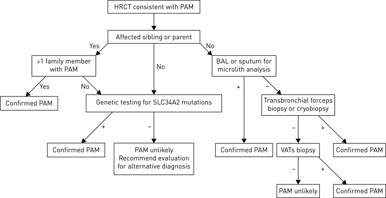
  • FIGURE 4
    • Download figure
    • Open in new tab
    • Download powerpoint
    FIGURE 4

    Diagnostic algorithm for patients with suspected pulmonary alveolar microlithiasis (PAM). Obtaining a family and genetic testing history is key. Thereafter, diagnostic modalities progress from least invasive to most invasive. HRCT: high-resolution computed tomography; BAL: bronchoalveolar lavage; VAT: video-assisted thoracoscopic biopsy.

  • FIGURE 5
    • Download figure
    • Open in new tab
    • Download powerpoint
    FIGURE 5

    Other examples of diffuse lung calcification. Computed tomography depicting examples of a, d) amyloidosis with calcified masses, lymphadenopathy and rare cysts, b, e) silicosis with peri-lymphatic calcified nodules, eggshell lymph node calcifications and conglomerate fibrosis, c) metastatic pulmonary calcification in chronic renal failure characterised by lobular ground-glass opacity with interlobular septal sparing and f) healed granulomas due to histoplasmosis with maximum intensity projection reformats.

Tables

  • Figures
  • TABLE 1

    Pathogenetic mutations

    ExonSequence involvedDefectFirst author [ref.]
    1–13195 kb deletionTruncation without synthesis (deletion)Stokman [15]
    1c.-6773_-6588delTruncation without synthesis (deletion)Corut [9]
    2–65.5 kb deletionTruncation (deletion)Ishihara [16]
    2insT (not specified area)Truncation (frameshift)Dogan [17]
    3c.114delATruncation (deletion)Corut [9]
    3c.212_224delTruncation (deletion)Vismara [18]
    3c.226 C>TSubstitutionCorut [9]
    4c.316 G>ASubstitution (missense)Jonsson [19]
    5c.IVS4+1452_IVS5+660delTruncation (deletion) of entire exon 5Dandan [20]
    6c.560 G>ASubstitution (missense)Jonsson [19]
    6c.575 C>ASubstitutionMa [21]
    7c.560 G>ASubstitution (nonsense)Jonsson [19]
    7insdel857-871Insertion/deletion with truncationHuqun [8]
    8IVS8+1 G>ATruncation by splicing failureHuqun [8]
    8c.906 G>ASubstitution (nonsense)Jonsson [19]
    8c.910 A>TTruncationZhong [22]
    10c.1136 G>ASubstitution (missense)Jonsson [19]
    11c.1238 G>ASubstitution (nonsense)Jonsson [19]
    11c.1327delCTruncation (deletion)Jonsson [19]
    11c.1328delTTruncation (deletion)Corut [9]
    12c.1333+1 G>ASubstitution (nonsense, frameshift splicing)Jonsson [19]
    12c.1342delGTruncation (deletion)Corut [9]
    12c.1363 T>CSubstitutionWang [23]
    12c.1390 G>CUnclear resultIzumi [24]
    12c.1393-1404delACCTruncation, threonine deletionJonsson [25]
    12c.1402-1404delACCTruncation, threonine deletionJonsson [25]
    12c.1456 C>TTruncationProesmans [26]
  • TABLE 2

    Lung transplantation in cases of pulmonary alveolar microlithiasis

    Single versus doubleAge at transplant yearsSexOutcomeComplication(s)First author [ref.]
    Single32MaleDeath, NRPGD, haemodynamic instabilityShadmehr [106]
    Single47FemaleAlive, 12 monthsPossible acute rejection, bronchial strictureRaffa [87]
    Single53MaleAlive, 12 monthsBronchial anastomosis granulation with stenosis, bacterial infectionRen [34]
    Single53FemaleAlive, 90 monthsNoneJackson [107]
    Single64FemaleAlive, 60 monthsNoneBorrelli [108]
    Double32FemaleDeath, 11 daysPGD, sepsisKlikovits [35]
    Double32MaleAlive, 18 monthsBronchial artery bleed, post-operative tracheostomy, CMV/fungal infectionsStamatis [27]
    Double34MaleAlive, 67 monthsNoneKlikovits [35]
    Double36FemaleAlive, 32 monthsNoneEdelman [33]
    Double45MaleAlive, 12 monthsMild PGDAlrossais [36]
    Double46FemaleDeath, 20 monthsBronchiolitis obliteransBonnette [109]
    Double48MaleAlive, 12 monthsPGD, haemodynamic instability, ARFSamano [57]
    Double49FemaleDeath, 3 monthsInfectionCoulibaly [110]
    Double52FemaleAlive, 35 monthsNoneKlikovits [35]
    Double52FemaleAlive, 74 monthsPGD, atrial fibrillationKlikovits [35]
    Double53FemaleAlive, 12 monthsNoneGucyetmez [111]
    Double54FemaleAlive, 12 monthsNoneJindal [112]
    Double56MaleDeath, 5 daysPost-operative bleedingEdelman [33]
    Double62FemaleAlive, 29 monthsAtrial fibrillationKlikovits [35]
    Double63FemaleAlive, 24 monthsNoneShigemura [113]

    NR: not reported; PGD: primary graft dysfunction; CMV: cytomegalovirus; ARF: acute renal failure.

    PreviousNext
    Back to top
    View this article with LENS
    Vol 29 Issue 158 Table of Contents
    • Table of Contents
    • Index by author
    Email

    Thank you for your interest in spreading the word on European Respiratory Society .

    NOTE: We only request your email address so that the person you are recommending the page to knows that you wanted them to see it, and that it is not junk mail. We do not capture any email address.

    Enter multiple addresses on separate lines or separate them with commas.
    Pulmonary alveolar microlithiasis
    (Your Name) has sent you a message from European Respiratory Society
    (Your Name) thought you would like to see the European Respiratory Society web site.
    CAPTCHA
    This question is for testing whether or not you are a human visitor and to prevent automated spam submissions.
    Print
    Citation Tools
    Pulmonary alveolar microlithiasis
    Patrick Kosciuk, Cristopher Meyer, Kathryn A. Wikenheiser-Brokamp, Francis X. McCormack
    European Respiratory Review Dec 2020, 29 (158) 200024; DOI: 10.1183/16000617.0024-2020

    Citation Manager Formats

    • BibTeX
    • Bookends
    • EasyBib
    • EndNote (tagged)
    • EndNote 8 (xml)
    • Medlars
    • Mendeley
    • Papers
    • RefWorks Tagged
    • Ref Manager
    • RIS
    • Zotero

    Share
    Pulmonary alveolar microlithiasis
    Patrick Kosciuk, Cristopher Meyer, Kathryn A. Wikenheiser-Brokamp, Francis X. McCormack
    European Respiratory Review Dec 2020, 29 (158) 200024; DOI: 10.1183/16000617.0024-2020
    del.icio.us logo Digg logo Reddit logo Technorati logo Twitter logo CiteULike logo Connotea logo Facebook logo Google logo Mendeley logo
    Full Text (PDF)

    Jump To

    • Article
      • Abstract
      • Abstract
      • Introduction
      • Epidemiology
      • Pathogenesis
      • Signs and symptoms
      • Pulmonary function tests
      • Diagnostics
      • Radiology
      • Pathology
      • Extrapulmonary manifestations
      • Comorbidities
      • Diagnosis
      • Differential diagnosis
      • Pharmacological treatment options
      • Prognosis
      • Future directions
      • Footnotes
      • References
    • Figures & Data
    • Info & Metrics
    • PDF

    Subjects

    • Interstitial and orphan lung disease
    • Tweet Widget
    • Facebook Like
    • Google Plus One

    More in this TOC Section

    Series

    • The role of vaccination in COPD
    • Pulmonary rehabilitation and physical interventions
    • The role of diet and nutrition in the management of COPD
    Show more Series

    Ultra-rare lung disease

    • Ultra-rare cystic disease
    • Pulmonary capillary haemangiomatosis
    Show more Ultra-rare lung disease

    Related Articles

    Navigate

    • Home
    • Current issue
    • Archive

    About the ERR

    • Journal information
    • Editorial board
    • Press
    • Permissions and reprints
    • Advertising
    • Sponsorship

    The European Respiratory Society

    • Society home
    • myERS
    • Privacy policy
    • Accessibility

    ERS publications

    • European Respiratory Journal
    • ERJ Open Research
    • European Respiratory Review
    • Breathe
    • ERS books online
    • ERS Bookshop

    Help

    • Feedback

    For authors

    • Instructions for authors
    • Publication ethics and malpractice
    • Submit a manuscript

    For readers

    • Alerts
    • Subjects
    • RSS

    Subscriptions

    • Accessing the ERS publications

    Contact us

    European Respiratory Society
    442 Glossop Road
    Sheffield S10 2PX
    United Kingdom
    Tel: +44 114 2672860
    Email: journals@ersnet.org

    ISSN

    Print ISSN: 0905-9180
    Online ISSN: 1600-0617

    Copyright © 2023 by the European Respiratory Society