
Main menu

  • Home
  • Current issue
  • Past issues
  • Authors/reviewers
    • Instructions for authors
    • Submit a manuscript
    • COVID-19 submission information
    • Institutional open access agreements
    • Peer reviewer login
  • Alerts
  • Subscriptions
  • ERS Publications
    • European Respiratory Journal
    • ERJ Open Research
    • European Respiratory Review
    • Breathe
    • ERS Books
    • ERS publications home

User menu

  • Log in
  • Subscribe
  • Contact Us
  • My Cart

Search

  • Advanced search
  • ERS Publications
    • European Respiratory Journal
    • ERJ Open Research
    • European Respiratory Review
    • Breathe
    • ERS Books
    • ERS publications home

Login

European Respiratory Society

Advanced Search

  • Home
  • Current issue
  • Past issues
  • Authors/reviewers
    • Instructions for authors
    • Submit a manuscript
    • COVID-19 submission information
    • Institutional open access agreements
    • Peer reviewer login
  • Alerts
  • Subscriptions

Airway mechanical compression: its role in asthma pathogenesis and progression

Punnam Chander Veerati, Jennifer A. Mitchel, Andrew T. Reid, Darryl A. Knight, Nathan W. Bartlett, Jin-Ah Park, Chris L. Grainge
European Respiratory Review 2020 29: 190123; DOI: 10.1183/16000617.0123-2019
Punnam Chander Veerati
1School of Medicine and Public Health, University of Newcastle, Callaghan, Australia
2Priority Research Centre for Healthy Lungs, Hunter Medical Research Institute, University of Newcastle, New Lambton Heights, Australia
  • Find this author on Google Scholar
  • Find this author on PubMed
  • Search for this author on this site
  • ORCID record for Punnam Chander Veerati
Jennifer A. Mitchel
3Molecular and Integrative Physiological Sciences Program, Dept of Environmental Health, Harvard T.H. Chan School of Public Health, Boston, MA, USA
  • Find this author on Google Scholar
  • Find this author on PubMed
  • Search for this author on this site
Andrew T. Reid
1School of Medicine and Public Health, University of Newcastle, Callaghan, Australia
2Priority Research Centre for Healthy Lungs, Hunter Medical Research Institute, University of Newcastle, New Lambton Heights, Australia
  • Find this author on Google Scholar
  • Find this author on PubMed
  • Search for this author on this site
Darryl A. Knight
2Priority Research Centre for Healthy Lungs, Hunter Medical Research Institute, University of Newcastle, New Lambton Heights, Australia
4School of Biomedical Sciences and Pharmacy, University of Newcastle, Callaghan, Australia
5Dept of Anesthesiology, Pharmacology and Therapeutics, University of British Columbia, Vancouver, Canada
6Research and Academic Affairs, Providence Health Care Research Institute, Vancouver, Canada
  • Find this author on Google Scholar
  • Find this author on PubMed
  • Search for this author on this site
Nathan W. Bartlett
2Priority Research Centre for Healthy Lungs, Hunter Medical Research Institute, University of Newcastle, New Lambton Heights, Australia
4School of Biomedical Sciences and Pharmacy, University of Newcastle, Callaghan, Australia
  • Find this author on Google Scholar
  • Find this author on PubMed
  • Search for this author on this site
  • ORCID record for Nathan W. Bartlett
Jin-Ah Park
3Molecular and Integrative Physiological Sciences Program, Dept of Environmental Health, Harvard T.H. Chan School of Public Health, Boston, MA, USA
  • Find this author on Google Scholar
  • Find this author on PubMed
  • Search for this author on this site
Chris L. Grainge
1School of Medicine and Public Health, University of Newcastle, Callaghan, Australia
2Priority Research Centre for Healthy Lungs, Hunter Medical Research Institute, University of Newcastle, New Lambton Heights, Australia
7Dept of Respiratory and Sleep Medicine, John Hunter Hospital, Newcastle, Australia
  • Find this author on Google Scholar
  • Find this author on PubMed
  • Search for this author on this site
  • For correspondence: christopher.grainge@health.nsw.gov.au
  • Article
  • Figures & Data
  • Info & Metrics
  • PDF
Loading

Abstract

The lung is a mechanically active organ, but uncontrolled or excessive mechanical forces disrupt normal lung function and can contribute to the development of disease. In asthma, bronchoconstriction leads to airway narrowing and airway wall buckling. A growing body of evidence suggests that pathological mechanical forces induced by airway buckling alone can perpetuate disease processes in asthma. Here, we review the data obtained from a variety of experimental models, including in vitro, ex vivo and in vivo approaches, which have been used to study the impact of mechanical forces in asthma pathogenesis. We review the evidence showing that mechanical compression alters the biological and biophysical properties of the airway epithelium, including activation of the epidermal growth factor receptor pathway, overproduction of asthma-associated mediators, goblet cell hyperplasia, and a phase transition of epithelium from a static jammed phase to a mobile unjammed phase. We also define questions regarding the impact of mechanical forces on the pathology of asthma, with a focus on known triggers of asthma exacerbations such as viral infection.

Abstract

Bronchoconstriction in asthma is not only a symptom but is also a disease modifier. Targeting downstream mediators of mechanical force induction may provide opportunities for novel therapies for asthma and its exacerbations. http://bit.ly/2UktQDj

Introduction

The lung is a mechanically active organ; in utero, lung development is dependent on the presence of controlled mechanical forces [1–5] and, following birth, lung movement continues unabated until death. This ever moving and changing nature of the lungs has been apparent since before Heraclitus, although it has only recently become apparent that alterations in the mechanical forces present in the lungs can generate and propagate lung disease [6–10].

During lung growth, mechanical forces are critical to alveologenesis and branching morphogenesis [1–5]. Even in utero, the fetal lung is on the move; breathing movements generated by diaphragmatic contractions leading to cyclic mechanical stretch are fundamental to maintaining airway luminal liquid volume, facilitating cellular proliferation and driving fetal lung expansion [11, 12]. At birth, fluid within the alveoli clears as the lung is aerated and expanded, developing alveolar surface tension [4], and from that moment lung elastic recoil is balanced by negative intrapleural pressure; a combination of mechanical forces at this point leads to postpartum lung maturation [13, 14]. Absence of stretch and mechanical force generation within the lung leads to abnormal alveolar growth and differentiation, especially of type II alveolar epithelial cells, reducing surfactant release and impairing gas exchange [15, 16].

For respiration to take place, the mature lung has to dynamically inflate and deflate; these repeated cycles of inflation and deflation generate mechanical forces, which are required for normal lung function. While maintenance of normal physiological force is essential for respiration, abnormal or uncontrolled mechanical forces generated in the airways can compromise normal lung function and result in disease pathogenesis [10, 17]. Although the impact of mechanical forces is becoming more understood in a variety of lung diseases, this review focuses on their impact in asthma.

Uncontrolled mechanical forces in lung pathophysiology

Airway narrowing (bronchoconstriction) occurs in asthma due to the contraction of airway smooth muscle (ASM) [18], leading to many asthma symptoms, including shortness of breath, chest tightness, cough and wheeze [19, 20]. As a result of bronchoconstriction, the airway wall in asthmatics had been thought to fold, buckle and form rosette patterns, and although this had been modelled extensively in silico and in vitro [6, 21, 22] it has only recently been observed in vivo [23]. Using optical coherence tomography (OCT) via a fibreoptic bronchoscope in human asthmatic and non-asthmatic volunteers, Adams et al. [23] documented airway folding following bronchoconstriction induced by experimental segmental allergen instillation, not only immediately following allergen instillation, but also at later time-points. The airway folding demonstrated by OCT showed remarkably similar airway changes to those previously predicted by in silico modelling and those found in pathological specimens of airways from fatal episodes of asthma [22, 24]. As the airway wall folds, the epithelium experiences multiple mechanical forces, including compression (as adjacent cells push against each other), stretch (of cells at the apex of epithelial folds) and shear stress (as air velocity increases in conjunction with a reduction in airway diameter) [5, 22]. Of these forces, compression has been most studied using a number of in vitro models which allow cultured epithelial cells to be subjected to compressive stress mimicking the mechanical effect of bronchoconstriction [6, 25–28]. These studies have demonstrated that mechanical forces can contribute to changes within the epithelial and other structural cells of the airway, changes that are consistent with features seen in asthma despite the absence of inflammatory stimuli. Application of mechanical compression in vitro recapitulates key features of asthmatic airway remodelling, including goblet cell hyperplasia, collagen deposition [6, 9, 29], and hyperplasia and hypercontraction of ASM cells [30], as well as inducing in normal cells a gene signature normally associated with asthmatic cells [31]. These studies offer a potential alternate or parallel pathway of asthma pathogenesis outside the accepted dogma of inflammation of a variety of subtypes (i.e. T-helper (Th)2 high, Th2 low etc.) driving disease progression, whereas bronchoconstriction results in a cascade of disease-initiating or disease-amplifying events, which may act in concert with, or independent of, inflammatory cells [32–35].

Modelling mechanical compression

Although the magnitude of the mechanical forces exerted on the epithelium during bronchoconstriction cannot be directly measured, it can be calculated using a finite element method which integrates both material properties and geometry of the airways [22]. This model uses two layers to describe the airway, representing an inner basement membrane combined with epithelium and an outer ASM layer (figure 1a). Constriction of the ASM layer leads to inner layer folding and, by increasing the thickness of the inner basement layer, mimicking the thickened sub-epithelial collagen in asthmatic airways, results in larger epithelial folds and airway occlusion [22]. As mentioned previously, this folding and occlusion appear similar to that seen both in airways of patients who suffer fatal asthma attacks (figure 1b) [24], and recently observed to a lesser degree via OCT (figure 1c) [23].

FIGURE 1
  • Download figure
  • Open in new tab
  • Download powerpoint
FIGURE 1

a) Schematic diagram showing buckling of airways with epithelial folding. Reproduced and modified from [22] with permission. b) Pathological specimen from fatal asthma with epithelial folding and luminal closure. Reproduced and modified from [24] with permission. c) Bronchoconstriction-induced airway wall folding following allergen challenge observed using optical coherence tomography. Reproduced and modified from [23] with permission.

The estimated apical pressure that is exerted on airway epithelial cells during bronchoconstriction is approximately 30 cmH2O pressure, which is an order of magnitude higher than transpulmonary pressure during tidal breathing (∼5 cmH2O) [22, 36]. Studies using canine airways also reported a similar transmural bronchial pressure in both large and small airways following acetylcholine-induced bronchoconstriction [37] and, as epithelial responses to apical compression plateau at 30 cmH2O pressure [6, 29], this force has been widely used to recapitulate the effects of asthmatic bronchoconstriction in vitro [7–9, 28, 38–41].

Experimental models for mechanical compression

In vitro model: air–liquid interface cultures

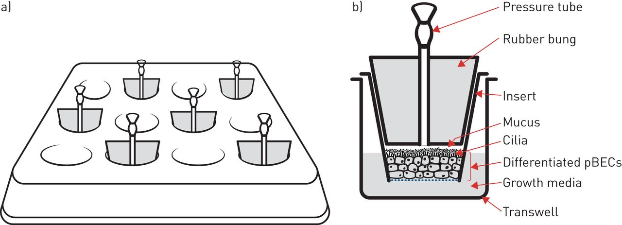
In vitro investigation of airway mechanical forces has primarily utilised human primary bronchial epithelial cells (pBECs) cultured at the air–liquid interface. Here, pBECs differentiate from a monolayer of basal cells to a pseudostratified structure containing goblet- and ciliated-cells to closely replicate the mature bronchial epithelium [42–44]. This well-differentiated airway epithelium is then exposed to a transcellular pressure gradient up to 30 cmH2O pressure (figure 2) to recapitulate the forces experienced during bronchoconstriction [22]. In addition to this apical stress model, another, more technically complex apparatus utilising lateral compressive stress has reported similar responses to apical compression [26, 27].

FIGURE 2
  • Download figure
  • Open in new tab
  • Download powerpoint
FIGURE 2

Apical compression system in vitro. a) Schematic of apical compression set-up on a 12-well transwell plate. b) Schematic of a frontal plane section of a single transwell insert enclosed by a bung with a pressure tube. pBEC: primary bronchial epithelial cell.

Mechanical compression of well-differentiated pBECs induces the release of growth factors into the lateral intercellular space between cells [38]. At the same time, mechanical compression collapses the lateral intercellular space without altering cell volume, this reduction in the volume of the lateral intercellular space results in an increase in the local concentration of epidermal growth factor (EGF) with consequent activation of the EGF receptor (EGFR) [38]. This pathway appears to mediate some of the changes, such as mucus secretion, that occur following modelled bronchoconstriction.

Ex vivo: precision-cut lung slices and whole lung perfusion models

In addition to pBEC monolayer in vitro models, epithelial compression during bronchoconstriction has been studied ex vivo using precision-cut lung slices (PCLS) and whole lung perfusion models, providing additional evidence in support of in vitro data. In PCLS, normal tissue architecture is maintained along with cellular communication between different cell types, albeit in thin lung slices; these slices can be generated from many animals, including humans [45–52]. Airways in PCLS exhibit bronchoconstriction and formation of a buckled epithelium following stimulation with agents such as methacholine or histamine [53] in a similar manner to isolated whole lung perfusion, where isolated lungs are generally perfused with a methacholine solution by puncturing the lung apex with multiple holes [38]. Though only a handful of studies have used PCLS or whole lung perfusion to examine the effects of bronchoconstriction, these techniques provide similar results to those from pBEC in vitro work (as discussed in the section Mechanical compression activates disease-associated pathways in the airway epithelium), supporting their conclusions.

In vivo: bronchoprovocation/allergen challenge models

Despite data from well-established in vitro and ex vivo models of airway compressive stress, until recently the question remained whether bronchoconstriction-induced epithelial buckling and mechanical stress actually occur in the human airway during an asthma exacerbation. It appears that these changes are likely to occur in real-world asthma; in addition to real-time OCT data [23], biopsies from patients with mild asthma undergoing repeated bronchoconstriction show pathological airway remodelling (structural changes) such as goblet cell hyperplasia and increased collagen deposition, in the absence of additional airway inflammation [10]. These remodelling changes were prevented by the administration of bronchodilators and inhaled corticosteroids during airway challenges [54, 55]. The threshold or number of bronchoconstriciton events required to induce any long-term changes in the airway is unknown. Participants with mild asthma in clinical studies who underwent a variety of bronchoprovocation tests, over a period of in some cases several years, did not show any unexpected fall in their forced expiratory volume in 1 s [56]. This could be that the frequency of challenge was too low, and the background asthma too mild to induce long-term changes, or that the changes observed after repeated allergen or methacholine challenge are transient, especially in otherwise well-controlled patients.

Data from animal models have also contradicted some of the human data. For example, mice exposed to a 30 min methacholine challenge at 48 h intervals over 6 weeks demonstrated airway remodelling only in terms of goblet cell hyperplasia and some mild macrophage-induced airway inflammatory response [57]. There was no effect on respiratory mechanics or the contractile capacity of ASM. These species differences mean that animal data must be interpreted with care when investigating the role of mechanical forces in human airway diseases.

Mechanical compression activates disease-associated pathways in the airway epithelium

Mechanical compression mimicking the mechanical environment of constricted airways recapitulates features of asthmatic airways, in addition to the activation of multiple signalling pathways leading to the production of asthma-associated mediators (figure 3). These pathways and mediators include the activation of EGFR, transforming growth factor (TGF)-β protein release, as well as procoagulant factors, and less well-understood compounds including YKL-40 and maspin.

FIGURE 3
  • Download figure
  • Open in new tab
  • Download powerpoint
FIGURE 3

Schematic image of the effects of airway mechanical compression on airway epithelial cells and remodelling. Dotted lines indicate the mediator's effect on airway remodelling components. ASM: airway smooth muscle; EGFR: epidermal growth factor receptor; EGR-1: early growth response-1; HB-EGF: heparin-binding epidermal growth factor-like growth factor; PAI-1: plasminogen activator inhibitor-1; PAR: plasminogen activator receptor; TGF-α: transforming growth factor-α; TGF-β2: transforming growth factor-β2; uPA: urokinase plasminogen activator.

EGFR activation

Mechanical compression of well-differentiated epithelial cells results in the activation of EGFR and downstream signalling pathways [38, 58, 59]. For example, compressive stress induces phosphorylation of extracellular-regulated kinase (ERK) which upregulates heparin-binding EGF-like growth factor (HB-EGF) gene expression [60]; HB-EGF induction is EGFR-dependent, being activated via an autocrine positive feedback mechanism [41]. In mouse tracheal epithelial cells, activation of EGFR is mediated by EGF and induces ERK and protein kinase B (Akt) phosphorylation while this activation further induces the expression of the EGFR family of ligands, such as HB-EGF, TGF-α, amphiregulin, epiregulin and betacellulin. These findings are supported by in vivo studies where methacholine-induced airway constriction of mouse lungs also leads to the release of EGFR family ligands, with later data demonstrating that mechanical activation of EGFR requires active TNF-α converting enzyme (TACE) [38, 59].

TGF-β

TGF-β is a pleotropic cytokine with three different mammalian isoforms (TGF-β1–3) which plays an important role in regulating inflammation, cell growth and differentiation [61]. All three isoforms are expressed in the airways, but localise to specific cell types: TGF-β1: ASM, fibroblasts and endothelial cells; TGF-β2: airway epithelial cells; and TGF-β3: macrophages [62–68]. In many studies of asthma, TGF-β expression is elevated in the airways in comparison to healthy controls and induces airway hyperresponsiveness, as well as airway remodelling by increasing the number of goblet cells, mucus secretion and thickened sub-epithelial basement membrane by collagen or extracellular matrix (ECM) deposition [67, 69]. In contrast, some studies highlight no difference in isoform-specific TGF-β expression in asthma and healthy controls, especially with respect to TGF-β1 expression [66, 68, 70, 71]. These observed differences may be due to dichotomous expression of TGF-β1 to TGF-β2 and TGF-β3 or potentially the specific asthma phenotype or techniques used to detect TGF-β1 expression.

Despite these caveats, it has been shown that following mechanical compression TGF-β is released from in vitro pBEC cultures and plays a pivotal role in goblet cell hyperplasia, sub-epithelial collagen deposition and fibrosis [7, 9, 10]. Initial work in differentiated rat tracheal epithelial cells demonstrated increased TGF-β1 gene expression in response to mechanical compression in a time- and pressure-dependent manner. In well-differentiated human pBECs, TGF-β2, but not TGF-β1, protein expression was increased in response to compression [7]. pBECs derived from patients with asthma released more TGF-β compared with healthy controls following apical mechanical compression [28].

In guinea pig PCLS, methacholine-induced bronchoconstriction-induced biologically active TGF-β release from airway epithelial cells, promoting expression of ASM contractile proteins (smooth muscle-α-actin, smooth muscle-myosin, calponin) [50]. The TGF-β response was inhibited by preventing bronchoconstriction using latrunculin A to prevent actin polymerisation, and was also reduced by inhibiting TGF-β receptor kinase or phosphodiesterase-4 and also by the administration of long-acting muscarinic receptor antagonists [50, 72]. Finally, in human studies, repeated bronchoconstriction of mild asthmatic patients showed elevated epithelial TGF-β expression compared with saline-treated individuals, along with changes in other mediators in bronchial biopsies [10]. Together, these studies demonstrate that TGF-β in airway epithelial cells is responsive to mechanical compression associated with bronchoconstriction as well as being linked to airway remodelling and airway hyperresponsiveness [73], and may provide a mechanistic connection between epithelial compression and asthma pathogenesis.

Procoagulant factors

Fibrin degradation products and the coagulation cascade are activated in the days preceding admission to hospital for an asthma exacerbation, and the degree of activation is associated with length of stay for those exacerbations [74, 75]. Coagulation cascades involving the activation of plasminogen systems, including urokinase plasminogen activator receptor, urokinase plasminogen activator, plasminogen activator inhibitor-1 and amphiregulin are upregulated after epithelial cell compression [8]. Tissue factor, a well-recognised coagulation initiator, is expressed in human airway epithelial cells; both gene expression and protein release are increased following compression [39]. Tissue factor protein is released via extracellular vesicles, and this release is augmented by interleukin (IL)-13, a type-2 cytokine, elevated in allergic asthma [76]. Tissue factor is expressed in pre-clinical models of allergic airways disease and is detected in bronchoalveolar lavage fluid from ovalbumin-challenged allergic mice. At baseline, tissue factor levels are higher in asthmatics compared with healthy controls and increase further in asthma in response to allergen challenge [39, 76]. It may be that bronchoconstriction, in combination with the underlying state of airway inflammation, directly leads to clotting cascade activation. It also appears that bronchoconstriction interacts with other disease processes in asthma and this interaction could provide a target for novel interventions.

YKL-40 and maspin

Genome-wide association studies have associated CHI3L1, the gene which encodes the YKL-40 protein, with increased asthma prevalence or susceptibility in multiple populations [77–80]. Higher expression of CHI3L1 in the lung correlates with higher concentrations of YKL-40 in serum and bronchoalveolar lavage fluid [81], which are themselves associated with reduced lung function [77]. More recent evidence suggested that YKL-40 plays a role in airway remodelling, including airway angiogenesis [82, 83] and ASM proliferation [84]. Compression of pBECs induces YKL-40 protein secretion, mediated through the activation of the EGFR-ERK pathway [40], although the relationship between CHI3L1, as an asthma susceptibility gene, and the secretion of YKL-40 protein from compressed airway epithelial cells is not yet clear. In addition to YKL-40, another protein which contributes to tissue remodelling, maspin, is also released from epithelial cells following apical compression or allergen exposure [85]. Maspin, a serine protease inhibitor, originally identified as a tumour suppressor protein in human mammary epithelial cells, contributes to tissue remodelling by inhibition of collagen degradation and alteration in cell–cell interactions [86–88]. The interaction of bronchoconstriction with genetic susceptibility to disease and proteins responsible for tissue remodelling will be a novel area for investigation.

Mechanical compression induces airway remodelling

Remodelling of the airways in asthma is characterised by increased goblet cell number, mucus hypersecretion, sub-epithelial collagen deposition and an increased ASM mass [18, 89, 90]. The driving factors behind, and clinical effect of, these changes are not clear, but are thought to be deleterious; mucus secretion leads to airway plugging and closure, collagen deposition contributes to fixed airway obstruction, and ASM hypertrophy contributes to dynamic airway narrowing and closure.

Goblet cell hyperplasia and MUC5AC protein expression

Goblet cells are non-ciliated secretory airway epithelial cells which secrete glycoprotein rich mucus into the airway lumen. This mucus forms a barrier to environmental insults and increases the airway's ability to clear pathogens or pollutants by mucociliary action [91–93]. In asthma, goblet cells increase in number and secrete more mucus; these increases are associated with an increase in asthma morbidity and mortality [89, 94–96]. This increased goblet cell number and mucus production is associated with increased Th2 cytokines (IL-4, IL-5 and IL-13) and human neutrophil elastase in the airways [97–99]. Goblet cell numbers are further increased in vivo following repeated experimentally induced bronchoconstriction in mild asthmatics and in vitro by apical compression of pBECs for only 1 h a day for 14 days [9, 10]. Compression applied in vitro leads to an increase in MUC5AC protein expression in addition to goblet cell hyperplasia which is decreased following pre-treatment of pBECs with an EGFR kinase inhibitor. EGFR inhibition in combination with a TGF-β2 neutralising antibody completely prevents this effect [9]. Although the mechanisms of goblet cell hyperplasia are not well understood, and may include the Notch3 pathway [100], this phenomenon has been shown to occur in the healthy mouse lung following repeated methacholine challenge [57].

Sub-epithelial collagen deposition

In addition to mucus production, thickening of ECM is another feature of asthmatic airway remodelling [69, 101]. ECM deposition occurs at the basolateral surface of the bronchial epithelium and consists, at least in part, of collagens (type I, III and IV) as well as fibronectin [69, 101–103]; asthmatic airways have measured ECM thicknesses of ∼9 µm in comparison to 5–6 µm for non-asthmatics [104]. Compressive stress promotes airway remodelling by inducing the deposition of sub-epithelial collagen and fibronectin [26, 29], this appears to be a result of epithelial cells responding to mechanical stress and releasing cellular factors which induce proliferation of fibroblasts and increased production of collagens (types III and V) [29]. In addition to the ECM deposition observed in this model, there was also an increase in matrix metalloproteinase-2 and -9 production [27, 29].

ASM proliferation and contraction

The ASM in asthmatic airways is characterised by increased mass and increased responsiveness to stimuli, which together result in excessive contraction [105–107]. Although the mechanism via which ASM becomes more responsive to contractile stimuli is unclear; it has been demonstrated that airway epithelial cells can augment ASM contraction and proliferation [108–110] via mediators such as endothelin (ET)-1 and YKL-40 [84, 111, 112]. Both these mediators (ET-1 and YKL-40) have been shown to be increased following the application of apical mechanical stress on epithelial cells in culture [7, 40] with condition media from those epithelial cells inducing ASM hypertrophy which was blocked by ET-1 antagonists, while ASM proliferation was also increased, but through an ET-1 independent mechanism [30]. These data indicate that mechanical compression of airway epithelial cells can elicit a response in the ASM and potentially generate a positive feedback loop that may be a critical therapeutic target for arresting asthma progression.

Mechanical compression induces epithelial cell unjamming

Collective cellular migration

It is widely recognised that airway epithelial cells migrate during wound repair; however, recently it has also been demonstrated that unwounded well-differentiated airway epithelial cells also can migrate in vitro [113]. Normally, in vitro, airway epithelial cells migrate relative to each other within the culture environment during their growth and differentiation phase, “locking” into a fixed arrangement as they reach maturity at the air–liquid interface [114]. Application of mechanical compression to well-differentiated “fixed” cells leads the epithelium to undergo a striking phase change, where the cells become motile and begin to flow collectively. This change from a static and solid-like jammed state to a fluid-like unjammed state has been defined as an unjamming transition [113, 114].

Cell jamming and cell shape

Jamming traditionally refers to a physical phenomenon whereby disordered materials, such as particulates, foams or colloids can acquire structural rigidity without the formation of crystal structure [115]. Jamming has been thoroughly investigated in inert materials as a function of density [116] but, recently, Bi et al. [117] sought to expand this paradigm to epithelial cellular systems where density may not change, but the cells within the system could exist in a solid-like or fluid-like state [117]. Their theory predicted that tissue behaviour would be determined by epithelial cell shape where a critical threshold exists in which cells “below” this cell shape threshold (more rounded or cobblestone-like) would display solid-like behaviour, whereas cells “above” this cell shape threshold (more elongated or anisotropic) would display fluid-like behaviour. This prediction was proved by Park and co-workers [113, 114], who showed that cells in the asthmatic epithelium jammed and unjammed dependent on cell shape. It has now been further shown that cell shapes and cell shape variability in a large range of epithelial systems are mutually constrained through a geometric relationship that is connected to jamming [118].

Taken together, this work has demonstrated that in the solid-like jammed phase, local rearrangements of cells do not occur due to the large energy barrier, whereas in the fluid-like unjammed phase, cellular rearrangements are possible or even favoured due to the vanished energy barrier. Epithelial cells exist in homeostatic conditions close to a jamming transition but can unjam, and therefore remodel, in response to many internal or external cues [114, 119].

Although the impact of collective cellular migration has not been established in vivo, previously published papers suggest a potential link between unjamming and asthma pathogenesis. For example, pBECs cultured from asthmatic donors show a delayed transition from the unjammed to jammed phase during maturation in air–liquid interface culture; a transition from a jammed to an unjammed phase is provoked by mechanical compression mimicking bronchoconstriction [113]. As epithelial cell unjamming is accompanied by prominent cell shape changes; these changes of cell shape infer changes in mechanics within the cells or in their local environment. Altered cellular mechanics through cell shape changes impact many cellular functions, including secretion of proteins [120, 121], relocation or activation of transcription factors, changes in downstream gene expression [122–125], and cell fate decisions [126]. As changes in cell shape and cell mechanics directly link to cell and tissue function [127], it is possible that compression and disease are linked through unjamming-related changes in cell shape.

As the study of cellular unjamming transitions is a relatively new field, the question of whether it is an epiphenomena or a factor in health or disease remains despite its demonstration in a variety of systems. Recent studies reveal the occurrence of this unjamming phenomenon during the development in a variety of systems. These include during ventral furrow formation during gastrulation in Drosophila [118], during elongation of the vertebrate body axis in the embryonic zebrafish [128], and during airway epithelial branching in the embryonic avian lung [129].

Although difficult to assess, the presence or absence of cellular jamming not only in asthma but also in other respiratory diseases in vivo could be assessed by an assessment of cellular shape. These approaches should be possible at bronchoscopy, either via biopsy, or perhaps by OCT, though were not investigated by Adams et al. [23] in their recent publication; this may establish a causal link between the unjammed phase of epithelium and disease. Once established, the role of asthma mediators and medications could be assessed for their influence on the unjamming transition.

Mechanical compression may impair epithelial anti-viral immunity

One of the fundamental roles of the airway epithelium is to protect against viral infection, including an innate immune response to viruses [130]. Patients with severe asthma are commonly found to exhibit reduced barrier function and as such have a greater susceptibility to lower respiratory tract illness particularly triggered by rhinovirus (RV) infections and have reduced epithelial defence responses [131–133]. Asthmatic airway epithelial cells also appear to have impaired innate immune responses following RV infection, including reduced production of interferons (IFNs) which often results in increased and prolonged RV replication [134–136]. This increased RV replication may translate to more severe viral infections in asthmatics [137].

Although there appears to be an intrinsic deficiency of IFN production from epithelial cells of asthmatics, this deficiency can be augmented by local mediators produced from epithelial cells in response to viral infection [138, 139]. In monolayer culture of epithelial cells from non-asthmatics infected with RV, the addition of TGF-β2 decreased release of IFN protein and increased viral replication; these effects were reversed by TGF-β neutralising antibodies. In the same study, cells cultured from asthmatics, where endogenous TGF-β2 concentrations are higher, also showed decreased IFN protein release, which was increased by the addition of anti-TGF-β antibody [138]. Also, viral infection may directly activate EGFR, leading to a reduction in IFN production from airway epithelial cells [139]. These data suggest that TGF-β and EGFR activation may further worsen anti-viral innate immune deficiencies in asthma.

Airway mechanical forces may influence glucocorticosteroid sensitivity

Inhaled corticosteroids (ICS) are fundamental to the treatment of asthma [140]. ICS treatment supresses type-2 inflammation and contributes to asthma control and a reduction in airway hyperresponsiveness [141–143]. However, there are groups of asthmatics in whom ICS treatment is less effective, including patients with non-eosinophilic asthma and more severe disease [35, 144, 145]. Even in the corticosteroid sensitive eosinophilic asthma phenotype, approximately 10–20% of patients respond sub-optimally to these drugs [146]. The airway epithelium is central to ICS resistance, and increasing resistance to ICS has been demonstrated during bronchoconstriction and RV infection through several pathways [147–149]. RV infection also modifies ICS activity and impairs the ability of ICS to inhibit release of the pro-inflammatory cytokines IL-1β and CXCL8, by reducing translocation of the glucocorticoid recpetors to the nucleus [150]. As both RV infection and bronchoconstriction interfere with ICS function, it may be that relative inhaled glucocorticoid resistance during viral exacerbations of asthma could be secondary to an interaction between viral infection and mechanical forces within the airway.

Conclusion

Bronchoconstriction induces airway structural changes in asthma even in the absence of additional airway inflammation. Airway distortion has now been shown to persist for an extended period after the induction of airway narrowing and may lead to the progression of asthma via airway structural changes, diminished responses to viral infections or decreased efficacy of commonly used medications. Targeting bronchoconstriction as a disease modifier as well as a symptom of asthma could lead to better outcomes for our patients, and it is possible that downstream mediators upregulated by mechanical force induction may provide opportunities for novel therapies for asthma.

Footnotes

  • Published online 4 Aug, 2020; republished 19 Sept, 2020 with amendments to figure 2.

  • Provenance: Submitted article, peer reviewed

  • Conflict of interest: P.C Veerati has nothing to disclose.

  • Conflict of interest: J.A. Mitchel has nothing to disclose.

  • Conflict of interest: A.T. Reid has nothing to disclose.

  • Conflict of interest: D.A. Knight has nothing to disclose.

  • Conflict of interest: N.W. Bartlett has nothing to disclose.

  • Conflict of interest: J-A. Park has nothing to disclose.

  • Conflict of interest: C. Grainge has nothing to disclose.

  • Received September 25, 2019.
  • Accepted January 30, 2020.
  • Copyright ©ERS 2020.
http://creativecommons.org/licenses/by-nc/4.0/

This article is open access and distributed under the terms of the Creative Commons Attribution Non-Commercial Licence 4.0.

References

  1. ↵
    1. Tang Z,
    2. Hu Y,
    3. Wang Z, et al.
    Mechanical forces program the orientation of cell division during airway tube morphogenesis. Dev Cell 2018; 44: 313–325.doi:10.1016/j.devcel.2017.12.013
    OpenUrlCrossRef
    1. Li J,
    2. Wang Z,
    3. Chu Q, et al.
    The strength of mechanical forces determines the differentiation of alveolar epithelial cells. Dev Cell 2018; 44: 297–312. doi:10.1016/j.devcel.2018.01.008
    OpenUrlCrossRef
    1. Wigglesworth JS,
    2. Desai R
    . Effect on lung growth of cervical cord section in the rabbit fetus. Early Hum Dev 1979; 3: 51–65. doi:10.1016/0378-3782(79)90020-3
    OpenUrlCrossRefPubMed
  2. ↵
    1. Hooper SB,
    2. Wallace MJ
    . Role of the physicochemical environment in lung development. Clin Exp Pharmacol Physiol 2006; 33: 273–279. doi:10.1111/j.1440-1681.2006.04358.x
    OpenUrlCrossRefPubMed
  3. ↵
    1. Wirtz HR,
    2. Dobbs LG
    . The effects of mechanical forces on lung functions. Respir Physiol 2000; 119: 1–17. doi:10.1016/S0034-5687(99)00092-4
    OpenUrlCrossRefPubMed
  4. ↵
    1. Tschumperlin DJ,
    2. Drazen JM
    . Mechanical stimuli to airway remodeling. Am J Respir Crit Care Med 2001; 164: S90–S94. doi:10.1164/ajrccm.164.supplement_2.2106060
    OpenUrlPubMed
  5. ↵
    1. Tschumperlin DJ,
    2. Shively JD,
    3. Kikuchi T, et al.
    Mechanical stress triggers selective release of fibrotic mediators from bronchial epithelium. Am J Respir Cell Mol Biol 2003; 28: 142–149. doi:10.1165/rcmb.2002-0121OC
    OpenUrlCrossRefPubMed
  6. ↵
    1. Chu EK,
    2. Cheng J,
    3. Foley JS, et al.
    Induction of the plasminogen activator system by mechanical stimulation of human bronchial epithelial cells. Am J Respir Cell Mol Biol 2006; 35: 628–638. doi:10.1165/rcmb.2006-0040OC
    OpenUrlCrossRefPubMed
  7. ↵
    1. Park JA,
    2. Tschumperlin DJ
    . Chronic intermittent mechanical stress increases MUC5AC protein expression. Am J Respir Cell Mol Biol 2009; 41: 459–466. doi:10.1165/rcmb.2008-0195OC
    OpenUrlCrossRefPubMed
  8. ↵
    1. Grainge CL,
    2. Lau LC,
    3. Ward JA, et al.
    Effect of bronchoconstriction on airway remodeling in asthma. N Engl J Med 2011; 364: 2006–2015. doi:10.1056/NEJMoa1014350
    OpenUrlCrossRefPubMed
  9. ↵
    1. Miller AA,
    2. Hooper SB,
    3. Harding R
    . Role of fetal breathing movements in control of fetal lung distension. J Appl Physiol 1993; 75: 2711–2717. doi:10.1152/jappl.1993.75.6.2711
    OpenUrlPubMed
  10. ↵
    1. Nardo L,
    2. Maritz G,
    3. Harding R, et al.
    Changes in lung structure and cellular division induced by tracheal obstruction in fetal sheep. Exp Lung Res 2000; 26: 105–119. doi:10.1080/019021400269907
    OpenUrlCrossRefPubMed
  11. ↵
    1. Siew ML,
    2. Wallace MJ,
    3. Kitchen MJ, et al.
    Inspiration regulates the rate and temporal pattern of lung liquid clearance and lung aeration at birth. J Appl Physiol 2009; 106: 1888–1895. doi:10.1152/japplphysiol.91526.2008
    OpenUrlCrossRefPubMed
  12. ↵
    1. Flecknoe SJ,
    2. Wallace MJ,
    3. Cock ML, et al.
    Changes in alveolar epithelial cell proportions during fetal and postnatal development in sheep. Am J Physiol Lung Cell Mol Physiol 2003; 285: L664–L670. doi:10.1152/ajplung.00306.2002
    OpenUrlCrossRefPubMed
  13. ↵
    1. Andreeva AV,
    2. Kutuzov MA,
    3. Voyno-Yasenetskaya TA
    . Regulation of surfactant secretion in alveolar type II cells. Am J Physiol Lung Cell Mol Physiol 2007; 293: L259–L271. doi:10.1152/ajplung.00112.2007
    OpenUrlCrossRefPubMed
  14. ↵
    1. Whitsett JA,
    2. Wert SE,
    3. Weaver TE
    . Alveolar surfactant homeostasis and the pathogenesis of pulmonary disease. Annu Rev Med 2010; 61: 105–119. doi:10.1146/annurev.med.60.041807.123500
    OpenUrlCrossRefPubMed
  15. ↵
    1. Tschumperlin DJ,
    2. Drazen JM
    . Chronic effects of mechanical force on airways. Annu Rev Physiol 2006; 68: 563–583. doi:10.1146/annurev.physiol.68.072304.113102
    OpenUrlCrossRefPubMed
  16. ↵
    1. Doeing DC,
    2. Solway J
    . Airway smooth muscle in the pathophysiology and treatment of asthma. J Appl Physiol 2013; 114: 834–843. doi:10.1152/japplphysiol.00950.2012
    OpenUrlCrossRefPubMed
  17. ↵
    1. Globe G,
    2. Martin M,
    3. Schatz M, et al.
    Symptoms and markers of symptom severity in asthma – content validity of the asthma symptom diary. Health Qual Life Outcomes 2015; 13: 21. doi:10.1186/s12955-015-0217-5
    OpenUrlCrossRefPubMed
  18. ↵
    1. Krishnan JA,
    2. Lemanske RF Jr.,
    3. Canino GJ, et al.
    Asthma outcomes: symptoms. J Allergy Clin Immunol 2012; 129: S124–S135. doi:10.1016/j.jaci.2011.12.981
    OpenUrlCrossRefPubMed
  19. ↵
    1. Yager D,
    2. Butler JP,
    3. Bastacky J, et al.
    Amplification of airway constriction due to liquid filling of airway interstices. J Appl Physiol 1989; 66: 2873–2884. doi:10.1152/jappl.1989.66.6.2873
    OpenUrlCrossRefPubMed
  20. ↵
    1. Wiggs BR,
    2. Hrousis CA,
    3. Drazen JM, et al.
    On the mechanism of mucosal folding in normal and asthmatic airways. J Appl Physiol 1997; 83: 1814–1821. doi:10.1152/jappl.1997.83.6.1814
    OpenUrlCrossRefPubMed
  21. ↵
    1. Adams DC,
    2. Miller AJ,
    3. Applegate MB, et al.
    Quantitative assessment of airway remodelling and response to allergen in asthma. Respirology 2019; 24: 1073–1080. doi:10.1111/resp.13521
    OpenUrl
  22. ↵
    1. Saetta M,
    2. Turato G
    . Airway pathology in asthma. Eur Respir J 2001; 18: Suppl, 18s–23s. doi:10.1183/09031936.01.00229501
    OpenUrlCrossRef
  23. ↵
    1. Ressler B,
    2. Lee RT,
    3. Randell SH, et al.
    Molecular responses of rat tracheal epithelial cells to transmembrane pressure. Am J Physiol Lung Cell Mol Physiol 2000; 278: L1264–L1272. doi:10.1152/ajplung.2000.278.6.L1264
    OpenUrlPubMed
  24. ↵
    1. Choe MM,
    2. Sporn PH,
    3. Swartz MA
    . An in vitro airway wall model of remodeling. Am J Physiol Lung Cell Mol Physiol 2003; 285: L427–L433. doi:10.1152/ajplung.00005.2003
    OpenUrlCrossRefPubMed
  25. ↵
    1. Choe MM,
    2. Sporn PH,
    3. Swartz MA
    . Extracellular matrix remodeling by dynamic strain in a three-dimensional tissue-engineered human airway wall model. Am J Respir Cell Mol Biol 2006; 35: 306–313. doi:10.1165/rcmb.2005-0443OC
    OpenUrlCrossRefPubMed
  26. ↵
    1. Grainge C,
    2. Dennison P,
    3. Lau L, et al.
    Asthmatic and normal respiratory epithelial cells respond differently to mechanical apical stress. Am J Respir Crit Care Med 2014; 190: 477–480. doi:10.1164/rccm.201401-0107LE
    OpenUrlCrossRefPubMed
  27. ↵
    1. Swartz MA,
    2. Tschumperlin DJ,
    3. Kamm RD, et al.
    Mechanical stress is communicated between different cell types to elicit matrix remodeling. Proc Natl Acad Sci USA 2001; 98: 6180–6185. doi:10.1073/pnas.111133298
    OpenUrlAbstract/FREE Full Text
  28. ↵
    1. Lan B,
    2. Mitchel JA,
    3. O'Sullivan MJ, et al.
    Airway epithelial compression promotes airway smooth muscle proliferation and contraction. Am J Physiol Lung Cell Mol Physiol 2018; 315: L645–L652. doi:10.1152/ajplung.00261.2018
    OpenUrl
  29. ↵
    1. Kilic A,
    2. Ameli A,
    3. Park JA, et al.
    Mechanical forces induce an asthma gene signature in healthy airway epithelial cells. Sci Rep 2020; 10: 966. doi:10.1038/s41598-020-57755-8
    OpenUrl
  30. ↵
    1. Fahy JV
    . Asthma was talking, but we weren't listening. Missed or ignored signals that have slowed treatment progress. Ann Am Thorac Soc 2016; 13: Suppl. 1, S78–S82.
    OpenUrl
    1. Kuruvilla ME,
    2. Lee FE,
    3. Lee GB
    . Understanding asthma phenotypes, endotypes, and mechanisms of disease. Clin Rev Allergy Immunol 2019; 56: 219–233. doi:10.1007/s12016-018-8712-1
    OpenUrl
    1. Svenningsen S,
    2. Nair P
    . Asthma endotypes and an overview of targeted therapy for asthma. Front Med (Lausanne) 2017; 4: 158. doi:10.3389/fmed.2017.00158
    OpenUrl
  31. ↵
    1. Robinson D,
    2. Humbert M,
    3. Buhl R, et al.
    Revisiting type 2-high and type 2-low airway inflammation in asthma: current knowledge and therapeutic implications. Clin Exp Allergy 2017; 47: 161–175. doi:10.1111/cea.12880
    OpenUrlCrossRef
  32. ↵
    1. Mead J,
    2. Takishima T,
    3. Leith D
    . Stress distribution in lungs: a model of pulmonary elasticity. J Appl Physiol 1970; 28: 596–608. doi:10.1152/jappl.1970.28.5.596
    OpenUrlCrossRefPubMed
  33. ↵
    1. Gunst SJ,
    2. Stropp JQ
    . Pressure-volume and length-stress relationships in canine bronchi in vitro. J Appl Physiol 1988; 64: 2522–2531. doi:10.1152/jappl.1988.64.6.2522
    OpenUrlPubMed
  34. ↵
    1. Tschumperlin DJ,
    2. Dai G,
    3. Maly IV, et al.
    Mechanotransduction through growth-factor shedding into the extracellular space. Nature 2004; 429: 83–86. doi:10.1038/nature02543
    OpenUrlCrossRefPubMed
  35. ↵
    1. Park JA,
    2. Sharif AS,
    3. Tschumperlin DJ, et al.
    Tissue factor-bearing exosome secretion from human mechanically stimulated bronchial epithelial cells in vitro and in vivo. J Allergy Clin Immunol 2012; 130: 1375–1383. doi:10.1016/j.jaci.2012.05.031
    OpenUrlCrossRefPubMed
  36. ↵
    1. Park JA,
    2. Drazen JM,
    3. Tschumperlin DJ
    . The chitinase-like protein YKL-40 is secreted by airway epithelial cells at base line and in response to compressive mechanical stress. J Biol Chem 2010; 285: 29817–29825. doi:10.1074/jbc.M110.103416
    OpenUrlAbstract/FREE Full Text
  37. ↵
    1. Chu EK,
    2. Foley JS,
    3. Cheng J, et al.
    Bronchial epithelial compression regulates epidermal growth factor receptor family ligand expression in an autocrine manner. Am J Respir Cell Mol Biol 2005; 32: 373–380. doi:10.1165/rcmb.2004-0266OC
    OpenUrlCrossRefPubMed
  38. ↵
    1. Fulcher ML,
    2. Gabriel S,
    3. Burns KA, et al.
    Well-differentiated human airway epithelial cell cultures. Methods Mol Med 2005; 107: 183–206.
    OpenUrlCrossRefPubMed
    1. Schwab UE,
    2. Fulcher ML,
    3. Randell SH, et al.
    Equine bronchial epithelial cells differentiate into ciliated and mucus producing cells in vitro. In Vitro Cell Dev Biol Anim 2010; 46: 102–106. doi:10.1007/s11626-009-9258-6
    OpenUrlCrossRefPubMed
  39. ↵
    1. Ross AJ,
    2. Dailey LA,
    3. Brighton LE, et al.
    Transcriptional profiling of mucociliary differentiation in human airway epithelial cells. Am J Respir Cell Mol Biol 2007; 37: 169–185. doi:10.1165/rcmb.2006-0466OC
    OpenUrlCrossRefPubMed
  40. ↵
    1. Wohlsen A,
    2. Martin C,
    3. Vollmer E, et al.
    The early allergic response in small airways of human precision-cut lung slices. Eur Respir J 2003; 21: 1024–1032. doi:10.1183/09031936.03.00027502
    OpenUrlAbstract/FREE Full Text
    1. Bergner A,
    2. Sanderson MJ
    . Acetylcholine-induced calcium signaling and contraction of airway smooth muscle cells in lung slices. J Gen Physiol 2002; 119: 187–198. doi:10.1085/jgp.119.2.187
    OpenUrlAbstract/FREE Full Text
    1. Held HD,
    2. Martin C,
    3. Uhlig S
    . Characterization of airway and vascular responses in murine lungs. Br J Pharmacol 1999; 126: 1191–1199. doi:10.1038/sj.bjp.0702394
    OpenUrlCrossRefPubMed
    1. Martin C,
    2. Uhlig S,
    3. Ullrich V
    . Videomicroscopy of methacholine-induced contraction of individual airways in precision-cut lung slices. Eur Respir J 1996; 9: 2479–2487. doi:10.1183/09031936.96.09122479
    OpenUrlAbstract
    1. Ressmeyer AR,
    2. Larsson AK,
    3. Vollmer E, et al.
    Characterisation of guinea pig precision-cut lung slices: comparison with human tissues. Eur Respir J 2006; 28: 603–611. doi:10.1183/09031936.06.00004206
    OpenUrlAbstract/FREE Full Text
  41. ↵
    1. Oenema TA,
    2. Maarsingh H,
    3. Smit M, et al.
    Bronchoconstriction induces TGF-beta release and airway remodelling in guinea pig lung slices. PLoS One 2013; 8: e65580. doi:10.1371/journal.pone.0065580
    OpenUrlCrossRefPubMed
    1. Kott KS,
    2. Pinkerton KE,
    3. Bric JM, et al.
    Methacholine responsiveness of proximal and distal airways of monkeys and rats using videomicrometry. J Appl Physiol 2002; 92: 989–996. doi:10.1152/japplphysiol.00415.2001
    OpenUrlCrossRefPubMed
  42. ↵
    1. Vietmeier J,
    2. Niedorf F,
    3. Baumer W, et al.
    Reactivity of equine airways – a study on precision-cut lung slices. Vet Res Commun 2007; 31: 611–619. doi:10.1007/s11259-007-3501-y
    OpenUrlCrossRefPubMed
  43. ↵
    1. Lavoie TL,
    2. Krishnan R,
    3. Siegel HR, et al.
    Dilatation of the constricted human airway by tidal expansion of lung parenchyma. Am J Respir Crit Care Med 2012; 186: 225–232. doi:10.1164/rccm.201202-0368OC
    OpenUrlCrossRefPubMed
  44. ↵
    1. Capraz F,
    2. Kunter E,
    3. Cermik H, et al.
    The effect of inhaled budesonide and formoterol on bronchial remodeling and HRCT features in young asthmatics. Lung 2007; 185: 89–96. doi:10.1007/s00408-006-0007-2
    OpenUrlPubMed
  45. ↵
    1. Bullone M,
    2. Vargas A,
    3. Elce Y, et al.
    Fluticasone/salmeterol reduces remodelling and neutrophilic inflammation in severe equine asthma. Sci Rep 2017; 7: 8843. doi:10.1038/s41598-017-09414-8
    OpenUrl
  46. ↵
    1. Janssen LJ,
    2. Gauvreau GM,
    3. Killian KJ, et al.
    The effects of repeated bronchoprovocation on FEV1 in subjects with asthma. Ann Am Thorac Soc 2015; 12: 1589–1591. doi:10.1513/AnnalsATS.201506-325LE
    OpenUrl
  47. ↵
    1. Mailhot-Larouche S,
    2. Deschenes L,
    3. Gazzola M, et al.
    Repeated airway constrictions in mice do not alter respiratory function. J Appl Physiol 2018; 124: 1483–1490. doi:10.1152/japplphysiol.01073.2017
    OpenUrl
  48. ↵
    1. Kojic N,
    2. Chung E,
    3. Kho AT, et al.
    An EGFR autocrine loop encodes a slow-reacting but dominant mode of mechanotransduction in a polarized epithelium. FASEB J 2010; 24: 1604–1615. doi:10.1096/fj.09-145367
    OpenUrlCrossRefPubMed
  49. ↵
    1. Shiomi T,
    2. Tschumperlin DJ,
    3. Park JA, et al.
    TNF-alpha-converting enzyme/a disintegrin and metalloprotease-17 mediates mechanotransduction in murine tracheal epithelial cells. Am J Respir Cell Mol Biol 2011; 45: 376–385. doi:10.1165/rcmb.2010-0234OC
    OpenUrlCrossRefPubMed
  50. ↵
    1. Tschumperlin DJ,
    2. Shively JD,
    3. Swartz MA, et al.
    Bronchial epithelial compression regulates MAP kinase signaling and HB-EGF-like growth factor expression. Am J Physiol Lung Cell Mol Physiol 2002; 282: L904–L911. doi:10.1152/ajplung.00270.2001
    OpenUrlCrossRefPubMed
  51. ↵
    1. Howell JE,
    2. McAnulty RJ
    . TGF-beta: its role in asthma and therapeutic potential. Curr Drug Targets 2006; 7: 547–565. doi:10.2174/138945006776818692
    OpenUrlCrossRefPubMed
  52. ↵
    1. Coker RK,
    2. Laurent GJ,
    3. Shahzeidi S, et al.
    Diverse cellular TGF-beta 1 and TGF-beta 3 gene expression in normal human and murine lung. Eur Respir J 1996; 9: 2501–2507. doi:10.1183/09031936.96.09122501
    OpenUrlAbstract
    1. Magnan A,
    2. Retornaz F,
    3. Tsicopoulos A, et al.
    Altered compartmentalization of transforming growth factor-beta in asthmatic airways. Clin Exp Allergy 1997; 27: 389–395. doi:10.1111/j.1365-2222.1997.tb00723.x
    OpenUrlCrossRefPubMed
    1. Vignola AM,
    2. Chanez P,
    3. Chiappara G, et al.
    Transforming growth factor-beta expression in mucosal biopsies in asthma and chronic bronchitis. Am J Respir Crit Care Med 1997; 156: 591–599. doi:10.1164/ajrccm.156.2.9609066
    OpenUrlCrossRefPubMed
    1. Coker RK,
    2. Laurent GJ,
    3. Jeffery PK, et al.
    Localisation of transforming growth factor beta1 and beta3 mRNA transcripts in normal and fibrotic human lung. Thorax 2001; 56: 549–556. doi:10.1136/thorax.56.7.549
    OpenUrlAbstract/FREE Full Text
  53. ↵
    1. Aubert JD,
    2. Dalal BI,
    3. Bai TR, et al.
    Transforming growth factor beta 1 gene expression in human airways. Thorax 1994; 49: 225–232. doi:10.1136/thx.49.3.225
    OpenUrlAbstract/FREE Full Text
  54. ↵
    1. Chu HW,
    2. Balzar S,
    3. Seedorf GJ, et al.
    Transforming growth factor-β2 induces bronchial epithelial mucin expression in asthma. Am J Pathol 2004; 165: 1097–1106. doi:10.1016/S0002-9440(10)63371-8
    OpenUrlCrossRefPubMed
  55. ↵
    1. Redington AE,
    2. Roche WR,
    3. Holgate ST, et al.
    Co-localization of immunoreactive transforming growth factor-beta 1 and decorin in bronchial biopsies from asthmatic and normal subjects. J Pathol 1998; 186: 410–415. doi:10.1002/(SICI)1096-9896(199812)186:4<410::AID-PATH198>3.0.CO;2-9
    OpenUrlCrossRefPubMed
  56. ↵
    1. Chu HW,
    2. Halliday JL,
    3. Martin RJ, et al.
    Collagen deposition in large airways may not differentiate severe asthma from milder forms of the disease. Am J Respir Crit Care Med 1998; 158: 1936–1944. doi:10.1164/ajrccm.158.6.9712073
    OpenUrlCrossRefPubMed
  57. ↵
    1. Balzar S,
    2. Chu HW,
    3. Silkoff P, et al.
    Increased TGF-beta2 in severe asthma with eosinophilia. J Allergy Clin Immunol 2005; 115: 110–117. doi:10.1016/j.jaci.2004.09.034
    OpenUrlCrossRefPubMed
  58. ↵
    1. Hoshino M,
    2. Nakamura Y,
    3. Sim JJ
    . Expression of growth factors and remodelling of the airway wall in bronchial asthma. Thorax 1998; 53: 21–27. doi:10.1136/thx.53.1.21
    OpenUrlAbstract/FREE Full Text
  59. ↵
    1. Kistemaker LEM,
    2. Oenema TA,
    3. Baarsma HA, et al.
    The PDE4 inhibitor CHF-6001 and LAMAs inhibit bronchoconstriction-induced remodeling in lung slices. Am J Physiol Lung Cell Mol Physiol 2017; 313: L507–L515. doi:10.1152/ajplung.00069.2017
    OpenUrlCrossRefPubMed
  60. ↵
    1. Ojiaku CA,
    2. Yoo EJ,
    3. Panettieri RA Jr.
    . Transforming growth factor β1 function in airway remodeling and hyperresponsiveness. The missing link? Am J Respir Cell Mol Biol 2017; 56: 432–442. doi:10.1165/rcmb.2016-0307TR
    OpenUrl
  61. ↵
    1. Brims FJ,
    2. Chauhan AJ,
    3. Higgins B, et al.
    Up-regulation of the extrinsic coagulation pathway in acute asthma – a case study. J Asthma 2010; 47: 695–698. doi:10.3109/02770901003682802
    OpenUrlCrossRefPubMed
  62. ↵
    1. Tattersfield AE,
    2. Postma DS,
    3. Barnes PJ, et al.
    Exacerbations of asthma: a descriptive study of 425 severe exacerbations. The FACET International Study Group. Am J Respir Crit Care Med 1999; 160: 594–599. doi:10.1164/ajrccm.160.2.9811100
    OpenUrlCrossRefPubMed
  63. ↵
    1. Mitchel JA,
    2. Antoniak S,
    3. Lee JH, et al.
    IL-13 augments compressive stress-induced tissue factor expression in human airway epithelial cells. Am J Respir Cell Mol Biol 2016; 54: 524–531. doi:10.1165/rcmb.2015-0252OC
    OpenUrl
  64. ↵
    1. Ober C,
    2. Tan Z,
    3. Sun Y, et al.
    Effect of variation in CHI3L1 on serum YKL-40 level, risk of asthma, and lung function. N Engl J Med 2008; 358: 1682–1691. doi:10.1056/NEJMoa0708801
    OpenUrlCrossRefPubMed
    1. Ober C,
    2. Chupp GL
    . The chitinase and chitinase-like proteins: a review of genetic and functional studies in asthma and immune-mediated diseases. Curr Opin Allergy Clin Immunol 2009; 9: 401–408. doi:10.1097/ACI.0b013e3283306533
    OpenUrlCrossRefPubMed
    1. Rathcke CN,
    2. Holmkvist J,
    3. Husmoen LL, et al.
    Association of polymorphisms of the CHI3L1 gene with asthma and atopy: a populations-based study of 6514 Danish adults. PLoS One 2009; 4: e6106. doi:10.1371/journal.pone.0006106
    OpenUrlCrossRefPubMed
  65. ↵
    1. Tsai Y,
    2. Ko Y,
    3. Huang M, et al.
    CHI3L1 polymorphisms associate with asthma in a Taiwanese population. BMC Med Genet 2014; 15: 86. doi:10.1186/1471-2350-15-86
    OpenUrlCrossRefPubMed
  66. ↵
    1. Chupp GL,
    2. Lee CG,
    3. Jarjour N, et al.
    A chitinase-like protein in the lung and circulation of patients with severe asthma. N Engl J Med 2007; 357: 2016–2027. doi:10.1056/NEJMoa073600
    OpenUrlCrossRefPubMed
  67. ↵
    1. Shao R,
    2. Hamel K,
    3. Petersen L, et al.
    YKL-40, a secreted glycoprotein, promotes tumor angiogenesis. Oncogene 2009; 28: 4456–4468. doi:10.1038/onc.2009.292
    OpenUrlCrossRefPubMed
  68. ↵
    1. Francescone RA,
    2. Scully S,
    3. Faibish M, et al.
    Role of YKL-40 in the angiogenesis, radioresistance, and progression of glioblastoma. J Biol Chem 2011; 286: 15332–15343. doi:10.1074/jbc.M110.212514
    OpenUrlAbstract/FREE Full Text
  69. ↵
    1. Bara I,
    2. Ozier A,
    3. Girodet PO, et al.
    Role of YKL-40 in bronchial smooth muscle remodeling in asthma. Am J Respir Crit Care Med 2012; 185: 715–722. doi:10.1164/rccm.201105-0915OC
    OpenUrlCrossRefPubMed
  70. ↵
    1. Kim SH,
    2. Mitchel JA,
    3. McGill M, et al.
    Increased extracellular maspin levels after mechanical compression in vitro or allergen challenge in vivo. J Allergy Clin Immunol 2019; 144: 1116–1118. doi:10.1016/j.jaci.2019.06.006
    OpenUrl
  71. ↵
    1. Bodenstine TM,
    2. Seftor RE,
    3. Khalkhali-Ellis Z, et al.
    Maspin: molecular mechanisms and therapeutic implications. Cancer Metastasis Rev 2012; 31: 529–551. doi:10.1007/s10555-012-9361-0
    OpenUrlCrossRefPubMed
    1. Blacque OE,
    2. Worrall DM
    . Evidence for a direct interaction between the tumor suppressor serpin, maspin, and types I and III collagen. J Biol Chem 2002; 277: 10783–10788. doi:10.1074/jbc.M110992200
    OpenUrlAbstract/FREE Full Text
  72. ↵
    1. Zou Z,
    2. Anisowicz A,
    3. Hendrix MJ, et al.
    Maspin, a serpin with tumor-suppressing activity in human mammary epithelial cells. Science 1994; 263: 526–529. doi:10.1126/science.8290962
    OpenUrlAbstract/FREE Full Text
  73. ↵
    1. Ordonez CL,
    2. Khashayar R,
    3. Wong HH, et al.
    Mild and moderate asthma is associated with airway goblet cell hyperplasia and abnormalities in mucin gene expression. Am J Respir Crit Care Med 2001; 163: 517–523. doi:10.1164/ajrccm.163.2.2004039
    OpenUrlCrossRefPubMed
  74. ↵
    1. Yamauchi K,
    2. Inoue H
    . Airway remodeling in asthma and irreversible airflow limitation-ECM deposition in airway and possible therapy for remodeling. Allergol Int 2007; 56: 321–329. doi:10.2332/allergolint.R-07-151
    OpenUrlCrossRefPubMed
  75. ↵
    1. Knowles MR,
    2. Boucher RC
    . Mucus clearance as a primary innate defense mechanism for mammalian airways. J Clin Invest 2002; 109: 571–577. doi:10.1172/JCI0215217
    OpenUrlCrossRefPubMed
    1. Williams OW,
    2. Sharafkhaneh A,
    3. Kim V, et al.
    Airway mucus: from production to secretion. Am J Respir Cell Mol Biol 2006; 34: 527–536. doi:10.1165/rcmb.2005-0436SF
    OpenUrlCrossRefPubMed
  76. ↵
    1. Rogers DF
    . Physiology of airway mucus secretion and pathophysiology of hypersecretion. Respir Care 2007; 52: 1134–1146.
    OpenUrlAbstract/FREE Full Text
  77. ↵
    1. Cohn L
    . Mucus in chronic airway diseases: sorting out the sticky details. J Clin Invest 2006; 116: 306–308. doi:10.1172/JCI27690
    OpenUrlCrossRefPubMed
    1. Curran DR,
    2. Cohn L
    . Advances in mucous cell metaplasia: a plug for mucus as a therapeutic focus in chronic airway disease. Am J Respir Cell Mol Biol 2010; 42: 268–275. doi:10.1165/rcmb.2009-0151TR
    OpenUrlCrossRefPubMed
  78. ↵
    1. Fahy JV,
    2. Dickey BF
    . Airway mucus function and dysfunction. N Engl J Med 2010; 363: 2233–2247. doi:10.1056/NEJMra0910061
    OpenUrlCrossRefPubMed
  79. ↵
    1. Bolmarcich J,
    2. Wilbert S,
    3. Jackson GR, et al.
    In vitro human airway models for study of goblet cell hyperplasia and mucus production: effects of Th2 cytokines, double-stranded RNA, and tobacco smoke. Appl In Vitro Toxicol 2018; 4: 332–346. doi:10.1089/aivt.2017.0001
    OpenUrl
    1. Dabbagh K,
    2. Takeyama K,
    3. Lee HM, et al.
    IL-4 induces mucin gene expression and goblet cell metaplasia in vitro and in vivo. J Immunol 1999; 162: 6233–6237.
    OpenUrlAbstract/FREE Full Text
  80. ↵
    1. Voynow JA,
    2. Fischer BM,
    3. Malarkey DE, et al.
    Neutrophil elastase induces mucus cell metaplasia in mouse lung. Am J Physiol Lung Cell Mol Physiol 2004; 287: L1293–L1302. doi:10.1152/ajplung.00140.2004
    OpenUrlCrossRefPubMed
  81. ↵
    1. Reid AT,
    2. Nichol KS,
    3. Chander-Veerati P, et al.
    Blocking Notch3 signaling abolishes MUC5AC production in airway epithelial cells from asthmatics. Am J Respir Cell Mol Biol 2020; 62: 513–523. doi:10.1165/rcmb.2019-0069OC
    OpenUrl
  82. ↵
    1. Roche WR,
    2. Beasley R,
    3. Williams JH, et al.
    Subepithelial fibrosis in the bronchi of asthmatics. Lancet 1989; 1: 520–524. doi:10.1016/S0140-6736(89)90067-6
    OpenUrlCrossRefPubMed
    1. Burgstaller G,
    2. Oehrle B,
    3. Gerckens M, et al.
    The instructive extracellular matrix of the lung: basic composition and alterations in chronic lung disease. Eur Respir J 2017; 50: 1601805. doi:10.1183/13993003.01805-2016
    OpenUrlAbstract/FREE Full Text
  83. ↵
    1. Royce SG,
    2. Tan L,
    3. Koek AA, et al.
    Effect of extracellular matrix composition on airway epithelial cell and fibroblast structure: implications for airway remodeling in asthma. Ann Allergy Asthma Immunol 2009; 102: 238–246. doi:10.1016/S1081-1206(10)60087-7
    OpenUrlCrossRefPubMed
  84. ↵
    1. Jeffery PK,
    2. Wardlaw AJ,
    3. Nelson FC, et al.
    Bronchial biopsies in asthma. An ultrastructural, quantitative study and correlation with hyperreactivity. Am Rev Respir Dis 1989; 140: 1745–1753. doi:10.1164/ajrccm/140.6.1745
    OpenUrlCrossRefPubMed
  85. ↵
    1. Ozier A,
    2. Allard B,
    3. Bara I, et al.
    The pivotal role of airway smooth muscle in asthma pathophysiology. J Allergy (Cairo) 2011; 2011: 742710.
    OpenUrlPubMed
    1. Dowell ML,
    2. Lavoie TL,
    3. Solway J, et al.
    Airway smooth muscle: a potential target for asthma therapy. Curr Opin Pulm Med 2014; 20: 66–72. doi:10.1097/MCP.0000000000000011
    OpenUrlCrossRefPubMed
  86. ↵
    1. Lauzon AM,
    2. Martin JG
    . Airway hyperresponsiveness; smooth muscle as the principal actor. F1000Res 2016; 5: F1000 Faculty Rev-306. doi:10.12688/f1000research.7422.1
    OpenUrl
  87. ↵
    1. Malavia NK,
    2. Raub CB,
    3. Mahon SB, et al.
    Airway epithelium stimulates smooth muscle proliferation. Am J Respir Cell Mol Biol 2009; 41: 297–304. doi:10.1165/rcmb.2008-0358OC
    OpenUrlCrossRefPubMed
    1. O'Sullivan MJ,
    2. Gabriel E,
    3. Panariti A, et al.
    Epithelial cells induce a cyclo-oxygenase-1-dependent endogenous reduction in airway smooth muscle contractile phenotype. Am J Respir Cell Mol Biol 2017; 57: 683–691. doi:10.1165/rcmb.2016-0427OC
    OpenUrl
  88. ↵
    1. Spina D
    . Epithelium smooth muscle regulation and interactions. Am J Respir Crit Care Med 1998; 158: S141–S145. doi:10.1164/ajrccm.158.supplement_2.13tac100a
    OpenUrlCrossRefPubMed
  89. ↵
    1. Chalmers GW,
    2. Little SA,
    3. Patel KR, et al.
    Endothelin-1-induced bronchoconstriction in asthma. Am J Respir Crit Care Med 1997; 156: 382–388. doi:10.1164/ajrccm.156.2.9702066
    OpenUrlCrossRefPubMed
  90. ↵
    1. Gregory LG,
    2. Jones CP,
    3. Mathie SA, et al.
    Endothelin-1 directs airway remodeling and hyper-reactivity in a murine asthma model. Allergy 2013; 68: 1579–1588. doi:10.1111/all.12271
    OpenUrlCrossRefPubMed
  91. ↵
    1. Park JA,
    2. Kim JH,
    3. Bi D, et al.
    Unjamming and cell shape in the asthmatic airway epithelium. Nat Mater 2015; 14: 1040–1048. doi:10.1038/nmat4357
    OpenUrlCrossRefPubMed
  92. ↵
    1. Park JA,
    2. Atia L,
    3. Mitchel JA, et al.
    Collective migration and cell jamming in asthma, cancer and development. J Cell Sci 2016; 129: 3375–3383. doi:10.1242/jcs.187922
    OpenUrlAbstract/FREE Full Text
  93. ↵
    1. Liu AJ,
    2. Nagel SR
    . The jamming transition and the marginally jammed solid. Annu Rev Condens Matter Phys 2010; 1: 347–369. doi:10.1146/annurev-conmatphys-070909-104045
    OpenUrlCrossRef
  94. ↵
    1. Keys AS,
    2. Abate AR,
    3. Glotzer SC, et al.
    Measurement of growing dynamical length scales and prediction of the jamming transition in a granular material. Nat Phys 2007; 3: 260–264. doi:10.1038/nphys572
    OpenUrlCrossRef
  95. ↵
    1. Bi DP,
    2. Lopez JH,
    3. Schwarz JM, et al.
    A density-independent rigidity transition in biological tissues. Nat Phys 2015; 11: 1074–1079. doi:10.1038/nphys3471
    OpenUrlCrossRef
  96. ↵
    1. Atia L,
    2. Bi D,
    3. Sharma Y, et al.
    Geometric constraints during epithelial jamming. Nat Phys 2018; 14: 613–620. doi:10.1038/s41567-018-0089-9
    OpenUrl
  97. ↵
    1. Fredberg JJ
    . Power steering, power brakes, and jamming: evolution of collective cell–cell interactions. Physiology (Bethesda) 2014; 29: 218–219.
    OpenUrlCrossRefPubMed
  98. ↵
    1. Hong HL,
    2. Brunette DM
    . Effect of cell shape on proteinase secretion by epithelial cells. J Cell Sci 1987; 87: 259–267.
    OpenUrlAbstract/FREE Full Text
  99. ↵
    1. Torgerson RR,
    2. McNiven MA
    . Agonist-induced changes in cell shape during regulated secretion in rat pancreatic acini. J Cell Physiol 2000; 182: 438–447. doi:10.1002/(SICI)1097-4652(200003)182:3<438::AID-JCP15>3.0.CO;2-N
    OpenUrlCrossRefPubMed
  100. ↵
    1. Sero JE,
    2. Sailem HZ,
    3. Ardy RC, et al.
    Cell shape and the microenvironment regulate nuclear translocation of NF-kappaB in breast epithelial and tumor cells. Mol Syst Biol 2015; 11: 790. doi:10.15252/msb.20145644
    OpenUrlAbstract/FREE Full Text
    1. Sailem HZ,
    2. Bakal C
    . Identification of clinically predictive metagenes that encode components of a network coupling cell shape to transcription by image-omics. Genome Res 2017; 27: 196–207. doi:10.1101/gr.202028.115
    OpenUrlAbstract/FREE Full Text
    1. Jain N,
    2. Iyer KV,
    3. Kumar A, et al.
    Cell geometric constraints induce modular gene-expression patterns via redistribution of HDAC3 regulated by actomyosin contractility. Proc Natl Acad Sci USA 2013; 110: 11349–11354. doi:10.1073/pnas.1300801110
    OpenUrlAbstract/FREE Full Text
  101. ↵
    1. Makhija E,
    2. Jokhun DS,
    3. Shivashankar GV
    . Nuclear deformability and telomere dynamics are regulated by cell geometric constraints. Proc Natl Acad Sci USA 2016; 113: E32–E40. doi:10.1073/pnas.1513189113
    OpenUrlAbstract/FREE Full Text
  102. ↵
    1. Kalaji R,
    2. Wheeler AP,
    3. Erasmus JC, et al.
    ROCK1 and ROCK2 regulate epithelial polarisation and geometric cell shape. Biol Cell 2012; 104: 435–451. doi:10.1111/boc.201100093
    OpenUrlCrossRefPubMed
  103. ↵
    1. Tao J,
    2. Li Y,
    3. Vig DK, et al.
    Cell mechanics: a dialogue. Rep Prog Phys 2017; 80: 036601. doi:10.1088/1361-6633/aa5282
    OpenUrlCrossRef
  104. ↵
    1. Mongera A,
    2. Rowghanian P,
    3. Gustafson HJ, et al.
    A fluid-to-solid jamming transition underlies vertebrate body axis elongation. Nature 2018; 561: 401–405. doi:10.1038/s41586-018-0479-2
    OpenUrlCrossRef
  105. ↵
    1. Spurlin JW,
    2. Siedlik MJ,
    3. Nerger BA, et al.
    Mesenchymal proteases and tissue fluidity remodel the extracellular matrix during airway epithelial branching in the embryonic avian lung. Development 2019; 146: dev175257. doi:10.1242/dev.175257
    OpenUrlAbstract/FREE Full Text
  106. ↵
    1. Vareille M,
    2. Kieninger E,
    3. Edwards MR, et al.
    The airway epithelium: soldier in the fight against respiratory viruses. Clin Microbiol Rev 2011; 24: 210–229. doi:10.1128/CMR.00014-10
    OpenUrlAbstract/FREE Full Text
  107. ↵
    1. Corne JM,
    2. Marshall C,
    3. Smith S, et al.
    Frequency, severity, and duration of rhinovirus infections in asthmatic and non-asthmatic individuals: a longitudinal cohort study. Lancet 2002; 359: 831–834. doi:10.1016/S0140-6736(02)07953-9
    OpenUrlCrossRefPubMed
    1. Bochkov YA,
    2. Hanson KM,
    3. Keles S, et al.
    Rhinovirus-induced modulation of gene expression in bronchial epithelial cells from subjects with asthma. Mucosal Immunol 2010; 3: 69–80. doi:10.1038/mi.2009.109
    OpenUrlCrossRefPubMed
  108. ↵
    1. Edwards MR,
    2. Regamey N,
    3. Vareille M, et al.
    Impaired innate interferon induction in severe therapy resistant atopic asthmatic children. Mucosal Immunol 2013; 6: 797–806. doi:10.1038/mi.2012.118
    OpenUrlCrossRefPubMed
  109. ↵
    1. Wark PA,
    2. Johnston SL,
    3. Bucchieri F, et al.
    Asthmatic bronchial epithelial cells have a deficient innate immune response to infection with rhinovirus. J Exp Med 2005; 201: 937–947. doi:10.1084/jem.20041901
    OpenUrlAbstract/FREE Full Text
    1. Lopez-Souza N,
    2. Favoreto S,
    3. Wong H, et al.
    In vitro susceptibility to rhinovirus infection is greater for bronchial than for nasal airway epithelial cells in human subjects. J Allergy Clin Immunol 2009; 123: 1384–1390. doi:10.1016/j.jaci.2009.03.010
    OpenUrlCrossRefPubMed
  110. ↵
    1. Contoli M,
    2. Message SD,
    3. Laza-Stanca V, et al.
    Role of deficient type III interferon-lambda production in asthma exacerbations. Nat Med 2006; 12: 1023–1026. doi:10.1038/nm1462
    OpenUrlCrossRefPubMed
  111. ↵
    1. Jackson DJ,
    2. Makrinioti H,
    3. Rana BM, et al.
    IL-33-dependent type 2 inflammation during rhinovirus-induced asthma exacerbations in vivo. Am J Respir Crit Care Med 2014; 190: 1373–1382. doi:10.1164/rccm.201406-1039OC
    OpenUrlCrossRefPubMed
  112. ↵
    1. Bedke N,
    2. Sammut D,
    3. Green B, et al.
    Transforming growth factor-beta promotes rhinovirus replication in bronchial epithelial cells by suppressing the innate immune response. PLoS One 2012; 7: e44580. doi:10.1371/journal.pone.0044580
    OpenUrlCrossRefPubMed
  113. ↵
    1. Ueki IF,
    2. Min-Oo G,
    3. Kalinowski A, et al.
    Respiratory virus-induced EGFR activation suppresses IRF1-dependent interferon lambda and antiviral defense in airway epithelium. J Exp Med 2013; 210: 1929–1936. doi:10.1084/jem.20121401
    OpenUrlAbstract/FREE Full Text
  114. ↵
    1. Barnes PJ
    . Inhaled corticosteroids. Pharmaceuticals (Basel) 2010; 3: 514–540. doi:10.3390/ph3030514
    OpenUrl
  115. ↵
    1. Erin EM,
    2. Zacharasiewicz AS,
    3. Nicholson GC, et al.
    Rapid effect of inhaled ciclesonide in asthma: a randomized, placebo-controlled study. Chest 2008; 134: 740–745. doi:10.1378/chest.07-2575
    OpenUrlCrossRefPubMed
    1. Ketchell RI,
    2. Jensen MW,
    3. Lumley P, et al.
    Rapid effect of inhaled fluticasone propionate on airway responsiveness to adenosine 5′-monophosphate in mild asthma. J Allergy Clin Immunol 2002; 110: 603–606. doi:10.1067/mai.2002.128486
    OpenUrlCrossRefPubMed
  116. ↵
    1. Gibson PG,
    2. Saltos N,
    3. Fakes K
    . Acute anti-inflammatory effects of inhaled budesonide in asthma: a randomized controlled trial. Am J Respir Crit Care Med 2001; 163: 32–36. doi:10.1164/ajrccm.163.1.9807061
    OpenUrlCrossRefPubMed
  117. ↵
    1. Barnes PJ
    . Corticosteroid resistance in patients with asthma and chronic obstructive pulmonary disease. J Allergy Clin Immunol 2013; 131: 636–645. doi:10.1016/j.jaci.2012.12.1564
    OpenUrlCrossRefPubMed
  118. ↵
    1. Adcock IM,
    2. Marwick J,
    3. Casolari P, et al.
    Mechanisms of corticosteroid resistance in severe asthma and chronic obstructive pulmonary disease (COPD). Curr Pharm Des 2010; 16: 3554–3573. doi:10.2174/138161210793797889
    OpenUrlCrossRefPubMed
  119. ↵
    1. Mukherjee M,
    2. Svenningsen S,
    3. Nair P
    . Glucocortiosteroid subsensitivity and asthma severity. Curr Opin Pulm Med 2017; 23: 78–88. doi:10.1097/MCP.0000000000000337
    OpenUrl
  120. ↵
    1. Keenan CR,
    2. Mok JS,
    3. Harris T, et al.
    Bronchial epithelial cells are rendered insensitive to glucocorticoid transactivation by transforming growth factor-beta1. Respir Res 2014; 15: 55. doi:10.1186/1465-9921-15-55
    OpenUrl
    1. Salem S,
    2. Harris T,
    3. Mok JS, et al.
    Transforming growth factor-beta impairs glucocorticoid activity in the A549 lung adenocarcinoma cell line. Br J Pharmacol 2012; 166: 2036–2048. doi:10.1111/j.1476-5381.2012.01885.x
    OpenUrlCrossRefPubMed
  121. ↵
    1. Rider CF,
    2. Miller-Larsson A,
    3. Proud D, et al.
    Modulation of transcriptional responses by poly(I:C) and human rhinovirus: effect of long-acting beta(2)-adrenoceptor agonists. Eur J Pharmacol 2013; 708: 60–67. doi:10.1016/j.ejphar.2013.02.056
    OpenUrlCrossRefPubMed
  122. ↵
    1. Papi A,
    2. Contoli M,
    3. Adcock IM, et al.
    Rhinovirus infection causes steroid resistance in airway epithelium through nuclear factor kappaB and c-Jun N-terminal kinase activation. J Allergy Clin Immunol 2013; 132: 1075–1085. doi:10.1016/j.jaci.2013.05.028
    OpenUrlCrossRefPubMed
PreviousNext
Back to top
View this article with LENS
Vol 29 Issue 157 Table of Contents
European Respiratory Review: 29 (157)
  • Table of Contents
  • Index by author
Email

Thank you for your interest in spreading the word on European Respiratory Society .

NOTE: We only request your email address so that the person you are recommending the page to knows that you wanted them to see it, and that it is not junk mail. We do not capture any email address.

Enter multiple addresses on separate lines or separate them with commas.
Airway mechanical compression: its role in asthma pathogenesis and progression
(Your Name) has sent you a message from European Respiratory Society
(Your Name) thought you would like to see the European Respiratory Society web site.
CAPTCHA
This question is for testing whether or not you are a human visitor and to prevent automated spam submissions.
Print
Citation Tools
Airway mechanical compression: its role in asthma pathogenesis and progression
Punnam Chander Veerati, Jennifer A. Mitchel, Andrew T. Reid, Darryl A. Knight, Nathan W. Bartlett, Jin-Ah Park, Chris L. Grainge
European Respiratory Review Sep 2020, 29 (157) 190123; DOI: 10.1183/16000617.0123-2019

Citation Manager Formats

  • BibTeX
  • Bookends
  • EasyBib
  • EndNote (tagged)
  • EndNote 8 (xml)
  • Medlars
  • Mendeley
  • Papers
  • RefWorks Tagged
  • Ref Manager
  • RIS
  • Zotero

Share
Airway mechanical compression: its role in asthma pathogenesis and progression
Punnam Chander Veerati, Jennifer A. Mitchel, Andrew T. Reid, Darryl A. Knight, Nathan W. Bartlett, Jin-Ah Park, Chris L. Grainge
European Respiratory Review Sep 2020, 29 (157) 190123; DOI: 10.1183/16000617.0123-2019
del.icio.us logo Digg logo Reddit logo Technorati logo Twitter logo CiteULike logo Connotea logo Facebook logo Google logo Mendeley logo
Full Text (PDF)

Jump To

  • Article
    • Abstract
    • Abstract
    • Introduction
    • Uncontrolled mechanical forces in lung pathophysiology
    • Modelling mechanical compression
    • Experimental models for mechanical compression
    • Mechanical compression activates disease-associated pathways in the airway epithelium
    • Mechanical compression induces airway remodelling
    • Mechanical compression induces epithelial cell unjamming
    • Mechanical compression may impair epithelial anti-viral immunity
    • Conclusion
    • Footnotes
    • References
  • Figures & Data
  • Info & Metrics
  • PDF

Subjects

  • Asthma and allergy
  • Tweet Widget
  • Facebook Like
  • Google Plus One

More in this TOC Section

  • Mucolytics for acute exacerbations of COPD: a meta-analysis
  • Targeting IL-33 and TSLP pathways for novel pulmonary therapeutics
  • Physical activity promotion interventions in chronic airways disease
Show more Reviews

Related Articles

Navigate

  • Home
  • Current issue
  • Archive

About the ERR

  • Journal information
  • Editorial board
  • Reviewers
  • Press
  • Permissions and reprints
  • Advertising
  • Sponsorship

The European Respiratory Society

  • Society home
  • myERS
  • Privacy policy
  • Accessibility

ERS publications

  • European Respiratory Journal
  • ERJ Open Research
  • European Respiratory Review
  • Breathe
  • ERS books online
  • ERS Bookshop

Help

  • Feedback

For authors

  • Instructions for authors
  • Publication ethics and malpractice
  • Submit a manuscript

For readers

  • Alerts
  • Subjects
  • RSS

Subscriptions

  • Accessing the ERS publications

Contact us

European Respiratory Society
442 Glossop Road
Sheffield S10 2PX
United Kingdom
Tel: +44 114 2672860
Email: journals@ersnet.org

ISSN

Print ISSN: 0905-9180
Online ISSN: 1600-0617

Copyright © 2023 by the European Respiratory Society