
Main menu

  • Home
  • Current issue
  • Past issues
  • Authors/reviewers
    • Instructions for authors
    • Submit a manuscript
    • COVID-19 submission information
    • Institutional open access agreements
    • Peer reviewer login
  • Alerts
  • Subscriptions
  • ERS Publications
    • European Respiratory Journal
    • ERJ Open Research
    • European Respiratory Review
    • Breathe
    • ERS Books
    • ERS publications home

User menu

  • Log in
  • Subscribe
  • Contact Us
  • My Cart

Search

  • Advanced search
  • ERS Publications
    • European Respiratory Journal
    • ERJ Open Research
    • European Respiratory Review
    • Breathe
    • ERS Books
    • ERS publications home

Login

European Respiratory Society

Advanced Search

  • Home
  • Current issue
  • Past issues
  • Authors/reviewers
    • Instructions for authors
    • Submit a manuscript
    • COVID-19 submission information
    • Institutional open access agreements
    • Peer reviewer login
  • Alerts
  • Subscriptions

Chitotriosidase: a marker and modulator of lung disease

De Chang, Lokesh Sharma, Charles S. Dela Cruz
European Respiratory Review 2020 29: 190143; DOI: 10.1183/16000617.0143-2019
De Chang
1The 3rd Medical Center of Chinese PLA General Hospital, Beijing, China
2Section of Pulmonary and Critical Care and Sleep Medicine, Dept of Medicine, Yale University School of Medicine, New Haven, CT, USA
3Both authors contributed equally
  • Find this author on Google Scholar
  • Find this author on PubMed
  • Search for this author on this site
Lokesh Sharma
2Section of Pulmonary and Critical Care and Sleep Medicine, Dept of Medicine, Yale University School of Medicine, New Haven, CT, USA
3Both authors contributed equally
  • Find this author on Google Scholar
  • Find this author on PubMed
  • Search for this author on this site
Charles S. Dela Cruz
2Section of Pulmonary and Critical Care and Sleep Medicine, Dept of Medicine, Yale University School of Medicine, New Haven, CT, USA
  • Find this author on Google Scholar
  • Find this author on PubMed
  • Search for this author on this site
  • For correspondence: charles.delacruz@yale.edu
  • Article
  • Figures & Data
  • Info & Metrics
  • PDF
Loading

Abstract

Chitotriosidase (CHIT1) is a highly conserved and regulated chitinase secreted by activated macrophages; it is a member of the 18-glycosylase family (GH18). CHIT1 is the most prominent chitinase in humans, can cleave chitin and participates in the body's immune response and is associated with inflammation, infection, tissue damage and remodelling processes. Recently, CHIT1 has been reported to be involved in the molecular pathogenesis of pulmonary fibrosis, bronchial asthma, COPD and pulmonary infections, shedding new light on the role of these proteins in lung pathophysiology. The potential roles of CHIT1 in lung diseases are reviewed in this article.

Abstract

This is the first review of chitotriosidase in lung disease http://bit.ly/2LpZUQI

Introduction

Chitinases are glycosylated hydrolytic enzymes that catalyse the degradation of chitin into N-acetylglucosamine, which is an important carbon and nitrogen source. These enzymes are expressed widely in plants, bacteria, fungi, actinomycetes, insects and animals including humans [1]. Chitin was first reported by Benecke [2] in 1905, who isolated Bacillus chitinivorous which dissolves chitin and utilises it as a nutrient (figure 1). 16 years later, Folpmers [3] observed hydrolytic circles in plates containing chitin after adding two strains of microorganisms isolated from the harbour water at Kiel, which further proved the existence of chitinases. Finally, in 1929, Karrer and Hofmann [4] extracted the enzyme from the land snail Helix pomatia. Based on the homology of amino acids in chitinases, they were divided into multiple gene families including 18-, 19- and 20-glycosylase families [1, 5, 6]. Subsequently, glycoside hydrolase families 23, 45 and 48 were reported [7–9]. Both families 18 and 19 comprise chitinases from viruses, bacteria, fungi, animals and certain plants. However, these families have been said to evolve from different ancestors due to the lack of amino acid sequence homology and three-dimensional structure among these chitinases. Family 20 glycosylase enzymes include the N-acetylglucosaminidases from bacteria and certain fungi [10].

FIGURE 1
  • Download figure
  • Open in new tab
  • Download powerpoint
FIGURE 1

Enzymatic activity of chitotriosidase (CHIT1) in organisms.

Chitinases play various roles in different organisms. Most bacteria produce chitinases to degrade chitin in order to supply carbon and nitrogen as an energy source for their growth and replication [3, 11, 12]. In plants they play a protective role against foreign pathogens such as fungi and insects [13]. Chitinases of insects are mainly related to their late embryonic development and ecdysis [14]. Although mammals, including humans, do not have any endogenous chitin, they express chitinases and chitinase-like proteins, which are members of the evolutionary conserved family of 18 glycosyl hydrolases with high homology in structure, and can be further divided into active chitinases (chitotriosidase (CHIT1) and acidic mammalian chitinase (AMCase)) and inactive chitinase or chitinase-like proteins (Chi3L1/YKL40, Chi3L2/YKL39, Chi3L3/YM1, Chi3L4/YM2, Chi3L5/YM3, Chi3L6/YM4, Chi3L7/Bclp1/Bclp2, SI-CLP, OVGP1) [15–18] (table 1). While both chitinases and chitinase-like proteins can bind to chitin with high affinity, only true chitinases can cleave it.

View this table:
  • View inline
  • View popup
TABLE 1

Representative members of the conserved family of 18 glycosyl hydrolases

Mammals have two true chitinases: CHIT1 and AMCase. CHIT1 is the most prominent chitinase in human lung and circulation, where all the chitinase activity is attributed to CHIT1, while the contribution of AMCase in humans is negligible [19–22]. CHIT1 gene is located in chromosome 1q32.1, consisting of 13 exons and spanning ∼20 kb to encode a 445 amino acid long protein [23, 24]. CHIT1 is believed to be secreted by activated macrophages, although even at baseline there is abundance of chitinase proteins in the lungs and intestine, suggesting some baseline secretion by resident macrophages or structural cells or both [25, 26]. CHIT1 has been shown to participate in the innate and acquired immune responses [18, 27–30], and have been reported to correlate with various diseases including cancer, diabetes, asthma, sarcoidosis, infections, inflammatory bowel disease, atherosclerosis and nervous system diseases [26, 31–36].

Biological roles of CHIT1

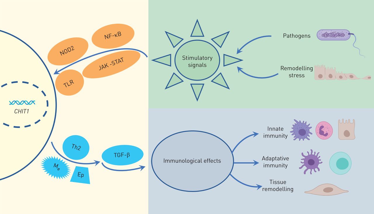
CHIT1 was the first chitinase to be discovered and characterised in mammals [23, 37]. Both immune cells, such as activated macrophages and neutrophils, and structural cells, such as epithelial cells can secrete CHIT1 [26, 37–41]. These cells constantly face environmental threats given their location in the lung, suggesting its role in innate immune system in pulmonary diseases [26, 37, 40, 42]. Macrophages and neutrophils release CHIT1 after stimulation with interferons, tumour necrosis factor (TNF) and granulocyte-macrophage colony stimulating factor through NF-κB, Jak-Stat, NOD2 and Toll-like receptor signalling pathways [38, 43–46], indicating the importance of this enzyme in immune response [38, 46]. Furthermore, CHIT1 is stored in ready-to-release granules in the immune cells along with other antimicrobial agents such as lysozyme, again emphasising the possible roles of CHIT1 in immunity [47] (figure 2).

FIGURE 2
  • Download figure
  • Open in new tab
  • Download powerpoint
FIGURE 2

Biological activity of chitotriosidase (CHIT1). TLR: Toll-like receptor; Th2: T-helper type 2 cell; TGF: transforming growth factor.

The basic biological function of chitinase is that it can hydrolyse chitin. Based on this property, it is believed to play an important role in biological defence against or digestion of pathogens containing chitin as part of their structure, such as fungus and arthropods. However, due to the evolutionary conserved nature of this protein, a wide variety of functions has been demonstrated across species for chitinases and related proteins [48]. For example, the CHIT1-homologous genes, such as imaginal disc growth factor (IDGF) from Drosophila, My-Clp1 from Japanese scallops and Cg-Clp1 and Cg-Clp2 from oysters have been shown to be in involved in wound healing, developmental tissue remodelling and immune defence [49–53]. In Drosophila, IDGF is essential for larval and adult moulting as it organises extracellular matrix formation at barrier tissues [54]. In Japanese scallops, the expression of My-Clp1 was observed in various developmental stages of metamorphosis, which suggest its roles in tissue degeneration and remodelling [55]. In oysters, Cg-Clp1 and Cg-Clp2 were reported to be involved in immune responses to bacterial lipopolysaccharide challenge [53].

However, the precise function of CHIT1 in mammals still remains unknown. CHIT1 does not appear to play a role during embryogenesis or early development in mammals as CHIT1-deficient mice have been developed successfully and do not show any overt signs of abnormalities [36]. Most of the clinical studies looking at the role of CHIT1 have largely been correlational. Basic science studies have shown several mechanistic insights into the role of CHIT1 during various disease models. In this review, we focus on the lung diseases and possible roles of CHIT1 (figure 3).

FIGURE 3
  • Download figure
  • Open in new tab
  • Download powerpoint
FIGURE 3

Chitotriosidase (CHIT1) in respiratory disorders. IL: interleukin; TGF: transforming growth factor.

CHIT1 and interstitial lung diseases

Interstitial lung diseases (ILDs) are a group of disorders with basic pathological changes including diffused parenchymal injury, alveolitis and fibrosis, with clinical manifestations such as shortness of breath, diffused shadow shown on chest radiography, restrictive ventilatory impairment, reduced diffusing capacity of the lung for carbon monoxide and hypoxaemia [56, 57]. The aetiology and pathogenesis of various ILDs are still not entirely known. The accumulation of collagen, secreted by myofibroblasts that are recruited in response to hypothesised microinjuries of the alveolar epithelium, leads to the development and sustainment of the fibrotic process [58]. Chitin is one of the abundantly present substances in the environment that can be inhaled into the lung and directly damage the alveolar epithelial cells [59, 60]. Chitins such as those present in the house dust mite can easily reach the lungs by inhalation. Van Dyken et al. [19] found that the amounts of chitin polymer content are increased in bronchoalveolar (BAL) fluid from patients with ILD compared to healthy controls, suggesting a possible role of impaired chitin clearance in the pathogenesis of pulmonary interstitial fibrosis. In other studies, CHIT1 concentrations in BAL were significantly increased in patients with sarcoidosis and idiopathic pulmonary fibrosis (IPF) compared to healthy BAL, although a consistent increase was only observed in the serum of patients with sarcoidosis, indicating compartment-specific regulation of CHIT1 and its possible role in causing pulmonary tissue remodelling and tissue damage in interstitial diseases [61–63]. However, studies have not yet reported whether there is a clear link between the amount of chitin in the lung and levels of chitinase, and if any of these two or both can possibly explain disease onset, progression and severity. In addition, recent studies have demonstrated increased CHIT1 expression in single-cell transcriptomes of an IPF-specific macrophage subpopulation [64, 65].

Mechanistic studies have shed important light on the role of CHIT1 in various cellular signalling pathways that can explain their contribution to ILDs. During the fibrotic process, CHIT1 enhanced transforming growth factor (TGF)-β-stimulated fibroblast proliferation and myofibroblast transformation through interaction with Tgfbrap1 and FoxO3a [66]. This led to the enhancement of TGF-β-stimulated Smad2/3 and MAPK/Erk activation and inhibition of TGF-β-stimulated Smad7 expression, which acts as a negative regulator of TGF-β signalling. At the same time, the expression of CHIT1 was increased in the lungs or blood of patients with IPF and scleroderma-associated ILD patients where CHIT1 expression was inversely associated with Smad7 expression [67]. Furthermore, bleomycin-induced pulmonary fibrosis was significantly reduced in CHIT1−/− mice, which may be due to CHIT1-mediated activation of fibroblasts by augmenting TGF-β1 and TGF-β1 receptors which induced the Smad and MAPK/ERK signalling pathways [40]. Therefore, it is conceivable that CHIT1 plays a role in the pathogenesis of IPF. A study by Dymek et al. [68] showed that CHIT1 was highly expressed in the macrophages of IPF patients. Based on this evidence, therapeutic inhibition of CHIT1 has been explored to alleviate lung fibrosis. In the bleomycin-induced pulmonary fibrosis model, the CHIT1 inhibitor OATD-01 had more significant therapeutic effects compared to treatment with nintedanib (an antifibrotic agent), while the therapeutic efficiency was comparable to pirfenidone. Both nintedanib and pirfenidone are currently approved for treatment of patients with IPF [68, 69]. If successful, these therapies will put chitinase in a strong spotlight as a modulator of human diseases.

CHIT1 and pulmonary inflammatory diseases

Pulmonary inflammatory diseases involve multiple inflammatory cells and are manifested as structural changes in the airway wall and/or parenchyma, including asthma, COPD, bronchopulmonary dysplasia and cystic fibrosis [70]. Studies have shown that chitinase plays an important role in airway inflammatory diseases by regulating the inflammatory response through degradation of chitin or pathways independent of chitin degradation [15, 70, 71]. COPD is caused by an abnormal inflammatory response to the inhalation of harmful substances, such as smoking. The upregulation of the CHIT1 gene, especially in macrophages, results in the higher levels of chitinase activity in smokers, which may be involved in COPD pathogenesis and progression of the disease [20]. In bronchial biopsies of smokers with COPD, more chitinase-positive cells and increased proportions of alveolar macrophages expressing CHIT1 were observed [72]. The expression level of chitinase in BAL fluid of patients with COPD were associated with airflow obstruction, emphysema, interleukin (IL)-1β, IL-8, TNF-α and its type II soluble receptor TNF-RII [72]. Thus, increased CHIT1 levels seem to be a risk factor for COPD. Agapov et al. [73] found that the level of plasma CHIT1 were associated with the degree of decline in lung function and increased significantly in severe and very severe COPD, based on which the CHIT1 was thought to be a marker for quantitative stratification of COPD. Genetic association studies suggested that the different CHIT1 genotypes were related with the rate of decline of the baseline forced expiratory volume in 1 s [74].

Similarly, CHIT1 has been implicated in the pathogenesis of asthma; however, the data have been largely conflicting. James et al. [75] reported that CHIT1 levels are significantly elevated during asthma. Furthermore, the level of CHIT1 correlated with disease severity [75]. In contrast, Seibold et al. [20] found that chitinase activity itself in the lung was mostly contributed by CHIT1 and was lower in subjects with asthma compared to the healthy controls. These differences may be due to different exposure profiles in the subjects. Similar to chitinase levels in asthma, conflicting results of genetic variants of CHIT1 and severity of the disease have been reported. In a small study, six cases of children with severe asthma had decreased chitinase expression and activity and all six patients were heterozygous for a 24-bp duplication in the CHIT1 gene [32]. However, Wu et al. [76] found that single nucleotide polymorphisms of the CHIT1 gene were not associated with asthma, asthma-related pulmonary physiological changes or allergen-related phenotypes, according to data from the Childhood Asthma Management Program. In spite of the conflicting evidence regarding the role of CHIT1 in the pathogenesis of airway inflammatory diseases, OAT-889, the inhibitor of CHIT1, has been tested in a mouse model of asthma [77]. These studies demonstrated that OAT-889 had profound anti-inflammatory effects in a chronic asthma model in mice [77]. Mechanistically, CHIT1 was reported to play a protective role in asthma by regulating expression of TGF-β and Foxp3+ regulatory T cells (Tregs). Decreased expression of TGF-β and a decreased number of Foxp3+ Tregs were observed in CHIT1-knockout mice in an asthma model [78]. TGF-β1 stimulation of Treg accumulation is regulated by the ability of CHIT1 to enhance the ability of TGF-β1 to stimulate T-cell TGF-β1 receptor expression. Based on these results, the authors concluded that CHIT1 decreased pulmonary inflammation through modulating T-helper type 2 cell (Th2) responses [78]. Some studies reported that AMCase rather than CHIT1 was involved in the pathogenesis of bronchial asthma in Caucasian children [79, 80]; however, given the fact that most chitinase activity in humans is contributed by CHIT1, the significance of AMCase in modulating diseases remains uncertain.

The significance of CHIT1 in patients with cystic fibrosis does not seem to be evident; however, some studies have indicated a possible relationship between chitinase and cystic fibrosis. Livnat et al. [81] investigated the relationship between CHIT1 duplication in cystic fibrosis patients and their predisposition to allergic bronchial pulmonary aspergillosis. They found there was no strong correlation between CHIT1 duplication (loss of chitinase activity) and W1282X cystic fibrosis transmembrane conductance regulator CFTR mutation [81]. Work by Hector et al. [82] demonstrated that CHIT1 levels were elevated in patients with cystic fibrosis, especially in patients with fungal infections. Additionally, this study demonstrated the susceptibility of CHIT1 to neutrophil elastase mediated cleavage. Given the persistent neutrophilic inflammation in cystic fibrosis lungs, it is not clear how this cleavage of CHIT1 alters its biological activity or its possible contribution to disease progression.

CHIT1 and pulmonary infections

Pulmonary infection manifests as inflammation in the lung caused by pathogenic microorganisms such as bacteria, viruses, fungi or parasites [83, 84]. Pulmonary infection, which affects ∼450 million people worldwide and kills ∼4 million people every year, is one of the leading causes of human deaths and of the ultimate cause of death for people with other serious chronic diseases [85, 86]. As the current population is ageing, infections pose a serious threat to humankind. Ageing leads to immunosuppression, and given the rise of drug-resistant strains of pathogens, the therapeutic options remain limited [87–90]. The role of CHIT1 in infection has been studied [21, 36, 91–93]. Anecdotal evidence has suggested important roles for CHIT1 in host immunity, given its high concentration in anatomical sites such as the lungs and gut where there is constant exposure to microorganisms [19, 94]. Furthermore, CHIT1 is released by activated macrophages and neutrophils, two prominent immune cells that contribute extensively to antiviral and antibacterial immunity [27, 95]. However, experimental evidence for their direct roles in pulmonary infections has only recently started to emerge.

Pulmonary tuberculosis, caused by Mycobacterium tuberculosis, is an infectious disease that attacks the lung [96, 97]. It has been shown that CHIT1 expression levels are increased and correlate with the severity of the disease. CHIT1 is released into the serum as a result of the pulmonary granuloma and its level is reduced with the regression of the inflammation, which may reflect the stage, severity and response to treatment of tuberculosis [63, 98, 99]. Similarly, elevated chitinase activity was observed in patients with cystic fibrosis, especially upon colonisation with Candida albicans. In contrast to chronic infections, acute infections such as Klebsiella pneumoniae or Pseudomonas aeruginosa led to the precipitous decrease of chitinase activity in the BAL. This effect was mediated by neutrophil elastase mediated cleavage of CHIT1 [36]. Similarly, in patients with cystic fibrosis it was observed that neutrophil elastase can cleave CHIT1 protein in a time-dependent manner [82].

As the breakdown of chitin is an obvious function that can work as a host defence mechanism against fungal infections, Gordon-Thomson et al. [100] studied the antifungal properties of chitinase in vitro by transfecting Chinese hamster ovary cells with the CHIT1 gene. The growth of Aspergillus niger, C. albicans and Cryptococcus neoformans can be inhibited by the supernatant of transfected cells. In the mouse model, this study demonstrated that the engraftment of microcapsules containing cells transfected with the chitinase gene have the potential to fight against infection caused by chitin-containing microbes by extending the delivery time of recombinant chitinase. Supporting these experimental models, Hector et al. [82] found that patients with CHIT1 deficiency due to genetic mutation had higher colonisation with C. albicans. However, this study found no association between chitinase genetic deficiency and colonisation of Aspergillus fumigatus, another chitin-containing fungal pathogen. Using a mouse model, Wiesner et al. [93] showed that CD11b+ conventional dendritic cells present chitin recognition via CHIT1 to induce Th2 differentiation during pulmonary cryptococcal infection, which led to pathogenic inflammation. CHIT1-deficient mice had improved survival in the face of cryptococcal infection in this study. These studies show the complexity of the regulation and role of chitinase during pulmonary fungal infections.

There are few studies on the relationship between CHIT1 and pulmonary infection with bacterial infections. We studied the role of CHIT1 during K. pneumoniae infection [36]. CHIT1-/- mice had decreased bacterial dissemination and increased survival of mice during pulmonary Klebsiella infection in the presence or absence of antibiotics [36]. The beneficial effect of CHIT1 deficiency on the host was associated with Akt pathway activation. Thus, targeting CHIT1 may be a new potential therapeutic target for defence against K. pneumoniae. However, these effects seem to be pathogen-specific, as CHIT1-/- mice were more susceptible to Pseudomonas lung infections. These studies indicate that role of CHIT1 is dependent on the type of bacterial infection.

CHIT1 and other lung diseases

Besides the aforementioned diseases, the role of CHIT1 has been indicated in many other lung diseases. Gołab et al. [101] reported that CHIT1 activity in lung cancer patients was higher compared to those with normal controls. However, similar elevations were found in patients with pulmonary exudates without cancer. They concluded that CHIT1 activity in patients' serum and exudate cannot be used as a parameter to distinguish lung cancer from inflammation [35, 101]. Obstructive sleep apnoea (OSA) is a sleep disorder caused by complete or partial obstruction of the upper airway and is characterised by recurrent nocturnal breathing cessation [102]. It has been reported that the prevalence of 24-bp duplication of the CHIT1 gene was higher in a severe sleep apnoea group than controls, and the concentration of CHIT1 was related to age, creatine and testosterone [103]. In a study by Teitsdottir et al. [104], plasma CHIT1 activity was measured before and after treatment with positive airway pressure in 97 OSA patients with an apnoea–hypopnoea index ≥15 events·h−1. The association between OSA severity and CHIT1 activity has not been confirmed. However, the level of CHI3L1 was decreased in severely obese OSA patients after positive airway pressure treatment, but not in mildly obese patients.

Evidence of limited roles of chitinases

CHIT1 is present at high concentrations in mammals at locations that are highly susceptible to invading pathogens, such as foodborne and airborne pathogens infecting the gut and lungs, respectively. Their presence in immune cells in these organs led to the hypothesis that they may have antibacterial properties. Hall et al. [105] explored the role of CHIT1 in killing a wide range of pathogens, either alone or in combination with lysozyme. Contrary to conventional belief, CHIT1 had no direct antibacterial properties against Gram-positive or Gram-negative pathogens, alone or in combination with lysozyme.

The most striking elevation of CHIT1 is observed in lysosomal storage diseases such as Gaucher's disease, where it serves as a strong diagnostic tool for monitoring disease severity and therapeutic effectiveness [42]. Gaucher's disease manifests as multiorgan disease where lung involvement is rare, but present in a subset of patients [106, 107]. However, not many systemic studies have explored directly the specific effects of CHIT1 on various organ phenotypes including in the lungs in these patients. The current knowledge implicating CHIT1 on a specific phenotype in Gaucher's disease is not adequate and requires further direct studies to determine how elevated levels of CHIT1 contribute to lung disease pathogenesis [108–111]. It is still possible that CHIT1 contributes to other organ effects in Gaucher's disease. One possibility is that it may contribute to liver fibrosis, as seen in some of the patients with the disease [112]. Contribution to fibrosis can be explained biologically given the ability of CHIT1 to modulate the TGF-β pathway; however, there are no direct studies to implicate CHIT1 in this phenomenon observed in Gaucher's disease. These observations are a strong reminder of the possible limitations of reverse genetics to study the impact of genes using mouse models where an alteration in one gene can change a cascade of effects, obscuring the effects of the gene per se in an unperturbed natural environment.

Furthermore, upregulation of CHIT1 in many diseases has been controversial and data have been shown to be conflicting. As mentioned earlier, studies have shown both upregulation and downregulation of CHIT1 activity during asthma [108, 113–115]. During fungal infection, upregulation was observed with Candida, but not with Aspergillus, although both fungal agents contain chitin as an integral part of their structure [116, 117]. The most accepted upregulation has been observed only in Gaucher's disease, where its role in disease pathogenesis or protection has not been identified.

Further major evidence suggesting the limited role of biological activity of CHIT1 is that a significant proportion of the human population is deficient in chitinase activity due to a 24-bp mutation in exon 10. The deficiency ranges from 5% to as high as 30% in some populations [110, 118, 119]. In spite of the widespread presence of this deficiency, no overt effect of CHIT1 deficiency has been noted in any epidemiological studies. Studies showing evidence of its role in diseases such as filarial disease are performed in a limited number of subjects and have not been validated extensively [120, 121]. This is surprising given the fact that CHIT1 is the only true chitinase present in the humans, and the homozygous genotype for 24-bp mutation has significantly reduced chitinase activity [122–124]. These observations are in stark contrast with the in vitro data showing that recombinant CHIT1 can digest the fungal cell wall and lead to the death of fungus [100, 125]. However, we should be aware that the true roles of chitinase may still be unknown. Similarly, it is possible that the role of chitinases can be observed only in the presence of certain stress or disease conditions, rather than at baseline. As described in the next section, there are probably more unknown than known functions in chitinase biology that merit further investigation.

Unknowns in chitinase biology

One prominent question that still remains unanswered is the precise role of CHIT1 in mammals including humans and whether these proteins have any major role in the host. It is not clear if the 24-bp mutation is indeed an evolutionary beginning of loss of this gene function due to its relative uselessness. The cost of producing the protein at such high levels must be justified with a significant survival advantage in the face of a major threat faced by humans. It is currently not clear if CHIT1 function depends on its enzymatic activity. This is an important aspect of chitinase biology, as a significant population with 24-bp mutation has no chitinase activity but does have the molecule. A precise receptor for CHIT1 remains unknown, but has been identified for the related chitinase-3 like protein 1 which binds to IL-13Ra2 and requires TMEM219 [125–128]. Another major lack of knowledge regarding chitinase biology is our lack of understanding of the internal substrate for the chitinase enzyme in mammals. This is another major direction for potential further investigations. In the absence of a known receptor, our knowledge of specific pathways remains unknown.

In summary, mammalian chitinase is an enzyme with chitinolytic activity. The upregulation of the CHIT1 gene results in increased chitinase activity in many lung diseases; however, conflicting reports often emerge. Increased CHIT1 activity has been shown to play important roles in chronic pulmonary inflammation, allergy, injury, healing and pathogen infection. The effects of CHIT1 appear to be influenced by many factors, and its distribution and expression very between diseases. Much more research is needed to understand the precise roles of CHIT1 in biological functions or to characterise it as an important biomarker for lung disease severity.

Acknowledgement

We acknowledge the contribution of Liang Liu (3rd Medical Center of Chinese PLA General Hospital, Beijing, China) for the critical reviews.

Footnotes

  • Submitted article, peer reviewed.

  • Conflict of interest: D. Chang has nothing to disclose.

  • Conflict of interest: L. Sharma has nothing to disclose.

  • Conflict of interest: C.S. Dela Cruz has nothing to disclose.

  • Support statement: The authors gratefully acknowledge financial support from the China Scholarship Council (DC; 201809112037), Beijing Nova Program (DC; Z171100001117012 and 191100001119021), National Institutes of Health National Heart, Lung, and Blood Institute (CDC; R01HL126094), American Legion Auxiliary (LS; 513385) and the American Thoracic Society (LS: 2018-18). Funding information for this article has been deposited with the Crossref Funder Registry.

  • Received October 29, 2019.
  • Accepted December 2, 2019.
  • Copyright ©ERS 2020.
http://creativecommons.org/licenses/by-nc/4.0/

This article is open access and distributed under the terms of the Creative Commons Attribution Non-Commercial Licence 4.0.

References

  1. ↵
    1. Rathore AS,
    2. Gupta RD
    . Chitinases from bacteria to human: properties, applications, and future perspectives. Enzyme Res 2015; 2015: 791907. doi:10.1155/2015/791907
    OpenUrl
  2. ↵
    1. Benecke W
    . Ueber Bacillus chitinoyqrous einen chitin zerzenden Spaltpilze [Bacillus chitinoyqrous: a chitin-splitting fungus]. Sottfeitung Ant I 1905; 63: 227–242.
    OpenUrl
  3. ↵
    1. Folpmers T
    . De ontleding van chitine en zijn splitsingsproduct, het glucosamine, door bacterien. [The decomposition of chitin and its cleavage product, the glucosamine, by bacteria]. Chem Weekbl 1921; 18: 249.
    OpenUrl
  4. ↵
    1. Karrer P,
    2. Hofmann A
    . Polysaccharide XXXIX. Über den enzymatischen Abbau von Chitin und Chitosan I [Polysaccharides XXXIX. About the enzymatic breakdown of chitin and chitosan I]. Helvetica Chimica Acta 1929; 12: 616–637. doi:10.1002/hlca.19290120167
    OpenUrlCrossRef
  5. ↵
    1. Patil RS,
    2. Ghormade VV,
    3. Deshpande MV
    . Chitinolytic enzymes: an exploration. Enzyme Microb Technol 2000; 26: 473–483. doi:10.1016/S0141-0229(00)00134-4
    OpenUrlCrossRefPubMed
  6. ↵
    1. Henrissat B
    . A classification of glycosyl hydrolases based on amino acid sequence similarities. Biochem J 1991; 280: 309–316. doi:10.1042/bj2800309
    OpenUrlAbstract/FREE Full Text
  7. ↵
    1. Arimori T,
    2. Kawamoto N,
    3. Shinya S, et al.
    Crystal structures of the catalytic domain of a novel glycohydrolase family 23 chitinase from Ralstonia sp. A-471 reveals a unique arrangement of the catalytic residues for inverting chitin hydrolysis. J Biol Chem 2013; 288: 18696–18706. doi:10.1074/jbc.M113.462135
    OpenUrlAbstract/FREE Full Text
    1. Fujita K,
    2. Shimomura K,
    3. Yamamoto K, et al.
    A chitinase structurally related to the glycoside hydrolase family 48 is indispensable for the hormonally induced diapause termination in a beetle. Biochem Biophys Res Commun 2006; 345: 502–507. doi:10.1016/j.bbrc.2006.04.126
    OpenUrlCrossRefPubMed
  8. ↵
    1. Henrissat B,
    2. Bairoch A
    . New families in the classification of glycosyl hydrolases based on amino acid sequence similarities. Biochem J 1993; 293: 781–788. doi:10.1042/bj2930781
    OpenUrlAbstract/FREE Full Text
  9. ↵
    1. Duan Y,
    2. Liu T,
    3. Zhou Y, et al.
    Glycoside hydrolase family 18 and 20 enzymes are novel targets of the traditional medicine berberine. J Biol Chem 2018; 293: 15429–15438. doi:10.1074/jbc.RA118.004351
    OpenUrlAbstract/FREE Full Text
  10. ↵
    1. Dahiya N,
    2. Tewari R,
    3. Hoondal GS
    . Biotechnological aspects of chitinolytic enzymes: a review. Appl Microbiol Biotechnol 2006; 71: 773–782. doi:10.1007/s00253-005-0183-7
    OpenUrlCrossRefPubMed
  11. ↵
    1. Cohen-Kupiec R,
    2. Chet I
    . The molecular biology of chitin digestion. Curr Opin Biotechnol 1998; 9: 270–277. doi:10.1016/S0958-1669(98)80058-X
    OpenUrlCrossRefPubMed
  12. ↵
    1. Gooday GW
    . Aggressive and defensive roles for chitinases. EXS 1999; 87: 157–169.
    OpenUrlPubMed
  13. ↵
    1. Zhang H,
    2. Liu M,
    3. Tian Y, et al.
    Comparative characterization of chitinases from silkworm (Bombyx mori) and bollworm (Helicoverpa armigera). Cell Biochem Biophys 2011; 61: 267–275. doi:10.1007/s12013-011-9196-2
    OpenUrlPubMed
  14. ↵
    1. Lee CG,
    2. Da Silva CA,
    3. Dela Cruz CS, et al.
    Role of chitin and chitinase/chitinase-like proteins in inflammation, tissue remodeling, and injury. Annu Rev Physiol 2011; 73: 479–501. doi:10.1146/annurev-physiol-012110-142250
    OpenUrlCrossRefPubMed
    1. Kzhyshkowska J,
    2. Gratchev A,
    3. Goerdt S
    . Human chitinases and chitinase-like proteins as indicators for inflammation and cancer. Biomark Insights 2007; 2: 128–146. doi:10.1177/117727190700200023
    OpenUrlPubMed
    1. Bussink AP,
    2. Speijer D,
    3. Aerts JM, et al.
    Evolution of mammalian chitinase(-like) members of family 18 glycosyl hydrolases. Genetics 2007; 177: 959–970. doi:10.1534/genetics.107.075846
    OpenUrlAbstract/FREE Full Text
  15. ↵
    1. Di Rosa M,
    2. Distefano G,
    3. Zorena K, et al.
    Chitinases and immunity: ancestral molecules with new functions. Immunobiology 2016; 221: 399–411. doi:10.1016/j.imbio.2015.11.014
    OpenUrlCrossRef
  16. ↵
    1. Van Dyken SJ,
    2. Liang HE,
    3. Naikawadi RP, et al.
    Spontaneous chitin accumulation in airways and age-related fibrotic lung disease. Cell 2017; 169: 497–509. doi:10.1016/j.cell.2017.03.044
    OpenUrlCrossRefPubMed
  17. ↵
    1. Seibold MA,
    2. Donnelly S,
    3. Solon M, et al.
    Chitotriosidase is the primary active chitinase in the human lung and is modulated by genotype and smoking habit. J Allergy Clin Immunol 2008; 122: 944–950. doi:10.1016/j.jaci.2008.08.023
    OpenUrlCrossRefPubMed
  18. ↵
    1. Vega K,
    2. Kalkum M
    . Chitin, chitinase responses, and invasive fungal infections. Int J Microbiol 2012; 2012: 920459. doi:10.1155/2012/920459
    OpenUrlCrossRefPubMed
  19. ↵
    1. Zhu Z,
    2. Zheng T,
    3. Homer RJ, et al.
    Acidic mammalian chitinase in asthmatic Th2 inflammation and IL-13 pathway activation. Science 2004; 304: 1678–1682. doi:10.1126/science.1095336
    OpenUrlAbstract/FREE Full Text
  20. ↵
    1. Boot RG,
    2. Renkema GH,
    3. Strijland A, et al.
    Cloning of a cDNA encoding chitotriosidase, a human chitinase produced by macrophages. J Biol Chem 1995; 270: 26252–26256. doi:10.1074/jbc.270.44.26252
    OpenUrlAbstract/FREE Full Text
  21. ↵
    1. Fadel F,
    2. Zhao Y,
    3. Cousido-Siah A, et al.
    X-ray crystal structure of the full length human chitotriosidase (CHIT1) reveals features of its chitin binding domain. PLoS One 2016; 11: e0154190. doi:10.1371/journal.pone.0154190
    OpenUrl
  22. ↵
    1. Boot RG,
    2. Bussink AP,
    3. Verhoek M, et al.
    Marked differences in tissue-specific expression of chitinases in mouse and man. J Histochem Cytochem 2005; 53: 1283–1292. doi:10.1369/jhc.4A6547.2005
    OpenUrlCrossRefPubMed
  23. ↵
    1. Kanneganti M,
    2. Kamba A,
    3. Mizoguchi E
    . Role of chitotriosidase (chitinase 1) under normal and disease conditions. J Epithel Biol Pharmacol 2012; 5: 1–9. doi:10.2174/1875044301205010001
    OpenUrlCrossRefPubMed
  24. ↵
    1. Di Rosa M,
    2. Brundo VM,
    3. Malaguarnera L
    . New insights on chitinases immunologic activities. World J Immunol 2016; 6: 96–104. doi:10.5411/wji.v6.i2.96
    OpenUrl
    1. Di Rosa M,
    2. Tibullo D,
    3. Cambria D, et al.
    Chitotriosidase expression during monocyte-derived dendritic cells differentiation and maturation. Inflammation 2015; 38: 2082–2091. doi:10.1007/s10753-015-0190-5
    OpenUrl
    1. Di Rosa M,
    2. De Gregorio C,
    3. Malaguarnera G, et al.
    Evaluation of AMCase and CHIT-1 expression in monocyte macrophages lineage. Mol Cell Biochem 2013; 374: 73–80. doi:10.1007/s11010-012-1506-5
    OpenUrl
  25. ↵
    1. Di Rosa M,
    2. Malaguarnera G,
    3. De Gregorio C, et al.
    Evaluation of CHI3L-1 and CHIT-1 expression in differentiated and polarized macrophages. Inflammation 2013; 36: 482–492. doi:10.1007/s10753-012-9569-8
    OpenUrl
  26. ↵
    1. Li F-F,
    2. Yan P,
    3. Zhao Z-X, et al.
    Polymorphisms in the CHIT1 gene: associations with colorectal cancer. Oncotarget 2016; 7: 39572–39581.
    OpenUrl
  27. ↵
    1. Vicencio AG,
    2. Chupp GL,
    3. Tsirilakis K, et al.
    CHIT1 mutations: genetic risk factor for severe asthma with fungal sensitization? Pediatrics 2010; 126: e982–e985. doi:10.1542/peds.2010-0321
    OpenUrlAbstract/FREE Full Text
    1. Kitamoto S,
    2. Egashira K,
    3. Ichiki T, et al.
    Chitinase inhibition promotes atherosclerosis in hyperlipidemic mice. Am J Pathol 2013; 183: 313–325. doi:10.1016/j.ajpath.2013.04.003
    OpenUrl
    1. Maver A,
    2. Medica I,
    3. Salobir B, et al.
    The role of chitotriosidase duplication gene polymorphism in the susceptibility to sarcoidosis. Slov Med J 2010; 79: 837–842.
    OpenUrl
  28. ↵
    1. Tercelj M,
    2. Salobir B,
    3. Simcic S, et al.
    Chitotriosidase activity in sarcoidosis and some other pulmonary diseases. Scand J Clin Lab Invest 2009; 69: 575–578. doi:10.1080/00365510902829362
    OpenUrlCrossRefPubMed
  29. ↵
    1. Sharma L,
    2. Amick AK,
    3. Vasudevan S, et al.
    Regulation and role of chitotriosidase during lung infection with Klebsiella pneumoniae. J Immunol 2018; 201: 615–626. doi:10.4049/jimmunol.1701782
    OpenUrlAbstract/FREE Full Text
  30. ↵
    1. Renkema GH,
    2. Boot RG,
    3. Muijsers AO, et al.
    Purification and characterization of human chitotriosidase, a novel member of the chitinase family of proteins. J Biol Chem 1995; 270: 2198–2202. doi:10.1074/jbc.270.5.2198
    OpenUrlAbstract/FREE Full Text
  31. ↵
    1. Malaguarnera L,
    2. Musumeci M,
    3. Di Rosa M, et al.
    Interferon-gamma, tumor necrosis factor-alpha, and lipopolysaccharide promote chitotriosidase gene expression in human macrophages. J Clin Lab Anal 2005; 19: 128–132. doi:10.1002/jcla.20063
    OpenUrlCrossRefPubMed
    1. van Eijk M,
    2. van Roomen CP,
    3. Renkema GH, et al.
    Characterization of human phagocyte-derived chitotriosidase, a component of innate immunity. Int Immunol 2005; 17: 1505–1512. doi:10.1093/intimm/dxh328
    OpenUrlCrossRefPubMed
  32. ↵
    1. Lee CG,
    2. Herzog EL,
    3. Ahangari F, et al.
    Chitinase 1 is a biomarker for and therapeutic target in scleroderma-associated interstitial lung disease that augments TGF-β1 signaling. J Immunol 2012; 189: 2635–2644. doi:10.4049/jimmunol.1201115
    OpenUrlAbstract/FREE Full Text
  33. ↵
    1. Di Rosa M,
    2. Musumeci M,
    3. Scuto A, et al.
    Effect of interferon-γ, interleukin-10, lipopolysaccharide and tumor necrosis factor-α on chitotriosidase synthesis in human macrophages. Clin Chem Lab Med 2005; 43: 499–502. doi:10.1515/CCLM.2005.088
    OpenUrlCrossRefPubMed
  34. ↵
    1. Sumarac Z,
    2. Suvajdžić N,
    3. Ignjatović S, et al.
    Biomarkers in Serbian patients with Gaucher disease. Clin Biochem 2011; 44: 950–954. doi:10.1016/j.clinbiochem.2011.05.016
    OpenUrlPubMed
  35. ↵
    1. Schroder K,
    2. Hertzog PJ,
    3. Ravasi T, et al.
    Interferon-gamma: an overview of signals, mechanisms and functions. J Leukoc Biol 2004; 75: 163–189. doi:10.1189/jlb.0603252
    OpenUrlCrossRefPubMed
    1. Sharif O,
    2. Bolshakov VN,
    3. Raines S, et al.
    Transcriptional profiling of the LPS induced NF-κB response in macrophages. BMC Immunol 2007; 8: 1. doi:10.1186/1471-2172-8-1
    OpenUrlCrossRefPubMed
    1. van Eijk M,
    2. Scheij SS,
    3. van Roomen CP, et al.
    TLR- and NOD2-dependent regulation of human phagocyte-specific chitotriosidase. FEBS Lett 2007; 581: 5389–5395. doi:10.1016/j.febslet.2007.10.039
    OpenUrlCrossRefPubMed
  36. ↵
    1. Boyle JP,
    2. Parkhouse R,
    3. Monie TP
    . Insights into the molecular basis of the NOD2 signalling pathway. Open Biol 2014; 4: 140178. doi:10.1098/rsob.140178
    OpenUrlCrossRefPubMed
  37. ↵
    1. Bouzas L,
    2. Carlos Guinarte J,
    3. Carlos Tutor J
    . Chitotriosidase activity in plasma and mononuclear and polymorphonuclear leukocyte populations. J Clin Lab Anal 2003; 17: 271–275. doi:10.1002/jcla.10108
    OpenUrlCrossRefPubMed
  38. ↵
    1. Patel S,
    2. Goyal A
    . Chitin and chitinase: role in pathogenicity, allergenicity and health. Int J Biol Macromol 2017; 97: 331–338. doi:10.1016/j.ijbiomac.2017.01.042
    OpenUrl
  39. ↵
    1. Sutherland TE
    . Chitinase-like proteins as regulators of innate immunity and tissue repair: helpful lessons for asthma? Biochem Soc Trans 2018; 46: 141–151. doi:10.1042/BST20170108
    OpenUrlAbstract/FREE Full Text
    1. Broz V,
    2. Kucerova L,
    3. Rouhova L, et al.
    Drosophila imaginal disc growth factor 2 is a trophic factor involved in energy balance, detoxification, and innate immunity. Sci Rep 2017; 7: 43273. doi:10.1038/srep43273
    OpenUrlCrossRef
    1. Kucerova L,
    2. Broz V,
    3. Arefin B, et al.
    The Drosophila chitinase-like protein IDGF3 is involved in protection against nematodes and in wound healing. J Innate Immun 2016; 8: 199–210. doi:10.1159/000442351
    OpenUrlCrossRef
    1. Zhu Q,
    2. Arakane Y,
    3. Beeman RW, et al.
    Characterization of recombinant chitinase-like proteins of Drosophila melanogaster and Tribolium castaneum. Insect Biochem Mol Biol 2008; 38: 467–477. doi:10.1016/j.ibmb.2007.06.011
    OpenUrlCrossRefPubMed
  40. ↵
    1. Badariotti F,
    2. Lelong C,
    3. Dubos MP, et al.
    Characterization of chitinase-like proteins (Cg-Clp1 and Cg-Clp2) involved in immune defence of the mollusc Crassostrea gigas. FEBS J 2007; 274: 3646–3654. doi:10.1111/j.1742-4658.2007.05898.x
    OpenUrlCrossRefPubMed
  41. ↵
    1. Pesch YY,
    2. Riedel D,
    3. Patil KR, et al.
    Chitinases and imaginal disc growth factors organize the extracellular matrix formation at barrier tissues in insects. Sci Rep 2016; 6: 18340. doi:10.1038/srep18340
    OpenUrlCrossRefPubMed
  42. ↵
    1. Gao L,
    2. Xu GJ,
    3. Su H, et al.
    Identification and expression analysis of cDNA encoding chitinase-like protein (CLP) gene in Japanese scallop Mizuhopecten yessoensis. Genet Mol Res 2014; 13: 10727–10740. doi:10.4238/2014.December.18.14
    OpenUrlCrossRef
  43. ↵
    1. DS Basow
    1. King T,
    2. Flaherty K,
    3. Hollingsworth H
    . Approach to the adult with interstitial lung disease: clinical evaluation. In: DS Basow, ed. UpToDate. Waltham, MA, UpToDate, 2010.
  44. ↵
    1. Leslie K
    . My approach to interstitial lung disease using clinical, radiological and histopathological patterns. J Clin Pathol 2009; 62: 387–401. doi:10.1136/jcp.2008.059782
    OpenUrlAbstract/FREE Full Text
  45. ↵
    1. Sgalla G,
    2. Iovene B,
    3. Calvello M, et al.
    Idiopathic pulmonary fibrosis: pathogenesis and management. Respir Res 2018; 19: 32. doi:10.1186/s12931-018-0730-2
    OpenUrl
  46. ↵
    1. Reese TA,
    2. Liang HE,
    3. Tager AM, et al.
    Chitin induces accumulation in tissue of innate immune cells associated with allergy. Nature 2007; 447: 92–96. doi:10.1038/nature05746
    OpenUrlCrossRefPubMed
  47. ↵
    1. Van Dyken SJ,
    2. Garcia D,
    3. Porter P, et al.
    Fungal chitin from asthma-associated home environments induces eosinophilic lung infiltration. J Immunol 2011; 187: 2261–2267. doi:10.4049/jimmunol.1100972
    OpenUrlAbstract/FREE Full Text
  48. ↵
    1. Bargagli E,
    2. Margollicci M,
    3. Perrone A, et al.
    Chitotriosidase analysis in bronchoalveolar lavage of patients with sarcoidosis. Sarcoidosis Vasc Diffuse Lung Dis 2007; 24: 59–64.
    OpenUrlPubMed
    1. Bargagli E,
    2. Margollicci M,
    3. Luddi A, et al.
    Chitotriosidase activity in patients with interstitial lung diseases. Respir Med 2007; 101: 2176–2181. doi:10.1016/j.rmed.2007.05.008
    OpenUrlCrossRefPubMed
  49. ↵
    1. Bargagli E,
    2. Margollicci M,
    3. Nikiforakis N, et al.
    Chitotriosidase activity in the serum of patients with sarcoidosis and pulmonary tuberculosis. Respiration 2007; 74: 548–552. doi:10.1159/000100555
    OpenUrlCrossRefPubMed
  50. ↵
    1. Reyfman PA,
    2. Walter JM,
    3. Joshi N, et al.
    Single-cell transcriptomic analysis of human lung provides insights into the pathobiology of pulmonary fibrosis. Am J Respir Crit Care Med 2019; 199: 1517–1536. doi:10.1164/rccm.201712-2410OC
    OpenUrl
  51. ↵
    1. Adams TS,
    2. Schupp JC,
    3. Poli S, et al.
    Single cell RNA-seq reveals ectopic and aberrant lung resident cell populations in idiopathic pulmonary fibrosis. BioRxiv 2019; 759902. 10.1101/759902.
  52. ↵
    1. Lee C-M,
    2. He C-H,
    3. Park JW, et al.
    Chitinase 1 regulates pulmonary fibrosis by modulating TGF-β/SMAD7 pathway via TGFBRAP1 and FOXO3. Life Sci Alliance 2019; 2: e201900350. doi:10.26508/lsa.201900350
    OpenUrlAbstract/FREE Full Text
  53. ↵
    1. Lee C,
    2. Park J,
    3. Lee J, et al.
    Chitotriosidase interacts with TGFbrap1 and FoxO3a, regulates TGF-β signaling and Smad7 expression in the pathogenesis of pulmonary fibrosis. Am J Respir Crit Care Med 2018; 197: A3870.
    OpenUrl
  54. ↵
    1. Dymek B,
    2. Sklepkiewicz P,
    3. Mlacki M, et al.
    OATD-01, a dual chitinase inhibitor, significantly ameliorates pulmonary fibrosis in the bleomycin-induced mouse model. Am J Respir Crit Care Med 2018; 197: A4346.
    OpenUrl
  55. ↵
    1. Dymek B,
    2. Sklepkiewicz P,
    3. Mlacki M, et al.
    CHIT1 is a novel therapeutic target in idiopathic pulmonary fibrosis (IPF): anti-fibrotic efficacy of OATD-01, a potent and selective chitinase inhibitor in the mouse model of pulmonary fibrosis. Eur Respir J 2018; 52: Suppl. 62, OA5361.
    OpenUrlCrossRef
  56. ↵
    1. Tiddens H,
    2. Silverman M,
    3. Bush A
    . The role of inflammation in airway disease: remodeling. Am J Respir Crit Care Med 2000; 162: S7–S10. doi:10.1164/ajrccm.162.supplement_1.maic-2
    OpenUrlPubMed
  57. ↵
    1. Lee CG
    . Chitin, chitinases and chitinase-like proteins in allergic inflammation and tissue remodeling. Yonsei Med J 2009; 50: 22–30. doi:10.3349/ymj.2009.50.1.22
    OpenUrlCrossRefPubMed
  58. ↵
    1. Létuvé S,
    2. Kozhich A,
    3. Humbles A, et al.
    Lung chitinolytic activity and chitotriosidase are elevated in chronic obstructive pulmonary disease and contribute to lung inflammation. Am J Pathol 2010; 176: 638–649. doi:10.2353/ajpath.2010.090455
    OpenUrlCrossRefPubMed
  59. ↵
    1. Agapov E,
    2. Battaile JT,
    3. Tidwell R, et al.
    Macrophage chitinase 1 stratifies chronic obstructive lung disease. Am J Respir Cell Mol Biol 2009; 41: 379–384. doi:10.1165/2009-0122R
    OpenUrlCrossRefPubMed
  60. ↵
    1. Aminuddin F,
    2. Akhabir L,
    3. Stefanowicz D, et al.
    Genetic association between human chitinases and lung function in COPD. Hum Genet 2012; 131: 1105–1114. doi:10.1007/s00439-011-1127-1
    OpenUrlPubMed
  61. ↵
    1. James AJ,
    2. Reinius LE,
    3. Verhoek M, et al.
    Increased YKL-40 and chitotriosidase in asthma and chronic obstructive pulmonary disease. Am J Respir Crit Care Med 2016; 193: 131–142. doi:10.1164/rccm.201504-0760OC
    OpenUrlCrossRefPubMed
  62. ↵
    1. Wu AC,
    2. Lasky-Su J,
    3. Rogers CA, et al.
    Polymorphisms of chitinases are not associated with asthma. J Allergy Clin Immunol 2010; 125: 754–757. doi:10.1016/j.jaci.2009.12.995
    OpenUrlCrossRef
  63. ↵
    1. Dymek B,
    2. Sklepkiewicz P,
    3. Salamon M, et al.
    The therapeutic efficacy of OAT-889 (dual AMCase/CHIT1 inhibitor) in comparison to montelukast in HDM-induced model of chronic airway inflammation in mice. Eur Respir J 2017; 50: Suppl. 61, PA4684.
    OpenUrl
  64. ↵
    1. Hong J,
    2. Kim M,
    3. Sol I, et al.
    Chitotriosidase inhibits allergic asthmatic airways via regulation of TGF-β expression and Foxp3+ Treg cells. Allergy 2018; 73: 1686–1699. doi:10.1111/all.13426
    OpenUrlCrossRef
  65. ↵
    1. Bierbaum S,
    2. Superti-Furga A,
    3. Heinzmann A
    . Genetic polymorphisms of chitotriosidase in Caucasian children with bronchial asthma. Int J Immunogenet 2006; 33: 201–204. doi:10.1111/j.1744-313X.2006.00597.x
    OpenUrlCrossRefPubMed
  66. ↵
    1. Sohn MH,
    2. Lee JH,
    3. Kim KW, et al.
    Genetic variation in the promoter region of chitinase 3-like 1 is associated with atopy. Am J Respir Crit Care Med 2009; 179: 449–456. doi:10.1164/rccm.200809-1422OC
    OpenUrlCrossRefPubMed
  67. ↵
    1. Livnat G,
    2. Bar-Yoseph R,
    3. Mory A, et al.
    Duplication in CHIT1 gene and the risk for Aspergillus lung disease in CF patients. Pediatr Pulmonol 2014; 49: 21–27. doi:10.1002/ppul.22749
    OpenUrl
  68. ↵
    1. Hector A,
    2. Chotirmall SH,
    3. Lavelle GM, et al.
    Chitinase activation in patients with fungus-associated cystic fibrosis lung disease. J Allergy Clin Immunol 2016; 138: 1183–1189.
    OpenUrl
  69. ↵
    1. Kunihiro Y,
    2. Tanaka N,
    3. Kawano R, et al.
    Differential diagnosis of pulmonary infections in immunocompromised patients using high-resolution computed tomography. Eur Radiol 2019; 29: 6089–6099. doi:10.1007/s00330-019-06235-3
    OpenUrl
  70. ↵
    1. Qazi S,
    2. Aboubaker S,
    3. MacLean R, et al.
    Ending preventable child deaths from pneumonia and diarrhoea by 2025. Development of the integrated Global Action Plan for the Prevention and Control of Pneumonia and Diarrhoea. Arch Dis Child 2015; 100: Suppl. 1, S23–S28. doi:10.1136/archdischild-2013-305429
    OpenUrlAbstract/FREE Full Text
  71. ↵
    1. Cilloniz C,
    2. Martin-Loeches I,
    3. Garcia-Vidal C, et al.
    Microbial etiology of pneumonia: epidemiology, diagnosis and resistance patterns. Int J Mol Sci 2016; 17: E2120. doi:10.3390/ijms17122120
    OpenUrl
  72. ↵
    1. Ruuskanen O,
    2. Lahti E,
    3. Jennings LC, et al.
    Viral pneumonia. Lancet 2011; 377: 1264–1275. doi:10.1016/S0140-6736(10)61459-6
    OpenUrlCrossRefPubMed
  73. ↵
    1. Weyand CM,
    2. Goronzy JJ
    . Aging of the immune system. Mechanisms and therapeutic targets. Ann Am Thorac Soc 2016; 13: Suppl. 5, S422–S428. doi:10.1513/AnnalsATS.201602-095AW
    OpenUrl
    1. Li B,
    2. Webster TJ
    . Bacteria antibiotic resistance: new challenges and opportunities for implant-associated orthopedic infections. J Orthop Res 2018; 36: 22–32.
    OpenUrlCrossRef
    1. Salsgiver EL,
    2. Fink AK,
    3. Knapp EA, et al.
    Changing epidemiology of the respiratory bacteriology of patients with cystic fibrosis. Chest 2016; 149: 390–400. doi:10.1378/chest.15-0676
    OpenUrlCrossRefPubMed
  74. ↵
    1. Sharma S,
    2. Gupta P,
    3. Gupta N, et al.
    Pulmonary infections in immunocompromised patients: the role of image-guided fine needle aspiration cytology. Cytopathology 2017; 28: 46–54. doi:10.1111/cyt.12359
    OpenUrl
  75. ↵
    1. Gao C,
    2. Cai X,
    3. Zhang Y, et al.
    Characterization and expression analysis of chitinase genes (CHIT1, CHIT2 and CHIT3) in turbot (Scophthalmus maximus L.) following bacterial challenge. Fish Shellfish Immunol 2017; 64: 357–366. doi:10.1016/j.fsi.2017.03.019
    OpenUrl
    1. Logue EC,
    2. Neff CP,
    3. Mack DG, et al.
    Upregulation of chitinase 1 in alveolar macrophages of HIV-infected smokers. J Immunol 2019; 202: 1363–1372. doi:10.4049/jimmunol.1801105
    OpenUrlAbstract/FREE Full Text
  76. ↵
    1. Wiesner DL,
    2. Specht CA,
    3. Lee CK, et al.
    Chitin recognition via chitotriosidase promotes pathologic type-2 helper T cell responses to cryptococcal infection. PLoS Pathog 2015; 11: e1004701. doi:10.1371/journal.ppat.1004701
    OpenUrlCrossRefPubMed
  77. ↵
    1. Cozzarini E,
    2. Bellin M,
    3. Norberto L, et al.
    CHIT1 and AMCase expression in human gastric mucosa: correlation with inflammation and Helicobacter pylori infection. Eur J Gastroenterol Hepatol 2009; 21: 1119–1126. doi:10.1097/MEG.0b013e328329742a
    OpenUrlCrossRefPubMed
  78. ↵
    1. Żurawska-Płaksej E,
    2. Ługowska A,
    3. Hetmańczyk K, et al.
    Neutrophils as a source of chitinases and chitinase-like proteins in type 2 diabetes. PLoS One 2015; 10: e0141730. doi:10.1371/journal.pone.0141730
    OpenUrlCrossRefPubMed
  79. ↵
    1. Leung AN
    . Pulmonary tuberculosis: the essentials. Radiology 1999; 210: 307–322. doi:10.1148/radiology.210.2.r99ja34307
    OpenUrlCrossRefPubMed
  80. ↵
    World Health Organization (WHO). Global Tuberculosis Report 2018. Geneva, WHO, 2018.
  81. ↵
    1. Tasci C,
    2. Tapan S,
    3. Ozkaya S, et al.
    Efficacy of serum chitotriosidase activity in early treatment of patients with active tuberculosis and a negative sputum smear. Ther Clin Risk Manag 2012; 8: 369–372. doi:10.2147/TCRM.S31752
    OpenUrlPubMed
  82. ↵
    1. Cakır G,
    2. Gumus S,
    3. Ucar E, et al.
    Serum chitotriosidase activity in pulmonary tuberculosis: response to treatment and correlations with clinical parameters. Ann Lab Med 2012; 32: 184–193.
    OpenUrlPubMed
  83. ↵
    1. Gordon-Thomson C,
    2. Kumari A,
    3. Tomkins L, et al.
    Chitotriosidase and gene therapy for fungal infections. Cell Mol Life Sci 2009; 66: 1116–1125. doi:10.1007/s00018-009-8765-7
    OpenUrlCrossRefPubMed
  84. ↵
    1. Gołab K,
    2. Passowicz-Muszyńska E,
    3. Jankowska R, et al.
    Aktywność chitotriozydazy oraz lizozymu i katepsyny H w surowicy chorych na raka płuca i chorych z wysiekiem zapalnym (doniesienie wstepne). [Serum activity of chitotriosidase, lysozyme and cathepsin H in patients with lung cancer and patients with inflammatory exudate (preliminary report)]. Pol Merkur Lekarski 2009; 26: 194–197.
    OpenUrl
  85. ↵
    1. Spicuzza L,
    2. Caruso D,
    3. Di Maria G
    . Obstructive sleep apnoea syndrome and its management. Ther Adv Chronic Dis 2015; 6: 273–285. doi:10.1177/2040622315590318
    OpenUrlCrossRefPubMed
  86. ↵
    1. Tamanaha P,
    2. D'Almeida V,
    3. Calegare BFA, et al.
    24 bp duplication of CHIT1 gene and determinants of human chitotriosidase activity among participants of EPISONO, a population-based cross-sectional study, São Paulo, Brazil. Clin Biochem 2013; 46: 1084–1088. doi:10.1016/j.clinbiochem.2013.05.044
    OpenUrl
  87. ↵
    1. Teitsdottir UD,
    2. Arnardottir ES,
    3. Bjornsdottir E, et al.
    Obesity modulates the association between sleep apnea treatment and CHI3L1 levels but not CHIT1 activity in moderate to severe OSA: an observational study. Sleep Breath 2018; 22: 1101–1109. doi:10.1007/s11325-018-1731-6
    OpenUrl
  88. ↵
    1. Hall AJ,
    2. Morroll S,
    3. Tighe P, et al.
    Human chitotriosidase is expressed in the eye and lacrimal gland and has an antimicrobial spectrum different from lysozyme. Microbes Infect 2008; 10: 69–78. doi:10.1016/j.micinf.2007.10.007
    OpenUrlCrossRefPubMed
  89. ↵
    1. Santamaria F,
    2. Parenti G,
    3. Guidi G, et al.
    Pulmonary manifestations of Gaucher disease: an increased risk for L444P homozygotes? Am J Respir Crit Care Med 1998; 157: 985–989. doi:10.1164/ajrccm.157.3.9706057
    OpenUrlCrossRefPubMed
  90. ↵
    1. Goitein O,
    2. Elstein D,
    3. Abrahamov A, et al.
    Lung involvement and enzyme replacement therapy in Gaucher's disease. QJM 2001; 94: 407–415. doi:10.1093/qjmed/94.8.407
    OpenUrlCrossRefPubMed
  91. ↵
    1. Elias JA,
    2. Homer RJ,
    3. Hamid Q, et al.
    Chitinases and chitinase-like proteins in TH2 inflammation and asthma. J Allergy Clin Immunol 2005; 116: 497–500. doi:10.1016/j.jaci.2005.06.028
    OpenUrlCrossRefPubMed
    1. Woo KH,
    2. Lee BH,
    3. Heo SH, et al.
    Allele frequency of a 24 bp duplication in exon 10 of the CHIT1 gene in the general Korean population and in Korean patients with Gaucher disease. J Hum Genet 2014; 59: 276–279. doi:10.1038/jhg.2014.16
    OpenUrl
  92. ↵
    1. Grace ME,
    2. Balwani M,
    3. Nazarenko I, et al.
    Type 1 Gaucher disease: null and hypomorphic novel chitotriosidase mutations – implications for diagnosis and therapeutic monitoring. Hum Mutat 2007; 28: 866–873. doi:10.1002/humu.20524
    OpenUrlCrossRefPubMed
  93. ↵
    1. Irún P,
    2. Alfonso P,
    3. Aznarez S, et al.
    Chitotriosidase variants in patients with Gaucher disease. Implications for diagnosis and therapeutic monitoring. Clin Biochem 2013; 46: 1804–1807. doi:10.1016/j.clinbiochem.2013.09.006
    OpenUrl
  94. ↵
    1. Lachmann RH,
    2. Wight DG,
    3. Lomas DJ, et al.
    Massive hepatic fibrosis in Gaucher's disease: clinico-pathological and radiological features. QJM 2000; 93: 237–244. doi:10.1093/qjmed/93.4.237
    OpenUrlCrossRefPubMed
  95. ↵
    1. Ober C,
    2. Chupp GL
    . The chitinase and chitinase-like proteins: a review of genetic and functional studies in asthma and immune-mediated diseases. Curr Opin Allergy Clin Immunol 2009; 9: 401–408. doi:10.1097/ACI.0b013e3283306533
    OpenUrlCrossRefPubMed
    1. Shuhui L,
    2. Mok YK,
    3. Wong WS
    . Role of mammalian chitinases in asthma. Int Arch Allergy Immunol 2009; 149: 369–377. doi:10.1159/000205583
    OpenUrlCrossRefPubMed
  96. ↵
    1. Sutherland TE,
    2. Maizels RM,
    3. Allen JE
    . Chitinases and chitinase-like proteins: potential therapeutic targets for the treatment of T-helper type 2 allergies. Clin Exp Allergy 2009; 39: 943–955. doi:10.1111/j.1365-2222.2009.03243.x
    OpenUrlCrossRefPubMed
  97. ↵
    1. Gao N,
    2. Yu FS
    . Chitinase 3-like 1 promotes Candida albicans killing and preserves corneal structure and function by controlling host antifungal responses. Infect Immun 2015; 83: 4154–4164. doi:10.1128/IAI.00980-15
    OpenUrlAbstract/FREE Full Text
  98. ↵
    1. Garth JM,
    2. Mackel JJ,
    3. Reeder KM, et al.
    Acidic mammalian chitinase negatively affects immune responses during acute and chronic Aspergillus fumigatus exposure. Infect Immun 2018; 86: e00944-17. doi:10.1128/IAI.00944-17
    OpenUrlAbstract/FREE Full Text
  99. ↵
    1. Malaguarnera L,
    2. Simporè J,
    3. Prodi DA, et al.
    A 24-bp duplication in exon 10 of human chitotriosidase gene from the sub-Saharan to the Mediterranean area: role of parasitic diseases and environmental conditions. Genes Immun 2003; 4: 570–574. doi:10.1038/sj.gene.6364025
    OpenUrlCrossRefPubMed
  100. ↵
    1. Lee P,
    2. Waalen J,
    3. Crain K, et al.
    Human chitotriosidase polymorphisms G354R and A442V associated with reduced enzyme activity. Blood Cells Mol Dis 2007; 39: 353–360. doi:10.1016/j.bcmd.2007.06.013
    OpenUrlPubMed
  101. ↵
    1. Sanprasert V,
    2. Charuchaibovorn S,
    3. Nuchprayoon S
    . The genetic polymorphisms of 24 base pair duplication and point g102s of human chitotriosidase to bancroftian filariasis at the Thai–Myanmar border. Pathogens 2019; 8: E41. doi:10.3390/pathogens8010041
    OpenUrl
  102. ↵
    1. Tachu B,
    2. Pillai S,
    3. Lucius R, et al.
    Essential role of chitinase in the development of the filarial nematode Acanthocheilonema viteae. Infect Immun 2008; 76: 221–228. doi:10.1128/IAI.00701-07
    OpenUrlAbstract/FREE Full Text
  103. ↵
    1. Boot RG,
    2. Renkema GH,
    3. Verhoek M, et al.
    The human chitotriosidase gene nature of inherited enzyme deficiency. J Biol Chem 1998; 273: 25680–25685. doi:10.1074/jbc.273.40.25680
    OpenUrlAbstract/FREE Full Text
    1. Pant S,
    2. Agarwal J,
    3. Goel A, et al.
    Does 24bp duplication of human CHIT1 gene (chitotriosidase1) predispose to filarial chyluria? A case-control study. J Clin Diagn Res 2016; 10: PC01–PC04. doi:10.1111/crj.12367
    OpenUrl
  104. ↵
    1. Choi EH,
    2. Zimmerman PA,
    3. Foster CB, et al.
    Genetic polymorphisms in molecules of innate immunity and susceptibility to infection with Wuchereria bancrofti in South India. Genes Immun 2001; 2: 248–253. doi:10.1038/sj.gene.6363767
    OpenUrlCrossRefPubMed
  105. ↵
    1. Ahangari F,
    2. Takyar S,
    3. Lee CG, et al.
    Chitotriosidase (Chitinase 1) plays an essential role in IL-13-induced inflammation, fibrosis and alternative (M2) macrophage activation. Am J Respir Crit Care Med 2011; 183: A2868.
    OpenUrl
    1. Correale J,
    2. Fiol M
    . Chitinase effects on immune cell response in neuromyelitis optica and multiple sclerosis. Mult Scler 2011; 17: 521–531. doi:10.1177/1352458510392619
    OpenUrlCrossRefPubMed
    1. He CH,
    2. Lee CG,
    3. Dela Cruz CS, et al.
    Chitinase 3-like 1 regulates cellular and tissue responses via IL-13 receptor α2. Cell Rep 2013; 4: 830–841. doi:10.1016/j.celrep.2013.07.032
    OpenUrlCrossRefPubMed
  106. ↵
    1. Lee C-M,
    2. He CH,
    3. Nour AM, et al.
    IL-13Rα2 uses TMEM219 in chitinase 3-like-1-induced signalling and effector responses. Nat Commun 2016; 7: 12752. doi:10.1038/ncomms12752
    OpenUrlCrossRef
PreviousNext
Back to top
View this article with LENS
Vol 29 Issue 156 Table of Contents
European Respiratory Review: 29 (156)
  • Table of Contents
  • Index by author
Email

Thank you for your interest in spreading the word on European Respiratory Society .

NOTE: We only request your email address so that the person you are recommending the page to knows that you wanted them to see it, and that it is not junk mail. We do not capture any email address.

Enter multiple addresses on separate lines or separate them with commas.
Chitotriosidase: a marker and modulator of lung disease
(Your Name) has sent you a message from European Respiratory Society
(Your Name) thought you would like to see the European Respiratory Society web site.
CAPTCHA
This question is for testing whether or not you are a human visitor and to prevent automated spam submissions.
Print
Citation Tools
Chitotriosidase: a marker and modulator of lung disease
De Chang, Lokesh Sharma, Charles S. Dela Cruz
European Respiratory Review Jun 2020, 29 (156) 190143; DOI: 10.1183/16000617.0143-2019

Citation Manager Formats

  • BibTeX
  • Bookends
  • EasyBib
  • EndNote (tagged)
  • EndNote 8 (xml)
  • Medlars
  • Mendeley
  • Papers
  • RefWorks Tagged
  • Ref Manager
  • RIS
  • Zotero

Share
Chitotriosidase: a marker and modulator of lung disease
De Chang, Lokesh Sharma, Charles S. Dela Cruz
European Respiratory Review Jun 2020, 29 (156) 190143; DOI: 10.1183/16000617.0143-2019
del.icio.us logo Digg logo Reddit logo Technorati logo Twitter logo CiteULike logo Connotea logo Facebook logo Google logo Mendeley logo
Full Text (PDF)

Jump To

  • Article
    • Abstract
    • Abstract
    • Introduction
    • Biological roles of CHIT1
    • CHIT1 and interstitial lung diseases
    • CHIT1 and pulmonary inflammatory diseases
    • CHIT1 and pulmonary infections
    • CHIT1 and other lung diseases
    • Evidence of limited roles of chitinases
    • Unknowns in chitinase biology
    • Acknowledgement
    • Footnotes
    • References
  • Figures & Data
  • Info & Metrics
  • PDF

Subjects

  • Pulmonary pharmacology and therapeutics
  • Tweet Widget
  • Facebook Like
  • Google Plus One

More in this TOC Section

  • Mucolytics for acute exacerbations of COPD: a meta-analysis
  • Targeting IL-33 and TSLP pathways for novel pulmonary therapeutics
  • Physical activity promotion interventions in chronic airways disease
Show more Reviews

Related Articles

Navigate

  • Home
  • Current issue
  • Archive

About the ERR

  • Journal information
  • Editorial board
  • Reviewers
  • Press
  • Permissions and reprints
  • Advertising
  • Sponsorship

The European Respiratory Society

  • Society home
  • myERS
  • Privacy policy
  • Accessibility

ERS publications

  • European Respiratory Journal
  • ERJ Open Research
  • European Respiratory Review
  • Breathe
  • ERS books online
  • ERS Bookshop

Help

  • Feedback

For authors

  • Instructions for authors
  • Publication ethics and malpractice
  • Submit a manuscript

For readers

  • Alerts
  • Subjects
  • RSS

Subscriptions

  • Accessing the ERS publications

Contact us

European Respiratory Society
442 Glossop Road
Sheffield S10 2PX
United Kingdom
Tel: +44 114 2672860
Email: journals@ersnet.org

ISSN

Print ISSN: 0905-9180
Online ISSN: 1600-0617

Copyright © 2023 by the European Respiratory Society