
Main menu

  • Home
  • Current issue
  • Past issues
  • Authors/reviewers
    • Instructions for authors
    • Submit a manuscript
    • COVID-19 submission information
    • Institutional open access agreements
    • Peer reviewer login
  • Alerts
  • Subscriptions
  • ERS Publications
    • European Respiratory Journal
    • ERJ Open Research
    • European Respiratory Review
    • Breathe
    • ERS Books
    • ERS publications home

User menu

  • Log in
  • Subscribe
  • Contact Us
  • My Cart

Search

  • Advanced search
  • ERS Publications
    • European Respiratory Journal
    • ERJ Open Research
    • European Respiratory Review
    • Breathe
    • ERS Books
    • ERS publications home

Login

European Respiratory Society

Advanced Search

  • Home
  • Current issue
  • Past issues
  • Authors/reviewers
    • Instructions for authors
    • Submit a manuscript
    • COVID-19 submission information
    • Institutional open access agreements
    • Peer reviewer login
  • Alerts
  • Subscriptions

Cellular mechanisms underlying steroid-resistant asthma

Ridhima Wadhwa, Kamal Dua, Ian M. Adcock, Jay C. Horvat, Richard Y. Kim, Philip M. Hansbro
European Respiratory Review 2019 28: 190096; DOI: 10.1183/16000617.0096-2019
Ridhima Wadhwa
1Centre for Inflammation, Centenary Institute, Sydney, Australia
2Discipline of Pharmacy, Graduate School of Health, University of Technology Sydney, Sydney, Australia
6Both authors contributed equally
  • Find this author on Google Scholar
  • Find this author on PubMed
  • Search for this author on this site
Kamal Dua
1Centre for Inflammation, Centenary Institute, Sydney, Australia
2Discipline of Pharmacy, Graduate School of Health, University of Technology Sydney, Sydney, Australia
3Priority Research Centre for Healthy Lungs, University of Newcastle and Hunter Medical Research Institute, Newcastle, Australia
6Both authors contributed equally
  • Find this author on Google Scholar
  • Find this author on PubMed
  • Search for this author on this site
Ian M. Adcock
4The Airways Disease Section, National Heart and Lung Institute, Imperial College London, London, UK
  • Find this author on Google Scholar
  • Find this author on PubMed
  • Search for this author on this site
Jay C. Horvat
3Priority Research Centre for Healthy Lungs, University of Newcastle and Hunter Medical Research Institute, Newcastle, Australia
  • Find this author on Google Scholar
  • Find this author on PubMed
  • Search for this author on this site
Richard Y. Kim
1Centre for Inflammation, Centenary Institute, Sydney, Australia
3Priority Research Centre for Healthy Lungs, University of Newcastle and Hunter Medical Research Institute, Newcastle, Australia
5Faculty of Science, University of Technology Sydney, Sydney, Australia
  • Find this author on Google Scholar
  • Find this author on PubMed
  • Search for this author on this site
Philip M. Hansbro
1Centre for Inflammation, Centenary Institute, Sydney, Australia
3Priority Research Centre for Healthy Lungs, University of Newcastle and Hunter Medical Research Institute, Newcastle, Australia
5Faculty of Science, University of Technology Sydney, Sydney, Australia
  • Find this author on Google Scholar
  • Find this author on PubMed
  • Search for this author on this site
  • Article
  • Figures & Data
  • Info & Metrics
  • PDF
Loading

This article has a correction. Please see:

  • “Cellular mechanisms underlying steroid-resistant asthma.” Ridhima Wadhwa, Kamal Dua, Ian M. Adcock, Jay C. Horvat, Richard Y. Kim and Philip M. Hansbro. Eur Respir Rev 2019; 28: 190021. - December 31, 2019

Abstract

Severe steroid-resistant asthma is clinically important, as patients with this form of the disease do not respond to mainstay corticosteroid therapies. The heterogeneity of this form of asthma and poor understanding of the pathological mechanisms involved hinder the identification of therapeutic targets and the development of more effective therapies. A major limiting factor in the understanding of severe steroid-resistant asthma is the existence of multiple endotypes represented by different immunological and inflammatory phenotypes, particularly in adults. Several clinical and experimental studies have revealed associations between specific respiratory infections and steroid-resistant asthma in adults. Here, we discuss recent findings from other authors as well as our own studies that have developed novel experimental models for interrogating the association between respiratory infections and severe steroid-resistant asthma. These models have enabled the identification of new therapies using macrolides, as well as several novel disease mechanisms, including the microRNA-21/phosphoinositide 3-kinase/histone deacetylase 2 axis and NLRP3 inflammasomes, and highlight the potential of these mechanisms as therapeutic targets.

Abstract

Severe steroid-resistant asthma is a significant clinical problem and recent advances in understanding the mechanisms of pathogenesis enable the identification of novel therapeutic approaches http://bit.ly/2mQpF3n

Introduction

The Global Initiative for Asthma (GINA) currently defines asthma as a heterogeneous disease that is characterised by inflammation of the airways and a clinical history of wheezing, cough, tightness of chest and shortness of breath varying with time and intensity, as well as expiratory airflow limitation [1]. Asthma is classically considered as a disease driven by increased type 2 immune responses and eosinophilic inflammation [2–5]. However, recent evidence shows that asthma can be associated with elevated type 1/17 immune responses that are associated with non-eosinophilic (i.e. neutrophilic) inflammation and tend to be present in adult patients with moderate to severe asthma [2, 6]. Importantly, gene–environmental interactions play a vital role in the progression of the disease, with specific insults to the respiratory epithelium [7] inducing the release of pro-inflammatory mediators that recruit inflammatory cells to the airways. Exogenous factors such as respiratory viral and bacterial infections, a high-fat diet and/or obesity, air pollution and cigarette smoke exposure have all been linked with severe forms of asthma in adults as well as with exacerbations of the disease [2, 8–20]. Here, we review the mechanisms of pathogenesis of severe steroid-resistant (SSR) asthma in adults. Children can also have severe asthma, which may have different pathomechanisms; these have been reviewed recently elsewhere [21–23].

Anti-inflammatory corticosteroids are the mainstay therapy for asthma and are often administered in conjunction with short- or long-acting β2-adrenoceptor agonists (bronchodilators) [2, 24]. These combination therapies allow most asthmatics to gain control of their disease symptoms by modulating type 2 cytokine responses and accompanying inflammation, although they have broad-spectrum, nonspecific anti-inflammatory activity. Importantly, however, these therapies are not cures, and long-term, high-dose corticosteroid therapy is associated with many side-effects [4, 24, 25]. Critically, 5–10% of asthmatics do not respond to steroid-based therapies, are more likely to have severe disease and, due to the paucity of effective treatment options, disproportionately account for 50–80% of all asthma-associated healthcare costs [26, 27]. SSR asthma is strongly associated with non-eosinophilic endotypes of the disease, including neutrophilic asthma, and often involves activation of innate immune responses, particularly those mediated by Toll-like receptor (TLR)2 and TLR4 responses, and NLRP3 (nucleotide-binding oligomerisation domain-like receptor family, pyrin domain-containing 3) inflammasome/interleukin (IL)-1β responses [28–32]. Schwartz et al. [33] first described steroid-resistant asthma in a small asthmatic subgroup with poor disease control that did not respond to high-dose oral steroid therapy. With progressive refinement of diagnostic criteria, SSR asthma in adults is now defined as ineffectiveness in achieving >15% improvement in forced expiratory volume in 1 s after 14 days of oral steroid treatment [27, 34]. SSR asthma is the major clinical issue in asthma management and there is an urgent need for therapies that more effectively suppress the symptoms of disease (figure 1).

FIGURE 1
  • Download figure
  • Open in new tab
  • Download powerpoint
FIGURE 1

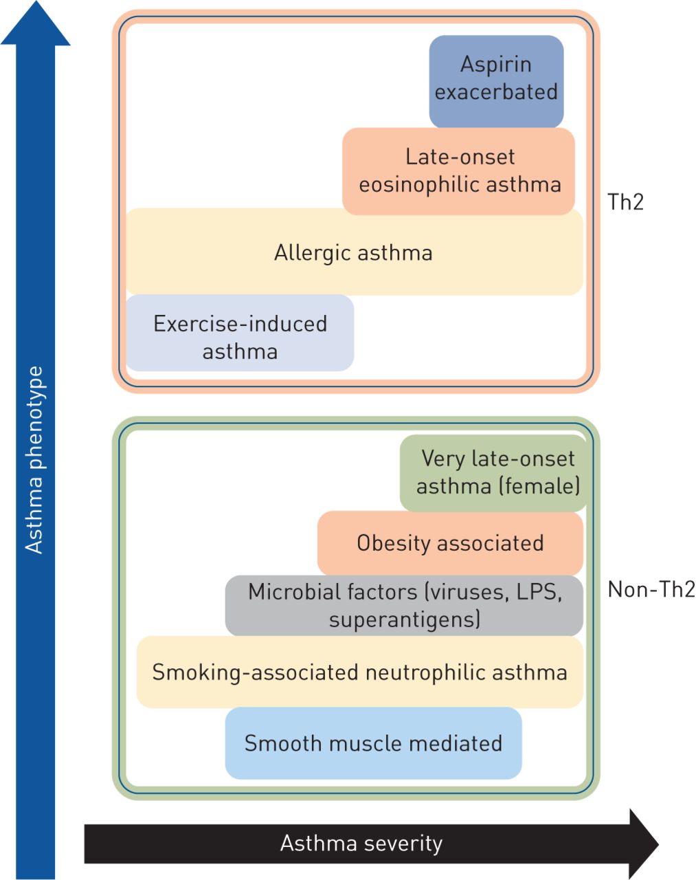
Classification of asthma phenotypes and disease severity based on T-helper cell type 2 (Th2) and non-Th2 characteristics. LPS: lipopolysaccharide.

Mechanisms of steroid resistance

Glucocorticoids (a class of anti-inflammatory corticosteroid) are the mainstay therapy for asthma and are administered through inhaled or oral formulations. Inhaled corticosteroids (ICSs) are synthetic, lipophilic glucocorticoids that, upon inhalation, readily diffuse into the airway tissue and bind and activate cytosolic glucocorticoid receptors (GRs; also called nuclear receptor subfamily 3, group C, member 1 (NR3C1)) [35, 36]. The anti-inflammatory effects are mediated by the GRα isoform via ligand-dependent transcription factors, while GRβ acts as a dominant inhibitor of GRα [37]. GRα homodimers are actively transported into the nucleus by importin-α and importin-13 and bind to a glucocorticoid response element (GRE) to regulate gene transcription [25, 36, 38]. The interaction with transcriptional co-activators, including CBP (cAMP response element binding (CREB)-binding protein), leads to the acetylation of core histones associated with anti-inflammatory glucocorticoid response genes and induction of RNA polymerase II-mediated gene transcription. This phenomenon is known as transactivation. Alternatively, activated GR interacts with CBP proteins complexed with promoter regions of pro-inflammatory genes, such as NF-κB and activator protein (AP)-1, causing suppression of pro-inflammatory gene transcription via inhibition of histone acetylation and preventing DNA access to RNA polymerase II [39–41]. This process is called transrepression.

A large body of clinical and experimental evidence shows that defective GRα expression and activity impairs the anti-inflammatory effects of glucocorticoids and plays important roles in the induction of steroid resistance. Interestingly, other studies show that the higher levels of IL-2, IL-4 and IL-13 gene expression result in local cytokine secretion and steroid resistance, which further alters GRα translocation [34, 42]. A study by Goleva et al. [43] reported that specific silencing of GRβ in bronchoalveolar lavage macrophages of individuals with SSR asthma resulted in improved GRα activity. In another study, Irusen et al. [44] reported that activation of p38 mitogen-activated protein kinase results in the phosphorylation and impaired function of GRs. Moreover, defective nuclear translocation of GRs results in reduced interaction with GREs and GR:GRE binding is affected by increased NF-κB, AP-1, c-Fos and c-Jun N-terminal kinase responses [34, 42, 45, 46]. Defective nuclear translocation of GRs reduces expression and activity of histone deacetylase (HDAC)2 in patients [47]. Importantly, many patients with SSR asthma have normal nuclear translocation of GR but reduced GR:GRE binding affinity [48, 49], suggesting that SSR asthma can also be induced by dysfunction of a different mechanism. Interestingly, reduced HDAC2 expression and activity is associated with steroid insensitivity and more severe disease in both asthma and COPD [50–52], suggesting that deficiencies in transcriptional co-repressor expression and activity may be important in the pathogenesis of SSR asthma.

Role of respiratory infections in SSR asthma

SSR asthma is a serious clinical and socioeconomic problem that is associated with lung function impairment, airway hyperresponsiveness (AHR) and more intense disease exacerbations leading to hospitalisation. Limited treatments are available, including corticosteroid therapy, but none are cures or broadly effective. A crucial drawback is that there are several subsets of SSR asthma with different immunological and inflammatory phenotypes that are likely to drive steroid resistance through different mechanisms [2, 5, 6, 53]. Increasing clinical and experimental evidence strongly implicates specific respiratory infections in the pathogenesis of SSR asthma. We have developed novel and unique animal models of respiratory infection-induced SSR asthma and used them to demonstrate that Chlamydia, Haemophilus influenzae, influenza A virus (IAV) and respiratory syncytial virus (RSV) respiratory infections induce steroid resistance of the key disease features [16–18, 30, 54–56].

We first established Chlamydia, Haemophilus, IAV and RSV respiratory infections and allergic airway disease (AAD) in BALB/c mice separately. Clinical studies show that infections drive more severe disease in established disease [30, 55, 56]. Thus, AAD was first established and then infection was induced. Mice were systemically sensitised with ovalbumin (Ova) and aluminium hydroxide intraperitoneally, followed 10–12 days later by inhaled Ova challenge to induce AAD. Other mouse models of allergic asthma widely use other allergens, such as house dust mite [57–59], Alternaria [60] or cockroach [61] antigen. Our mice were left for 20 days and were re-challenged on days 33–34 of the model, in order to model an allergen-induced exacerbation of disease. To induce severe steroid-resistant AAD (SSRAAD), we induced infections in allergic mice after AAD was initially established, to model the human scenario. Infections were resolved before allergen-induced exacerbation of AAD. To model ICS treatment and examine the response to steroid treatment, mice were administered the steroid dexamethasone (DEX) for the final 3 days of the protocol. Key disease features were assessed 1 day after the final Ova challenge, to assess the impact of resolved infection on the disease phenotype. Importantly, we found that infection drives a severe form of AAD that does not respond to steroid treatment. These protocols have resulted in novel experimental models of Chlamydia-, Haemophilus-, IAV- and RSV-induced SSR asthma that each represents unique endotypes of SSR asthma in humans (table 1).

View this table:
  • View inline
  • View popup
TABLE 1

Role of infections in the pathogenesis of severe steroid-resistant asthma and their response to steroid therapy

Chlamydia respiratory tract infection in asthma corresponds to increased airway neutrophils, which predicts the presence of this pathogen and of steroid resistance [55]. It has also been linked to severe asthma in children [62–64]. Chlamydia respiratory infection in mice with AAD results in increased type 1- and 17-associated immune responses, neutrophilic inflammation, suppression of type 2 and eosinophilic inflammatory responses [18, 30, 55, 56], and steroid-resistant inflammation and AHR. In early life, Chlamydia respiratory infections promote more severe experimental asthma in mice by switching type 1 phenotype and inducing IL-13 and TRAIL (tumour necrosis factor-related apoptosis-inducing ligand) responses [17, 65–67]. These models of early-life infection-induced severe asthma are also steroid resistant (P.M. Hansbro, unpublished data).

H. influenzae is frequently isolated from the airways of SSR asthma patients and correlates with oxidative stress markers, neutrophilic inflammation, more severe airflow obstruction and steroid resistance [19, 20]. Experimentally, H. influenzae respiratory infection drives type 17-associated immune responses, airway neutrophilia, steroid resistance and impaired macrophage phagocytosis [16, 27, 54–56].

Viral respiratory tract infections are strongly associated with the induction of asthma in early life and in exacerbations of asthma that are resistant to mainstay therapies. IAV and RSV infections in murine models of asthma result in exaggerated airway eosinophil influx and type 1 and 2 immune responses, mixed eosinophilic and neutrophilic airway inflammation, and steroid resistance of airway inflammation and AHR [56, 68, 69].

Thus, these mouse models are highly representative of the different immune and inflammatory phenotypes of SSR asthma in humans but in each case the inflammation and AHR are steroid resistant. We have interrogated these models to identify critical roles for microRNA (miR)-21/phosphatase and tensin homologue (PTEN)/phosphoinositide 3-kinase (PI3K)/HDAC2 and NLRP3 inflammasome/IL-1β responses in the pathogenesis of experimental and clinical SSR asthma [27, 55, 56]. Importantly, the infections can activate innate immune responses and induce neutrophilic inflammation and type 1 and 17 immune responses that are also associated with SSR asthma [2]. We have identified potential new treatments using macrolides, miR-21-specific antagomirs, and PI3K and NLRP3 inflammasome inhibitors [30, 55, 56]. Our models can be interrogated to identify potentially important relationships between microbiome load in pulmonary and extrapulmonary compartments and key disease features. Such analyses should be done rigorously to identify novel disease-causing factors.

Novel treatment strategies for SSR asthma

Macrolides

Macrolide antibiotics are a first-line treatment for bacterial infections and exert their effects by blocking bacterial protein synthesis, adherence, motility and biofilm formation [70, 71]. Importantly, recent clinical and experimental evidence shows that they also have anti-inflammatory properties. Macrolides have been shown to be immunomodulatory in severe asthma and can enhance the steroid response by suppressing inflammatory cell infiltration and AHR [72].

We recently used our novel mouse models of Chlamydia and Haemophilus respiratory infection-induced SSR asthma to examine the effects of treatment on key disease features with a macrolide (clarithromycin) or standard beta-lactam antibiotic (amoxicillin, which has no known anti-inflammatory properties) in the presence or absence of DEX [55]. Treatment with clarithromycin and amoxicillin had similar antimicrobial effects; however, only clarithromycin suppressed steroid-resistant airway inflammation and AHR in both eosinophilic and neutrophilic AAD. Importantly, clarithromycin exerted its beneficial effects by reducing type 2, tumour necrosis factor (TNF)-α and IL-17 responses. Treatment with amoxicillin had limited effects on key disease features but restored steroid sensitivity, probably by reducing bacterial load. By comparison, the effects of clarithromycin on suppressing features of AAD were independent of its antimicrobial effects. These data show that macrolide-based therapies may be effective in SSR asthma irrespective of the presence of respiratory infection.

A different study showed that treatment with the macrolide roxithromycin significantly improved lung function within 6 weeks in asthmatics with evidence of Chlamydia infection [73]. Similarly, in asthmatic children with evidence of Chlamydia infection, clarithromycin treatment reduced the risk and duration of wheezing [74]. Clarithromycin treatment of asthmatics with evidence of Chlamydia infection also reduced IL-5 levels in the airways [75].

In our study, we showed that treatment with clarithromycin reduced IL-17 responses and suppressed inflammation and AHR in both SSR and steroid-sensitive AAD [54, 55]. Treatment also reduced airway neutrophil count and the levels of sputum IL-8, neutrophil elastase and matrix metalloproteinase-9 [76]. These data demonstrate that clarithromycin treatment has antimicrobial as well as anti-inflammatory effects that suppress pro-inflammatory responses in different asthma endotypes with evidence of steroid-resistant disease features, and highlight its potential application in treating infection-induced SSR asthma.

The immunomodulatory effects of macrolides were elegantly demonstrated in a clinical study (AMAZES study) performed by Gibson et al. [77], who performed a randomised, double-blind, placebo-controlled parallel group trial to assess whether the macrolide azithromycin (500 mg, orally administered three times per week for up to 48 weeks) reduced asthma exacerbations in adults with symptomatic asthma. In this study, azithromycin was used as an add-on therapy to 1) ICS and long-acting β2-adrenoceptor agonist (n=345; 82%), 2) long-acting β2-adrenoceptor agonist with long-acting muscarinic antagonist (n=68; 16%), 3) long-acting muscarinic antagonist (n=5; 1.2%), or 4) theophylline (n=2; <1%). A total of 420 randomised asthma patients were assigned, 213 for an azithromycin group consisting of 144 (43%) patients with an eosinophilic endotype and 187 (57%) patients with a neutrophilic endotype, while 207 were treated with placebo control. Azithromycin reduced exacerbations down to 1.07 per patient-year compared to placebo with 1.86 per patient-year. Towards the end of the treatment regimen it was found through sputum culture that 37 patients (17 in the treatment and 20 in the placebo group) had detectable levels of potentially pathogenic microbes. Azithromycin significantly improved quality of life in asthma patients; however, its use was associated with diarrhoea. Nevertheless, these data show that the immunomodulatory effects of azithromycin may be an effective add-on therapy in severe asthma.

miR-21/PI3K/HDAC2 axis

The anti-inflammatory effects of corticosteroids are mediated by activation of GR/NR3C1 and deacetylation of histones by HDAC2, leading to suppression of pro-inflammatory gene transcription and/or induced expression of anti-inflammatory genes [36, 78]. Importantly, increased PI3K activity is known to decrease HDAC2 responses and diminish steroid responses [52]. Furthermore, increased PI3K activity induces type 17-associated immune responses and reduces interferon (IFN) responses, which allows entry and replication of pathogens in host cells, which can lead to further increases in PI3K activity [79, 80]. Several miRs have been implicated in the pathogenesis of asthma [81, 82] and, of these, miR-21 is known to be important in AAD pathogenesis. miR-21-deficient (miR-21−/−) mice with AAD have reduced eosinophilic inflammation and IL-4 levels, and elevated IFN-γ responses [81, 83]. Biological network-based transcriptome analysis of Ova-challenged miR-21−/− mice determined that dysregulation of IL-12/IFN-γ played an important role in the observed phenotype and implicated miR-21 as a key regulator of IFN-γ signalling and T-cell polarisation. Thus, the loss of miR-21 augments T-helper cell type 1 (Th1)-related delayed hypersensitivity [83]. Interestingly, miR-21 has also been shown to directly downregulate the expression of PTEN, which is an endogenous suppressor of PI3K activity [84]. A study by Kwak et al. [84] demonstrated that mice with Ova-induced AAD had reduced PTEN levels in the epithelial layer of bronchioles and that adenovirus-mediated overexpression of PTEN in AAD resulted in increased IL-4 and IL-5 levels in the airways and reduced AHR. Furthermore, intratracheal administration of pharmacological pan-PI3K inhibitors suppressed bronchial inflammation and AHR in mice with AAD, which highlights the potential for using PI3K inhibitors, such as wortmannin, in the treatment of severe asthma [85].

Recently, we examined the role of miR-21 in the pathogenesis of SSR asthma and assessed its potential for therapeutic targeting using our novel mouse models of Chlamydia, H. influenzae, influenza and RSV respiratory infection-induced SSR asthma [56]. We first profiled the expression of all known mouse miRs using microarray analysis and found that miR-21 was the most highly expressed in our mouse models. We confirmed this result by quantitative PCR and then showed that miR-21 expression in AAD is not reduced by steroid treatment. We measured the expression of several putative targets of miR-21 (identified by multiple target prediction algorithms) and found that PTEN was consistently downregulated in a miR-21-enriched environment and that this was associated with increased PI3K responses (elevated nuclear levels of phosphorylated AKT). Importantly, we showed that these effects were associated with reductions in both nuclear levels of HDAC2 and lung expression of NR3C1. This led us to hypothesise that a novel miR-21/PTEN/PI3K/HDAC2 pathway plays an important disease-causing role in SSRAAD (figure 2).

FIGURE 2
  • Download figure
  • Open in new tab
  • Download powerpoint
FIGURE 2

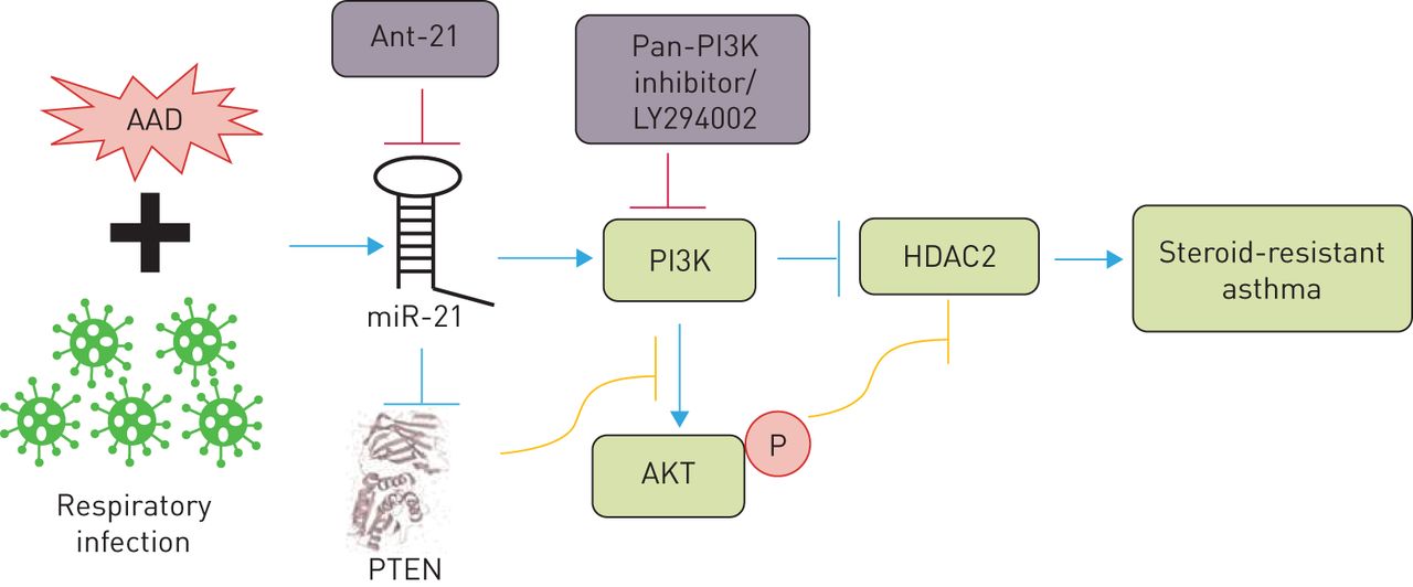
A microRNA (miR)-21/phosphoinositide 3-kinase (PI3K)/histone deacetylase (HDAC)2 axis drives severe steroid-resistant asthma and may be targeted using miR-21-specific inhibitors (such as antagomiR (Ant)-21) and/or PI3K inhibitors (such as LY294002). AAD: allergic airway disease; PTEN: phosphatase and tensin homologue.

We then demonstrated the functional role of this pathway and its potential for therapeutic targeting using our mouse models of infection-induced SSRAAD. Intranasal treatment with a miR-21 inhibitor, antagomiR-21, completely suppressed lung miR-21 expression, increased PTEN expression, reduced PI3K activity and increased nuclear levels of HDAC2. Importantly, treatment with antagomiR-21 effectively suppressed key disease features in SSRAAD. Treatment with antagomiR-21 alone had no effect on airway inflammation (i.e. total leukocytes, eosinophils, neutrophils, macrophages or lymphocytes) but restored sensitivity to steroid treatment in SSRAAD (numbers of all cells were reduced after DEX treatment). Interestingly, treatment with antagomiR-21 in the absence of steroids completely suppressed steroid-resistant AHR in SSRAAD. We also observed similar effects using the pan-PI3K inhibitor LY294002. Notably, all disease features were completely suppressed by antagomiR-21 or LY294002 back to baseline levels achieved with steroid treatment of steroid-sensitive AAD in the absence of infection. Furthermore, we observed similar effects in Haemophilus-, IAV- and RSV-induced SSRAAD.

These data are especially important because Chlamydia/Haemophilus respiratory infections are strongly implicated in type 1/17 immune response-associated, steroid-resistant, neutrophilic asthma, and IAV/RSV infections are strongly implicated in type 1-associated, steroid-resistant, eosinophilic asthma. Collectively, our data show that targeting miR-21 and/or PI3K is likely to be effective across several endotypes of SSR asthma (figure 2) [56].

NLRP3 inflammasome

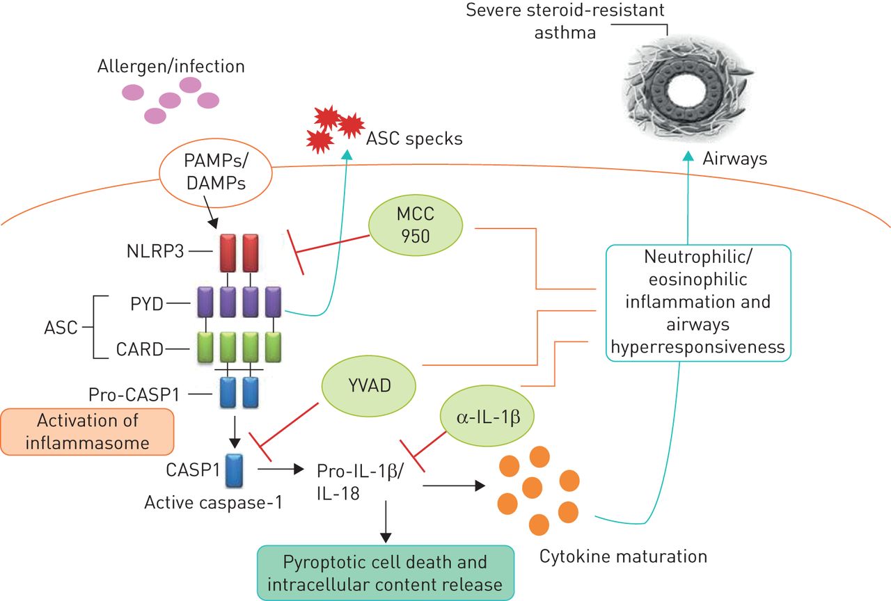
Inflammasomes are multiprotein signalling complexes that control the maturation and release of potent pro-inflammatory cytokines that are produced in response to endogenous, exogenous and pathogenic risk signals [28, 86]. The NLRP3 inflammasome is composed of an apoptosis-associated speck-like protein containing a pyrin domain, a caspase recruitment domain and a pro-caspase-1 domain [86]. The expression and assembly of inflammasomes occurs as a result of stimulation by microbe-derived pathogen-associated molecular patterns such as lipopolysaccharide and double-stranded DNA. A second signal in the form of damage-associated molecular patterns induces the activation of assembled inflammasomes that cleave and activate pro-caspase-1, which in turn cleaves pro-IL-1β and pro-IL-18 into active IL-1β and IL-18, respectively [28, 31, 86]. Thus, inflammasome activation is essential for caspase-1-dependent IL-1β processing, maturation and secretion (figure 3).

FIGURE 3
  • Download figure
  • Open in new tab
  • Download powerpoint
FIGURE 3

Role of the NLRP3 inflammasome in severe steroid-resistant asthma. An infection and/or allergen promotes NLRP3 inflammasome-mediated cleavage of pro-caspase-1 into caspase-1 (CASP1), which then cleaves pro-interleukin (IL)-1β into active IL-1β, which drives steroid-resistant asthma. Neutralising anti (α)-IL-1β antibodies, a CASP1-specific inhibitor (YVAD) and a potent and highly specific NLRP3 inflammasome inhibitor (MCC950) may have therapeutic utility in severe steroid-resistant asthma. PAMPs: pathogen-associated molecular patterns; DAMPs: damage-associated molecular patterns; ASC: adaptor protein apoptosis-associated speck-like containing a caspase recruitment domain; PYD: pyrin domain; CARD: caspase recruitment domain.

Neutrophilic asthma is associated with increased levels of NLRP3 and caspase-1 in the airways, exaggerated IL-1β responses, and Th17 differentiation and IL-17 production that are all implicated in AHR and more severe forms of asthma [30, 31]. Simpson et al. [87] first demonstrated that neutrophilic asthma is associated with increased sputum expression of TLR2, TLR4, IL-1β and IL-8 and increased levels of lipopolysaccharide. These observations are supported by another clinical study by Baines et al. [88], which used sputum RNA expression profiling to show that genes in the IL-1 and TNF-α/NF-κB signalling pathways were overexpressed and correlated with key clinical parameters and neutrophilic airway inflammation in asthma. Notably, Simpson et al. [89] showed that sputum macrophages are a major source of the elevated NLRP3, caspase-1, caspase-4, caspase-5 and IL-1β levels they observed. Furthermore, Darville et al. [90] observed that ATP-mediated P2X7R signalling is essential for immunity against Chlamydia, and Chlamydia, Haemophilus, IAV and RSV infections all induce NLRP3 inflammasome-induced caspase-1-mediated activation and release of IL-1β [91–94].

We recently showed that NLRP3, caspase-1 and IL-1β responses are increased in the lung in Chlamydia infection-induced experimental SSR asthma [30] and that sputum NLRP3 and IL-1β expression correlate with key clinical parameters of human SSR asthma, including increased percentage and absolute numbers of neutrophils in the airways, loss of asthma control and reduced lung function [30]. We then interrogated the functional roles and potential for therapeutic targeting of NLRP3, caspase-1 and IL-1β in experimental SSR asthma by intranasally administering MCC950 (a potent and highly selective NLRP3 inflammasome inhibitor [95]), ac-YVAD-cho (a selective caspase-1 inhibitor) or neutralising anti-IL-1β monoclonal antibody during Chlamydia and Haemophilus infection-induced SSRAAD. Increased lung IL-1β response was suppressed by all three treatments and this effect was associated with the suppression of steroid-resistant neutrophilic airway inflammation and AHR. We then demonstrated that the disease-causing effects of infection are driven by IL-1β and neutrophilic inflammation by recapitulating the key features of neutrophilic SSRAAD in mice with eosinophilic, steroid-sensitive AAD using recombinant mouse IL-1β and anti-Ly6G (blocks recruitment of neutrophils). We propose that specifically targeting the exaggerated NLRP3 inflammasome response component in SSR asthma is a novel and viable therapeutic approach and suggest that this strategy is clinically preferable because it allows for IL-1β processing through other mechanisms for protection against infections [8, 30].

Biologics

There is ample recent evidence that next generation biologics may be steroid sparing or potentially be treatments for SSR disease. IL-5 is well known to be a potent signal for the development, maturation and chemotaxis of eosinophils [4, 5]. Numerous studies suggest that mepolizumab (anti-IL-5 monoclonal antibody) treatment reduces the requirement for oral steroids in severe eosinophilic asthma [96, 97]. Furthermore, there are known links between innate type 2 inducers and mediators IL-33, IL-25 and TSLP (thymic stromal lymphopoietin), and type 2 innate lymphoid and T-helper cells and steroid resistance [98–100]. Antibodies against these factors may prove to be somewhat effective in severe disease [4, 99, 101].

Conclusions

SSR asthma is a clinical problem of significant concern and more effective, and potentially broadly applicable, therapies are urgently required. As a consequence of the underlying mechanisms of pathogenesis, patients do not respond to mainstay steroid-based therapies. Steroid resistance in asthma has been linked to bacterial and viral respiratory infections, infection-induced exacerbations, and a high-fat diet/obesity. Understanding the mechanisms of pathogenesis will enable the identification of novel therapeutic approaches and the development of new and effective treatments. Collectively, clinical and experimental studies have identified macrolides and inhibitors of miR-21, PI3K and inflammasomes as potential new treatments for SSR asthma that may be broadly effective across multiple SSR asthma endotypes. Importantly, our data suggest that aberrant miR-21 and IL-1β responses drive steroid-resistant airway inflammation and AHR in SSR asthma through mechanisms that are independent of the canonical steroid response pathway and are therefore not amenable to steroid-mediated suppression. The logical extension of these studies would be to identify and characterise the roles, and assess the potential for therapeutic targeting, of other miR-21 targets and downstream mediators of IL-1β responses in SSR asthma. Biologics that target type 2 responses may also have utility in SSR asthma.

Footnotes

  • Provenance: Commissioned article, peer reviewed.

  • Conflict of interest: R. Wadhwa has nothing to disclose.

  • Conflict of interest: K. Dua has nothing to disclose.

  • Conflict of interest: I.M. Adcock has nothing to disclose.

  • Conflict of interest: J.C. Horvat has nothing to disclose.

  • Conflict of interest: R.Y. Kim has nothing to disclose.

  • Conflict of interest: P.M. Hansbro reports grants to develop inflammasome inhibitors for therapeutic use.

  • Support statement: R.Y. Kim is supported by a Fellowship from Lung Foundation Australia and Boehringer Ingelheim. P.M. Hansbro is supported by a Fellowship from the National Health and Medical Research Council of Australia (NHMRC #1079187).

  • Received August 1, 2019.
  • Accepted September 19, 2019.
  • Copyright ©ERS 2019.
http://creativecommons.org/licenses/by-nc/4.0/

This article is open access and distributed under the terms of the Creative Commons Attribution Non-Commercial Licence 4.0.

References

  1. ↵
    1. Global Initiative for Asthma (GINA)
    . Global Strategy for Asthma Management and Prevention. 2018. Available from http://ginasthma.org/
  2. ↵
    1. Hansbro PM,
    2. Kim RY,
    3. Starkey MR, et al.
    Mechanisms and treatments for severe, steroid-resistant allergic airway disease and asthma. Immunol Rev 2017; 278: 41–62. doi:10.1111/imr.12543
    OpenUrlCrossRefPubMed
    1. Foster PS,
    2. Maltby S,
    3. Rosenberg HF, et al.
    Modeling TH2 responses and airway inflammation to understand fundamental mechanisms regulating the pathogenesis of asthma. Immunol Rev 2017; 278: 20–40. doi:10.1111/imr.12549
    OpenUrl
  3. ↵
    1. Hansbro PM,
    2. Scott GV,
    3. Essilfie AT, et al.
    Th2 cytokine antagonists: potential treatments for severe asthma. Expert Opin Investig Drugs 2013; 22: 49–69. doi:10.1517/13543784.2013.732997
    OpenUrlCrossRefPubMed
  4. ↵
    1. Hansbro PM,
    2. Kaiko GE,
    3. Foster PS
    . Cytokine/anti-cytokine therapy – novel treatments for asthma? Br J Pharmacol 2011; 163: 81–95. doi:10.1111/j.1476-5381.2011.01219.x
    OpenUrlCrossRefPubMed
  5. ↵
    1. Wenzel SE
    . Asthma phenotypes: the evolution from clinical to molecular approaches. Nat Med 2012; 18: 716–725. doi:10.1038/nm.2678
    OpenUrlCrossRefPubMed
  6. ↵
    1. Hallstrand TS,
    2. Hackett TL,
    3. Altemeier WA, et al.
    Airway epithelial regulation of pulmonary immune homeostasis and inflammation. Clin Immunol 2014; 151: 1–15. doi:10.1016/j.clim.2013.12.003
    OpenUrlCrossRefPubMed
  7. ↵
    1. Kim RY,
    2. Rae B,
    3. Neal R, et al.
    Elucidating novel disease mechanisms in severe asthma. Clin Transl Immunology 2016; 5: e91. doi:10.1038/cti.2016.37
    OpenUrl
    1. Moheimani F,
    2. Hsu AC,
    3. Reid AT, et al.
    The genetic and epigenetic landscapes of the epithelium in asthma. Respir Res 2016; 17: 119. doi:10.1186/s12931-016-0434-4
    OpenUrl
    1. Hansbro PM,
    2. Beagley KW,
    3. Horvat JC, et al.
    Role of atypical bacterial infection of the lung in predisposition/protection of asthma. Pharmacol Ther 2004; 101: 193–210. doi:10.1016/j.pharmthera.2003.10.007
    OpenUrlCrossRefPubMed
    1. Kaiko GE,
    2. Phipps S,
    3. Hickey DK, et al.
    Chlamydia muridarum infection subverts dendritic cell function to promote Th2 immunity and airways hyperreactivity. J Immunol 2008; 180: 2225–2232. doi:10.4049/jimmunol.180.4.2225
    OpenUrlAbstract/FREE Full Text
    1. Starkey MR,
    2. Jarnicki AG,
    3. Essilfie AT, et al.
    Murine models of infectious exacerbations of airway inflammation. Curr Opin Pharmacol 2013; 13: 337–344. doi:10.1016/j.coph.2013.03.005
    OpenUrlCrossRefPubMed
    1. Wood LG,
    2. Li Q,
    3. Scott HA, et al.
    Saturated fatty acids, obesity, and the nucleotide oligomerization domain-like receptor protein 3 (NLRP3) inflammasome in asthmatic patients. J Allergy Clin Immunol 2019; 143: 305–315. doi:10.1016/j.jaci.2018.04.037
    OpenUrl
    1. Rutting S,
    2. Xenaki D,
    3. Malouf M, et al.
    Short-chain fatty acids increase TNFα-induced inflammation in primary human lung mesenchymal cells through the activation of p38 MAPK. Am J Physiol Lung Cell Mol Physiol 2019; 316: L157–L174. doi:10.1152/ajplung.00306.2018
    OpenUrl
    1. Hirota JA,
    2. Gold MJ,
    3. Hiebert PR, et al.
    The nucleotide-binding domain, leucine-rich repeat protein 3 inflammasome/IL-1 receptor I axis mediates innate, but not adaptive, immune responses after exposure to particulate matter under 10 μm. Am J Respir Cell Mol Biol 2015; 52: 96–105. doi:10.1165/rcmb.2014-0158OC
    OpenUrlCrossRefPubMed
  8. ↵
    1. Essilfie AT,
    2. Simpson JL,
    3. Dunkley ML, et al.
    Combined Haemophilus influenzae respiratory infection and allergic airways disease drives chronic infection and features of neutrophilic asthma. Thorax 2012; 67: 588–599. doi:10.1136/thoraxjnl-2011-200160
    OpenUrlAbstract/FREE Full Text
  9. ↵
    1. Horvat JC,
    2. Starkey MR,
    3. Kim RY, et al.
    Early-life chlamydial lung infection enhances allergic airways disease through age-dependent differences in immunopathology. J Allergy Clin Immunol 2010; 125: 617–625. doi:10.1016/j.jaci.2009.10.018
    OpenUrlCrossRef
  10. ↵
    1. Horvat JC,
    2. Starkey MR,
    3. Kim RY, et al.
    Chlamydial respiratory infection during allergen sensitization drives neutrophilic allergic airways disease. J Immunol 2010; 184: 4159–4169. doi:10.4049/jimmunol.0902287
    OpenUrlAbstract/FREE Full Text
  11. ↵
    1. Wood LG,
    2. Simpson JL,
    3. Hansbro PM, et al.
    Potentially pathogenic bacteria cultured from the sputum of stable asthmatics are associated with increased 8-isoprostane and airway neutrophilia. Free Radic Res 2010; 44: 146–154. doi:10.3109/10715760903362576
    OpenUrlCrossRefPubMed
  12. ↵
    1. Pavord ID,
    2. Beasley R,
    3. Agusti A, et al.
    After asthma: redefining airways diseases. Lancet 2018; 391: 350–400. doi:10.1016/S0140-6736(17)30879-6
    OpenUrlCrossRefPubMed
  13. ↵
    1. Ahmed H,
    2. Turner S
    . Severe asthma in children – a review of definitions, epidemiology, and treatment options in 2019. Pediatr Pulmonol 2019; 54: 778–787. doi:10.1002/ppul.24317
    OpenUrl
    1. Marko A,
    2. Ross KR
    . Severe asthma in childhood. Immunol Allergy Clin North Am 2019; 39: 243–257. doi:10.1016/j.iac.2018.12.007
    OpenUrl
  14. ↵
    1. Saglani S,
    2. Fleming L,
    3. Sonnappa S, et al.
    Advances in the aetiology, management, and prevention of acute asthma attacks in children. Lancet Child Adolesc Health 2019; 3: 354–364. doi:10.1016/S2352-4642(19)30025-2
    OpenUrl
  15. ↵
    1. Rhen T,
    2. Cidlowski JA
    . Antiinflammatory action of glucocorticoids – new mechanisms for old drugs. N Engl J Med 2005; 353: 1711–1723. doi:10.1056/NEJMra050541
    OpenUrlCrossRefPubMed
  16. ↵
    1. Barnes PJ
    . Biochemical basis of asthma therapy. J Biol Chem 2011; 286: 32899–32905. doi:10.1074/jbc.R110.206466
    OpenUrlAbstract/FREE Full Text
  17. ↵
    1. Wang W,
    2. Li JJ,
    3. Foster PS, et al.
    Potential therapeutic targets for steroid-resistant asthma. Curr Drug Targets 2010; 11: 957–970. doi:10.2174/138945010791591412
    OpenUrlCrossRefPubMed
  18. ↵
    1. Barnes PJ,
    2. Adcock IM
    . Glucocorticoid resistance in inflammatory diseases. Lancet 2009; 373: 1905–1917. doi:10.1016/S0140-6736(09)60326-3
    OpenUrlCrossRefPubMed
  19. ↵
    1. Kim RY,
    2. Pinkerton JW,
    3. Gibson PG, et al.
    Inflammasomes in COPD and neutrophilic asthma. Thorax 2015; 70: 1199–1201. doi:10.1136/thoraxjnl-2014-206736
    OpenUrlFREE Full Text
    1. Green R,
    2. Brightling CE,
    3. Woltmann G, et al.
    Analysis of induced sputum in adults with asthma: identification of subgroup with isolated sputum neutrophilia and poor response to inhaled corticosteroids. Thorax 2002; 57: 875–879. doi:10.1136/thorax.57.10.875
    OpenUrlAbstract/FREE Full Text
  20. ↵
    1. Kim RY,
    2. Pinkerton JW,
    3. Essilfie AT, et al.
    Role for NLRP3 inflammasome-mediated, IL-1β-dependent responses in severe, steroid-resistant asthma. Am J Respir Crit Care Med 2017; 196: 283–297. doi:10.1164/rccm.201609-1830OC
    OpenUrlCrossRefPubMed
  21. ↵
    1. Pinkerton JW,
    2. Kim RY,
    3. Robertson AAB, et al.
    Inflammasomes in the lung. Mol Immunol 2017; 86: 44–55. doi:10.1016/j.molimm.2017.01.014
    OpenUrlCrossRef
  22. ↵
    1. Gold M,
    2. Hiebert PR,
    3. Park HY, et al.
    Mucosal production of uric acid by airway epithelial cells contributes to particulate matter-induced allergic sensitization. Mucosal Immunol 2016; 9: 809–820. doi:10.1038/mi.2015.104
    OpenUrlCrossRefPubMed
  23. ↵
    1. Schwartz HJ,
    2. Lowell FC,
    3. Melby JC
    . Steroid resistance in bronchial asthma. Ann Intern Med 1968; 68: 1141. doi:10.7326/0003-4819-68-5-1141_3
    OpenUrl
  24. ↵
    1. Adcock IM,
    2. Ford PA,
    3. Bhavsar P, et al.
    Steroid resistance in asthma: mechanisms and treatment options. Curr Allergy Asthma Rep 2008; 8: 171–178. doi:10.1007/s11882-008-0028-4
    OpenUrlCrossRefPubMed
  25. ↵
    1. Barnes PJ
    . Inhaled glucocorticoids for asthma. N Engl J Med 1995; 332: 868–875. doi:10.1056/NEJM199503303321307
    OpenUrlCrossRefPubMed
  26. ↵
    1. Ito K,
    2. Getting SJ,
    3. Charron CE
    . Mode of glucocorticoid actions in airway disease. ScientificWorldJournal 2006; 6: 1750–1769. doi:10.1100/tsw.2006.274
    OpenUrlCrossRefPubMed
  27. ↵
    1. Oakley RH,
    2. Jewell CM,
    3. Yudt MR, et al.
    The dominant negative activity of the human glucocorticoid receptor β isoform. Specificity and mechanisms of action. J Biol Chem 1999; 274: 27857–27866. doi:10.1074/jbc.274.39.27857
    OpenUrlAbstract/FREE Full Text
  28. ↵
    1. Tao T,
    2. Lan J,
    3. Lukacs GL, et al.
    Importin 13 regulates nuclear import of the glucocorticoid receptor in airway epithelial cells. Am J Respir Cell Mol Biol 2006; 35: 668–680. doi:10.1165/rcmb.2006-0073OC
    OpenUrlCrossRefPubMed
  29. ↵
    1. Horwitz K,
    2. Jackson TA,
    3. Bain DL, et al.
    Nuclear receptor coactivators and corepressors. Mol Endocrinol 1996; 10: 1167–1177.
    OpenUrlCrossRefPubMed
    1. Barnes PJ
    . Glucocorticosteroids: current and future directions. Br J Pharmacol 2011; 163: 29–43. doi:10.1111/j.1476-5381.2010.01199.x
    OpenUrlCrossRefPubMed
  30. ↵
    1. Hansbro PM,
    2. Starkey MR,
    3. Mattes J, et al.
    Pulmonary immunity during respiratory infections in early life and the development of severe asthma. Ann Am Thorac Soc 2014; 11: Suppl. 5, S297–S302. doi:10.1513/AnnalsATS.201402-086AW
    OpenUrl
  31. ↵
    1. Ito K,
    2. Chung KF,
    3. Adcock IM
    . Update on glucocorticoid action and resistance. J Allergy Clin Immunol 2006; 117: 522–543. doi:10.1016/j.jaci.2006.01.032
    OpenUrlCrossRefPubMed
  32. ↵
    1. Goleva E,
    2. Li LB,
    3. Eves PT, et al.
    Increased glucocorticoid receptor β alters steroid response in glucocorticoid-insensitive asthma. Am J Respir Crit Care Med 2006; 173: 607–616. doi:10.1164/rccm.200507-1046OC
    OpenUrlCrossRefPubMed
  33. ↵
    1. Irusen E,
    2. Matthews JG,
    3. Takahashi A, et al.
    p38 Mitogen-activated protein kinase-induced glucocorticoid receptor phosphorylation reduces its activity: role in steroid-insensitive asthma. J Allergy Clin Immunol 2002; 109: 649–657. doi:10.1067/mai.2002.122465
    OpenUrlCrossRefPubMed
  34. ↵
    1. Adcock I,
    2. Lane S
    . Mechanisms of steroid action and resistance in inflammation. Corticosteroid-insensitive asthma: molecular mechanisms. J Endocrinol 2003; 178: 347–355. doi:10.1677/joe.0.1780347
    OpenUrlAbstract
  35. ↵
    1. Loke TK,
    2. Mallett KH,
    3. Ratoff J, et al.
    Systemic glucocorticoid reduces bronchial mucosal activation of activator protein 1 components in glucocorticoid-sensitive but not glucocorticoid-resistant asthmatic patients. J Allergy Clin Immunol 2006; 118: 368–375. doi:10.1016/j.jaci.2006.04.055
    OpenUrlCrossRefPubMed
  36. ↵
    1. Gunawardhana L,
    2. Gibson PG,
    3. Simpson JL, et al.
    Activity and expression of histone acetylases and deacetylases in inflammatory phenotypes of asthma. Clin Exp Allergy 2014; 44: 47–57. doi:10.1111/cea.12168
    OpenUrlCrossRefPubMed
  37. ↵
    1. Cosio BG,
    2. Tsaprouni L,
    3. Ito K, et al.
    Theophylline restores histone deacetylase activity and steroid responses in COPD macrophages. J Exp Med 2004; 200: 689–695. doi:10.1084/jem.20040416
    OpenUrlAbstract/FREE Full Text
  38. ↵
    1. Adcock IM,
    2. Barnes PJ
    . Molecular mechanisms of corticosteroid resistance. Chest 2008; 134: 394–401. doi:10.1378/chest.08-0440
    OpenUrlCrossRefPubMed
  39. ↵
    1. Barnes PJ
    . Histone deacetylase-2 and airway disease. Ther Adv Respir Dis 2009; 3: 235–243. doi:10.1177/1753465809348648
    OpenUrlCrossRefPubMed
    1. Adcock IM,
    2. Ito K,
    3. Barnes PJ
    . Histone deacetylation: an important mechanism in inflammatory lung diseases. COPD 2005; 2: 445–455. doi:10.1080/15412550500346683
    OpenUrl
  40. ↵
    1. Marwick JA,
    2. Caramori G,
    3. Stevenson CS, et al.
    Inhibition of PI3Kδ restores glucocorticoid function in smoking-induced airway inflammation in mice. Am J Respir Crit Care Med 2009; 179: 542–548. doi:10.1164/rccm.200810-1570OC
    OpenUrlCrossRefPubMed
  41. ↵
    1. Gibson PG,
    2. Saltos N,
    3. Borgas T
    . Airway mast cells and eosinophils correlate with clinical severity and airway hyperresponsiveness in corticosteroid-treated asthma. J Allergy Clin Immunol 2000; 105: 752–759. doi:10.1067/mai.2000.105319
    OpenUrlCrossRefPubMed
  42. ↵
    1. Essilfie AT,
    2. Simpson JL,
    3. Horvat JC, et al.
    Haemophilus influenzae infection drives IL-17-mediated neutrophilic allergic airways disease. PLoS Pathog 2011; 7: e1002244. doi:10.1371/journal.ppat.1002244
    OpenUrlCrossRefPubMed
  43. ↵
    1. Essilfie AT,
    2. Horvat JC,
    3. Kim RY, et al.
    Macrolide therapy suppresses key features of experimental steroid-sensitive and steroid-insensitive asthma. Thorax 2015; 70: 458–467. doi:10.1136/thoraxjnl-2014-206067
    OpenUrlAbstract/FREE Full Text
  44. ↵
    1. Kim RY,
    2. Horvat JC,
    3. Pinkerton JW, et al.
    MicroRNA-21 drives severe, steroid-insensitive experimental asthma by amplifying phosphoinositide 3-kinase-mediated suppression of histone deacetylase 2. J Allergy Clin Immunol 2017; 139: 519–532. doi:10.1016/j.jaci.2016.04.038
    OpenUrlCrossRefPubMed
  45. ↵
    1. Liu G,
    2. Cooley MA,
    3. Jarnicki AG, et al.
    Fibulin-1 regulates the pathogenesis of tissue remodeling in respiratory diseases. JCI Insight 2016; 1: e86380.
    OpenUrl
    1. Thorburn AN,
    2. Foster PS,
    3. Gibson PG, et al.
    Components of Streptococcus pneumoniae suppress allergic airways disease and NKT cells by inducing regulatory T cells. J Immunol 2012; 188: 4611–4620. doi:10.4049/jimmunol.1101299
    OpenUrlAbstract/FREE Full Text
  46. ↵
    1. Liu G,
    2. Cooley MA,
    3. Nair PM, et al.
    Airway remodeling and inflammation in asthma are dependent on the extracellular matrix protein fibulin-1c. J Pathol 2017; 243: 510–523. doi:10.1002/path.4979
    OpenUrl
  47. ↵
    1. Valladao AC,
    2. Frevert CW,
    3. Koch LK, et al.
    STAT6 regulates the development of eosinophilic versus neutrophilic asthma in response to Alternaria alternata. J Immunol 2016; 197: 4541–4551. doi:10.4049/jimmunol.1600007
    OpenUrlAbstract/FREE Full Text
  48. ↵
    1. Gong D,
    2. Fei F,
    3. Lim M, et al.
    Abr, a negative regulator of Rac, attenuates cockroach allergen-induced asthma in a mouse model. J Immunol 2013; 191: 4514–4520. doi:10.4049/jimmunol.1202603
    OpenUrlAbstract/FREE Full Text
  49. ↵
    1. Hahn DL,
    2. Schure A,
    3. Patel K, et al.
    Chlamydia pneumoniae-specific IgE is prevalent in asthma and is associated with disease severity. PLoS One 2012; 7: e35945. doi:10.1371/journal.pone.0035945
    OpenUrlCrossRefPubMed
    1. Patel KK,
    2. Vicencio AG,
    3. Du Z, et al.
    Infectious Chlamydia pneumoniae is associated with elevated interleukin-8 and airway neutrophilia in children with refractory asthma. Pediatr Infect Dis J 2010; 29: 1093–1098. doi:10.1097/inf.0b013e3181eaebdc
    OpenUrlCrossRefPubMed
  50. ↵
    1. Patel KK,
    2. Webley WC
    . Evidence of infectious asthma phenotype: Chlamydia-induced allergy and pathogen-specific IgE in a neonatal mouse model. PLoS One 2013; 8: e83453. doi:10.1371/journal.pone.0083453
    OpenUrl
  51. ↵
    1. Horvat JC,
    2. Beagley KW,
    3. Wade MA, et al.
    Neonatal chlamydial infection induces mixed T-cell responses that drive allergic airway disease. Am J Respir Crit Care Med 2007; 176: 556–564. doi:10.1164/rccm.200607-1005OC
    OpenUrlCrossRefPubMed
    1. Starkey MR,
    2. Essilfie AT,
    3. Horvat JC, et al.
    Constitutive production of IL-13 promotes early-life Chlamydia respiratory infection and allergic airway disease. Mucosal Immunol 2013; 6: 569–579. doi:10.1038/mi.2012.99
    OpenUrlCrossRefPubMed
  52. ↵
    1. Starkey MR,
    2. Nguyen DH,
    3. Essilfie AT, et al.
    Tumor necrosis factor-related apoptosis-inducing ligand translates neonatal respiratory infection into chronic lung disease. Mucosal Immunol 2014; 7: 478–488. doi:10.1038/mi.2013.65
    OpenUrlCrossRef
  53. ↵
    1. Beale J,
    2. Jayaraman A,
    3. Jackson DJ, et al.
    Rhinovirus-induced IL-25 in asthma exacerbation drives type 2 immunity and allergic pulmonary inflammation. Sci Transl Med 2014; 6: 256ra134. doi:10.1126/scitranslmed.3009124
    OpenUrlAbstract/FREE Full Text
  54. ↵
    1. Hansbro NG,
    2. Horvat JC,
    3. Wark PA, et al.
    Understanding the mechanisms of viral induced asthma: new therapeutic directions. Pharmacol Ther 2008; 117: 313–353. doi:10.1016/j.pharmthera.2007.11.002
    OpenUrlCrossRefPubMed
  55. ↵
    1. Crosbie P,
    2. Woodhead M
    . Long-term macrolide therapy in chronic inflammatory airway diseases. Eur Respir J 2009; 33: 171–181. doi:10.1183/09031936.00042208
    OpenUrlAbstract/FREE Full Text
  56. ↵
    1. Xepapadaki P,
    2. Koutsoumpari I,
    3. Papaevagelou V, et al.
    Atypical bacteria and macrolides in asthma. Allergy Asthma Clin Immunol 2008; 4: 111. doi:10.1186/1710-1492-4-3-111
    OpenUrlCrossRefPubMed
  57. ↵
    1. Spahn JD,
    2. Fost DA,
    3. Covar R, et al.
    Clarithromycin potentiates glucocorticoid responsiveness in patients with asthma: results of a pilot study. Ann Allergy Asthma Immunol 2001; 87: 501–505. doi:10.1016/S1081-1206(10)62264-8
    OpenUrlCrossRefPubMed
  58. ↵
    1. Black PN,
    2. Blasi F,
    3. Jenkins CR, et al.
    Trial of roxithromycin in subjects with asthma and serological evidence of infection with Chlamydia pneumoniae. Am J Respir Crit Care Med 2001; 164: 536–541. doi:10.1164/ajrccm.164.4.2011040
    OpenUrlCrossRefPubMed
  59. ↵
    1. Esposito S,
    2. Blasi F,
    3. Bosis F
    . Efficacy of clarithromycin for the treatment of acute episodes of bronchospasm in children with a history of recurrent wheezing. Tampere, 22nd Annual Meeting of European Society for Paediatric Infectious Diseases (ESPID), 2004.
  60. ↵
    1. Kraft M,
    2. Cassell GH,
    3. Pak J, et al.
    Mycoplasma pneumoniae and Chlamydia pneumoniae in asthma: effect of clarithromycin. Chest 2002; 121: 1782–1788. doi:10.1378/chest.121.6.1782
    OpenUrlCrossRefPubMed
  61. ↵
    1. Simpson JL,
    2. Powell H,
    3. Boyle MJ, et al.
    Clarithromycin targets neutrophilic airway inflammation in refractory asthma. Am J Respir Crit Care Med 2008; 177: 148–155. doi:10.1164/rccm.200707-1134OC
    OpenUrlCrossRefPubMed
  62. ↵
    1. Gibson PG,
    2. Yang IA,
    3. Upham JW, et al.
    Effect of azithromycin on asthma exacerbations and quality of life in adults with persistent uncontrolled asthma (AMAZES): a randomised, double-blind, placebo-controlled trial. Lancet 2017; 390: 659–668. doi:10.1016/S0140-6736(17)31281-3
    OpenUrlCrossRef
  63. ↵
    1. Ito K,
    2. Ito M,
    3. Elliott WM, et al.
    Decreased histone deacetylase activity in chronic obstructive pulmonary disease. N Engl J Med 2005; 352: 1967–1976. doi:10.1056/NEJMoa041892
    OpenUrlCrossRefPubMed
  64. ↵
    1. Hsu AC,
    2. Starkey MR,
    3. Hanish I, et al.
    Targeting PI3K-p110α suppresses influenza virus infection in chronic obstructive pulmonary disease. Am J Respir Crit Care Med 2015; 191: 1012–1023. doi:10.1164/rccm.201501-0188OC
    OpenUrlCrossRefPubMed
  65. ↵
    1. Haylock-Jacobs S,
    2. Comerford I,
    3. Bunting M, et al.
    PI3Kδ drives the pathogenesis of experimental autoimmune encephalomyelitis by inhibiting effector T cell apoptosis and promoting Th17 differentiation. J Autoimmun 2011; 36: 278–287. doi:10.1016/j.jaut.2011.02.006
    OpenUrlCrossRefPubMed
  66. ↵
    1. Foster PS,
    2. Plank M,
    3. Collison A, et al.
    The emerging role of microRNAs in regulating immune and inflammatory responses in the lung. Immunol Rev 2013; 253: 198–215. doi:10.1111/imr.12058
    OpenUrlCrossRefPubMed
  67. ↵
    1. Plank MW,
    2. Maltby S,
    3. Tay HL, et al.
    MicroRNA expression is altered in an ovalbumin-induced asthma model and targeting miR-155 with antagomirs reveals cellular specificity. PLoS One 2015; 10: e0144810. doi:10.1371/journal.pone.0144810
    OpenUrl
  68. ↵
    1. Lu TX,
    2. Munitz A,
    3. Rothenberg ME
    . MicroRNA-21 is up-regulated in allergic airway inflammation and regulates IL-12p35 expression. J Immunol 2009; 182: 4994–5002. doi:10.4049/jimmunol.0803560
    OpenUrlAbstract/FREE Full Text
  69. ↵
    1. Kwak YG,
    2. Song CH,
    3. Yi HK, et al.
    Involvement of PTEN in airway hyperresponsiveness and inflammation in bronchial asthma. J Clin Invest 2003; 111: 1083–1092. doi:10.1172/JCI16440
    OpenUrlCrossRefPubMed
  70. ↵
    1. Ezeamuzie CI,
    2. Sukumaran J,
    3. Philips E
    . Effect of wortmannin on human eosinophil responses in vitro and on bronchial inflammation and airway hyperresponsiveness in guinea pigs in vivo. Am J Respir Crit Care Med 2001; 164: 1633–1639. doi:10.1164/ajrccm.164.9.2101104
    OpenUrlCrossRefPubMed
  71. ↵
    1. Schroder K,
    2. Tschopp J
    . The inflammasomes. Cell 2010; 140: 821–832. doi:10.1016/j.cell.2010.01.040
    OpenUrlCrossRefPubMed
  72. ↵
    1. Simpson JL,
    2. Grissell TV,
    3. Douwes J, et al.
    Innate immune activation in neutrophilic asthma and bronchiectasis. Thorax 2007; 62: 211–218. doi:10.1136/thx.2006.061358
    OpenUrlAbstract/FREE Full Text
  73. ↵
    1. Baines KJ,
    2. Simpson JL,
    3. Wood LG, et al.
    Transcriptional phenotypes of asthma defined by gene expression profiling of induced sputum samples. J Allergy Clin Immunol 2011; 127: 153–160. doi:10.1016/j.jaci.2010.10.024
    OpenUrlCrossRef
  74. ↵
    1. Simpson JL,
    2. Phipps S,
    3. Baines KJ, et al.
    Elevated expression of the NLRP3 inflammasome in neutrophilic asthma. Eur Respir J 2014; 43: 1067–1076. doi:10.1183/09031936.00105013
    OpenUrlAbstract/FREE Full Text
  75. ↵
    1. Darville T,
    2. Welter-Stahl L,
    3. Cruz C, et al.
    Effect of the purinergic receptor P2X7 on Chlamydia infection in cervical epithelial cells and vaginally infected mice. J Immunol 2007; 179: 3707–3714. doi:10.4049/jimmunol.179.6.3707
    OpenUrlAbstract/FREE Full Text
  76. ↵
    1. He X,
    2. Mekasha S,
    3. Mavrogiorgos N, et al.
    Inflammation and fibrosis during Chlamydia pneumoniae infection is regulated by IL-1 and the NLRP3/ASC inflammasome. J Immunol 2010; 184: 5743–5754. doi:10.4049/jimmunol.0903937
    OpenUrlAbstract/FREE Full Text
    1. Rotta Detto Loria J,
    2. Rohmann K,
    3. Droemann D, et al.
    Nontypeable Haemophilus influenzae infection upregulates the NLRP3 inflammasome and leads to caspase-1-dependent secretion of interleukin-1β – a possible pathway of exacerbations in COPD. PLoS One 2013; 8: e66818. doi:10.1371/journal.pone.0066818
    OpenUrlCrossRefPubMed
    1. Ichinohe T,
    2. Pang IK,
    3. Iwasaki A
    . Influenza virus activates inflammasomes via its intracellular M2 ion channel. Nat Immunol 2010; 11: 404–410. doi:10.1038/ni.1861
    OpenUrlCrossRefPubMed
  77. ↵
    1. Segovia J,
    2. Sabbah A,
    3. Mgbemena V, et al.
    TLR2/MyD88/NF-κB pathway, reactive oxygen species, potassium efflux activates NLRP3/ASC inflammasome during respiratory syncytial virus infection. PLoS One 2012; 7: e29695. doi:10.1371/journal.pone.0029695
    OpenUrlCrossRefPubMed
  78. ↵
    1. Coll RC,
    2. Robertson AA,
    3. Chae JJ, et al.
    A small-molecule inhibitor of the NLRP3 inflammasome for the treatment of inflammatory diseases. Nat Med 2015; 21: 248–255. doi:10.1038/nm.3806
    OpenUrlCrossRefPubMed
  79. ↵
    1. Bel EH,
    2. Wenzel SE,
    3. Thompson PJ, et al.
    Oral glucocorticoid-sparing effect of mepolizumab in eosinophilic asthma. N Engl J Med 2014; 371: 1189–1197. doi:10.1056/NEJMoa1403291
    OpenUrlCrossRefPubMed
  80. ↵
    1. Ortega HG,
    2. Liu MC,
    3. Pavord ID, et al.
    Mepolizumab treatment in patients with severe eosinophilic asthma. N Engl J Med 2014; 371: 1198–1207. doi:10.1056/NEJMoa1403290
    OpenUrlCrossRefPubMed
  81. ↵
    1. Starkey MR,
    2. McKenzie AN,
    3. Belz GT, et al.
    Group 2 innate lymphoid cells: surprises and challenges. Mucosal Immunol 2019; 12: 299–311. doi:10.1038/s41385-018-0130-4
    OpenUrl
  82. ↵
    1. Robinson D,
    2. Humbert M,
    3. Buhl R, et al.
    Revisiting Type 2-high and Type 2-low airway inflammation in asthma: current knowledge and therapeutic implications. Clin Exp Allergy 2017; 47: 161–175. doi:10.1111/cea.12880
    OpenUrlCrossRef
  83. ↵
    1. Christianson CA,
    2. Goplen NP,
    3. Zafar I, et al.
    Persistence of asthma requires multiple feedback circuits involving type 2 innate lymphoid cells and IL-33. J Allergy Clin Immunol 2015; 136: 59–68.e14. doi:10.1016/j.jaci.2014.11.037
    OpenUrlCrossRef
  84. ↵
    1. Pepper AN,
    2. Renz H,
    3. Casale TB, et al.
    Biologic therapy and novel molecular targets of severe asthma. J Allergy Clin Immunol Pract 2017; 5: 909–916. doi:10.1016/j.jaip.2017.04.038
    OpenUrl
PreviousNext
Back to top
View this article with LENS
Vol 28 Issue 153 Table of Contents
European Respiratory Review: 28 (153)
  • Table of Contents
  • Index by author
Email

Thank you for your interest in spreading the word on European Respiratory Society .

NOTE: We only request your email address so that the person you are recommending the page to knows that you wanted them to see it, and that it is not junk mail. We do not capture any email address.

Enter multiple addresses on separate lines or separate them with commas.
Cellular mechanisms underlying steroid-resistant asthma
(Your Name) has sent you a message from European Respiratory Society
(Your Name) thought you would like to see the European Respiratory Society web site.
CAPTCHA
This question is for testing whether or not you are a human visitor and to prevent automated spam submissions.
Print
Citation Tools
Cellular mechanisms underlying steroid-resistant asthma
Ridhima Wadhwa, Kamal Dua, Ian M. Adcock, Jay C. Horvat, Richard Y. Kim, Philip M. Hansbro
European Respiratory Review Sep 2019, 28 (153) 190096; DOI: 10.1183/16000617.0096-2019

Citation Manager Formats

  • BibTeX
  • Bookends
  • EasyBib
  • EndNote (tagged)
  • EndNote 8 (xml)
  • Medlars
  • Mendeley
  • Papers
  • RefWorks Tagged
  • Ref Manager
  • RIS
  • Zotero

Share
Cellular mechanisms underlying steroid-resistant asthma
Ridhima Wadhwa, Kamal Dua, Ian M. Adcock, Jay C. Horvat, Richard Y. Kim, Philip M. Hansbro
European Respiratory Review Sep 2019, 28 (153) 190096; DOI: 10.1183/16000617.0096-2019
del.icio.us logo Digg logo Reddit logo Technorati logo Twitter logo CiteULike logo Connotea logo Facebook logo Google logo Mendeley logo
Full Text (PDF)

Jump To

  • Article
    • Abstract
    • Abstract
    • Introduction
    • Mechanisms of steroid resistance
    • Role of respiratory infections in SSR asthma
    • Novel treatment strategies for SSR asthma
    • Conclusions
    • Footnotes
    • References
  • Figures & Data
  • Info & Metrics
  • PDF

Subjects

  • Asthma and allergy
  • Tweet Widget
  • Facebook Like
  • Google Plus One

More in this TOC Section

  • Particulate matter and the airway epithelium
  • The microbiome in bronchiectasis
Show more Lung Science Conference Review

Related Articles

Navigate

  • Home
  • Current issue
  • Archive

About the ERR

  • Journal information
  • Editorial board
  • Reviewers
  • Press
  • Permissions and reprints
  • Advertising
  • Sponsorship

The European Respiratory Society

  • Society home
  • myERS
  • Privacy policy
  • Accessibility

ERS publications

  • European Respiratory Journal
  • ERJ Open Research
  • European Respiratory Review
  • Breathe
  • ERS books online
  • ERS Bookshop

Help

  • Feedback

For authors

  • Instructions for authors
  • Publication ethics and malpractice
  • Submit a manuscript

For readers

  • Alerts
  • Subjects
  • RSS

Subscriptions

  • Accessing the ERS publications

Contact us

European Respiratory Society
442 Glossop Road
Sheffield S10 2PX
United Kingdom
Tel: +44 114 2672860
Email: journals@ersnet.org

ISSN

Print ISSN: 0905-9180
Online ISSN: 1600-0617

Copyright © 2023 by the European Respiratory Society