


Figures
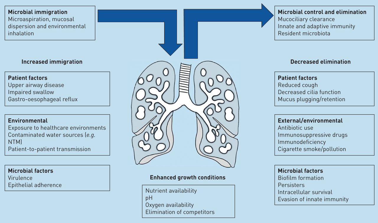
- FIGURE 1
Illustration of the factors modifying the healthy and diseased lung microbiome. The arrows illustrate the immigration of bacteria to the lungs and the balanced removal of bacteria by mucociliary clearance and immune clearance. Multiple factors have been described that increase immigration of bacteria and decrease clearance, leading to dysbiosis. The list is not intended to be comprehensive. NTM: nontuberculous mycobacteria.
- FIGURE 2
The microbiology of bronchiectasis. a) Culture-based microbiology results from a European cohort study [44]; b) culture-based microbiology results from the US Bronchiectasis Registry [43]; c) a representative microbiome profile for the bronchiectasis population (hypothetical data based on published work) [45] (low-abundance genera (<1%) such as Leptotrichia are not shown); d) simplified concept of microbiota dysbiosis and loss of diversity in the bronchiectasis lung. NTM: nontuberculous mycobacteria.

Tables
- TABLE 1
Commonly used terminology in microbiome studies
Microbiota The whole collection of microorganisms within a specific location (e.g. the lung), includes bacteria, fungi and viruses Microbiome The genetic information and inferred physiochemical properties of the gene products of the microbiota. In most articles the term is used to mean the taxonomic classification of bacteria present in a sample and is used as such in this review Bacteriome The genetic information and inferred physiochemical properties of the gene products of the bacteria present in a specific location Mycobiome The genetic information and inferred physiochemical properties of the gene products of the fungi present in a specific location Virome The genetic information and inferred physiochemical properties of the gene products of the viruses present in a specific location Metagenomics Shotgun random sequencing of total DNA in a sample, including DNA from host and microbe origin, which is analysed, organised and identified using sequence databases and computational tools 16S rRNA gene Component of the 30S small subunit of prokaryotic ribosomes. It is used in molecular studies owing to its extremely slow rate of evolution and the presence of both variable and constant regions OTUs Clusters of similar 16S rRNA gene sequences. Each OTU represents a taxonomic unit of a bacteria family or genus depending on the sequence similarity threshold. Identification to the species level is not usually possible α-diversity α-diversity is a measure of how diverse a sample is based on how many species there are (richness) and how abundant each species is (evenness) within that sample β-diversity β-diversity is used to show how different samples are from each other, based on differences in bacterial presence, abundance or a phylogenetic tree rRNA: ribosomal RNA; OTUs: operational taxonomic units.
- TABLE 2
Summary of bronchiectasis microbiome studies
First author, [ref.] Year Sample size n Sample type Patient characteristics DNA extraction 16S region Sequencing method Data analysis Taxa found Byun [41] 2017 14 Sputum, BAL Stable and exacerbated MG Blood Genomic DNA Extraction SV kit (MGmed, Seoul, Republic of Korea) V2,4,8 V3,6–7,9 Ion 318 v2 chip, Ion PGM Sequencing 400 kit (Life Technologies, Carlsbad, CA, USA) QIIME [37] and R packages Haemophilus, Pseudomonas, Moraxella, Klebsiella, Prevotella and Veillonella Cox [47] 2017 76 Sputum Stable and exacerbated samples FastDNA Spin Kit for Soil (MP Biomedicals, Santa Ana, CA, USA) V3-V5 Roche 454 FLX sequencer (Basel, Switzerland) QIIME and R packages Haemophilus, Pseudomonas and Staphylococcus Maughan [73] 2012 31 Lung tissue Surgical resection/lung transplantation Direct PCR DNA Extraction System (Viagen Biotech, Los Angeles, CA, USA) V1-V4 Applied Biosystems (Foster City, CA, USA) 3130XL sequencer QIIME Pseudomonas, Stenotrophomonas, Staphylococcus, Burkholderia and Haemophilus Purcell [74] 2014 70 Sputum 20 exacerbated, 50 non-exacerbated MoBio Ultraclean Microbial DNA isolation kit (MoBio, CA, USA) V3 Pyrosequencing QIIME Pasteurellaceae, Streptococceae, Pseudomonadaceae, Prevotellaceae, Veillonellaceae and Actinomycetaceae Rogers (BLESS) [42] 2013 41 Sputum, BAL Clinically stable patients with at least two infective exacerbations in the previous 12 months Laboratory-specific protocol including a bead-beating step and a phenol/chloroform precipitation V1-V3 Pyrosequencing Custom C# software in MicrosoftH.NET environment Pseudomonas, Haemophilus, Streptococcus, Prevotella, Veillonella and Neisseria Rogers (BLESS) [54] 2014 96 Sputum Patients required to be clinically stable, not receiving systemic corticosteroids and macrolide-naive Laboratory-specific protocol including a bead-beating step and a phenol/chloroform precipitation V1-V3 Pyrosequencing Custom C# software in MicrosoftH.NET environment Haemophilus, Pseudomonas, Veillonella and Streptococcus Rogers (BLESS) [46] 2014 96 Sputum As above As above V1-V3 Tag-encoded FLX amplicon pyrosequencing (bTEFAP) QIIME N/A Rogers (BLESS) [75] 2015 60 Sputum Stable As above V1-V3 Pyrosequencing PAST (palaeontological statistics) Patients stratified as Haemophilus- or Pseudomonas-dominated microbiomes Taylor (BLESS) [50] 2015 86 Sputum Patients required to be clinically stable, not receiving systemic corticosteroids and macrolide naive Laboratory-specific protocol including a bead-beating step and a phenol/chloroform precipitation V1-V3 Bacterial tag- endocoded FLX amplicon pyrosequencing Custom C# software in MicrosoftH.NET environment Patients stratified as Haemophilus- or Pseudomonas- or other-dominated microbiomes Tunney [43] 2013 29 Sputum Stable and exacerbated (pre- and post-antibiotics) FastDNA Spin Kit MP Biomedicals, Santa Ana, CA, USA) and OneStep™ PCR Inhibitor Removal Kit (Zymo Research, Irvine, CA, USA) V1-V3 Pyrosequencing QIIME Haemophilus, Streptococcus, Pseudomonas and Achromobacter Van der Gast (BLESS) [76] 2014 19 children 38 adults Sputum, BAL Stable Laboratory-specific protocol including a bead-beating step and a phenol/chloroform precipitation V1-V3 Tag-encoded FLX-titanium pyrosequencing Mothur, BLASTn Haemophilus, Pseudomonas, Veillonella, Streptococcus and Prevotella BAL: bronchoalveolar lavage; QIIME: Quantitative Insights Into Microbial Ecology.


