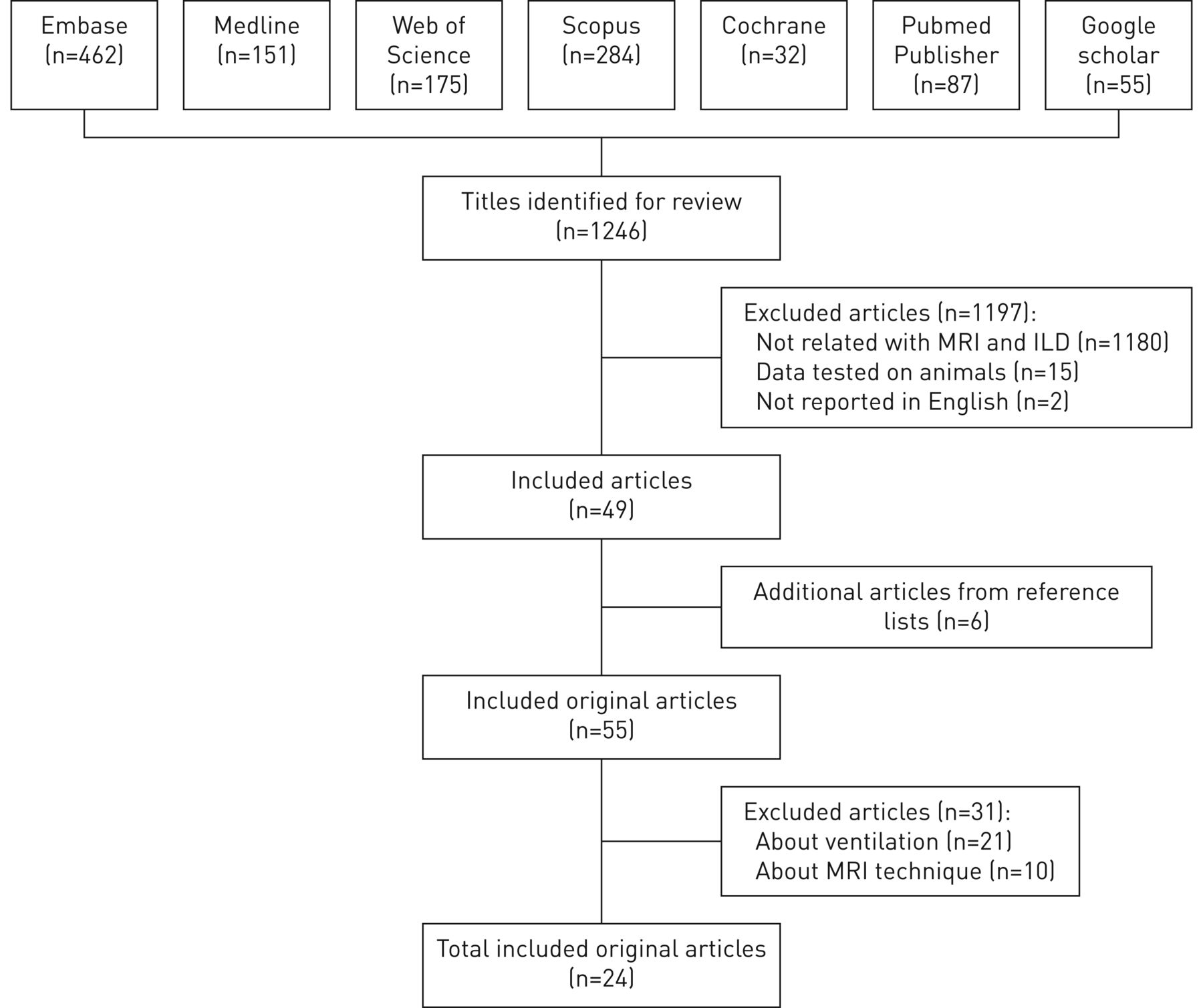
Main menu

  • Home
  • Current issue
  • Past issues
  • For authors
    • Instructions for authors
    • Submit a manuscript
    • ERS author centre
    • COVID-19 submission information
  • Alerts
  • Subscriptions
  • ERS Publications
    • European Respiratory Journal
    • ERJ Open Research
    • European Respiratory Review
    • Breathe
    • ERS Books
    • ERS publications home

User menu

  • Log in
  • Subscribe
  • Contact Us
  • My Cart

Search

  • Advanced search
  • ERS Publications
    • European Respiratory Journal
    • ERJ Open Research
    • European Respiratory Review
    • Breathe
    • ERS Books
    • ERS publications home

Login

European Respiratory Society

Advanced Search

  • Home
  • Current issue
  • Past issues
  • For authors
    • Instructions for authors
    • Submit a manuscript
    • ERS author centre
    • COVID-19 submission information
  • Alerts
  • Subscriptions

The use of chest magnetic resonance imaging in interstitial lung disease: a systematic review

Chiara Romei, Laura Turturici, Laura Tavanti, Jelle Miedema, Sara Fiorini, Massimo Marletta, Piotr Wielopolski, Harm Tiddens, Fabio Falaschi, Pierluigi Ciet
European Respiratory Review 2018 27: 180062; DOI: 10.1183/16000617.0062-2018
Chiara Romei
12nd Radiology Unit, Azienda Ospedaliera Universitaria Pisana, Pisa, Italy
  • Find this author on Google Scholar
  • Find this author on PubMed
  • Search for this author on this site
Laura Turturici
2Radiology, Azienda USL Toscana nord ovest Sede di Viareggio, Viareggio, Italy
  • Find this author on Google Scholar
  • Find this author on PubMed
  • Search for this author on this site
Laura Tavanti
3Dept of Surgical, Medical, Molecular Pathology and Critical Care, Azienda Ospedaliera Universitaria Pisana, Pisa, Italy
  • Find this author on Google Scholar
  • Find this author on PubMed
  • Search for this author on this site
Jelle Miedema
4Dept of Respiratory Medicine, Erasmus MC, University Medical Centre Rotterdam, Rotterdam, The Netherlands
  • Find this author on Google Scholar
  • Find this author on PubMed
  • Search for this author on this site
Sara Fiorini
51st Radiology Unit, Azienda Ospedaliero Universitaria Pisana, Pisa, Italy
  • Find this author on Google Scholar
  • Find this author on PubMed
  • Search for this author on this site
Massimo Marletta
51st Radiology Unit, Azienda Ospedaliero Universitaria Pisana, Pisa, Italy
  • Find this author on Google Scholar
  • Find this author on PubMed
  • Search for this author on this site
Piotr Wielopolski
6Dept of Radiology and Nuclear Medicine, Erasmus MC, University Medical Center Rotterdam, Rotterdam, The Netherlands
  • Find this author on Google Scholar
  • Find this author on PubMed
  • Search for this author on this site
Harm Tiddens
6Dept of Radiology and Nuclear Medicine, Erasmus MC, University Medical Center Rotterdam, Rotterdam, The Netherlands
7Dept of Pediatric Pulmonology and Allergology, Erasmus MC, University Medical Center Rotterdam, Rotterdam, The Netherlands
  • Find this author on Google Scholar
  • Find this author on PubMed
  • Search for this author on this site
Fabio Falaschi
12nd Radiology Unit, Azienda Ospedaliera Universitaria Pisana, Pisa, Italy
  • Find this author on Google Scholar
  • Find this author on PubMed
  • Search for this author on this site
Pierluigi Ciet
6Dept of Radiology and Nuclear Medicine, Erasmus MC, University Medical Center Rotterdam, Rotterdam, The Netherlands
7Dept of Pediatric Pulmonology and Allergology, Erasmus MC, University Medical Center Rotterdam, Rotterdam, The Netherlands
  • Find this author on Google Scholar
  • Find this author on PubMed
  • Search for this author on this site
  • Article
  • Figures & Data
  • Info & Metrics
  • PDF
Loading

Figures

  • Supplementary Materials
  • FIGURE 1
    • Download figure
    • Open in new tab
    • Download powerpoint
    FIGURE 1

    Flow chart of all the harvested papers. MRI: magnetic resonance imaging; ILD: interstitial lung disease.
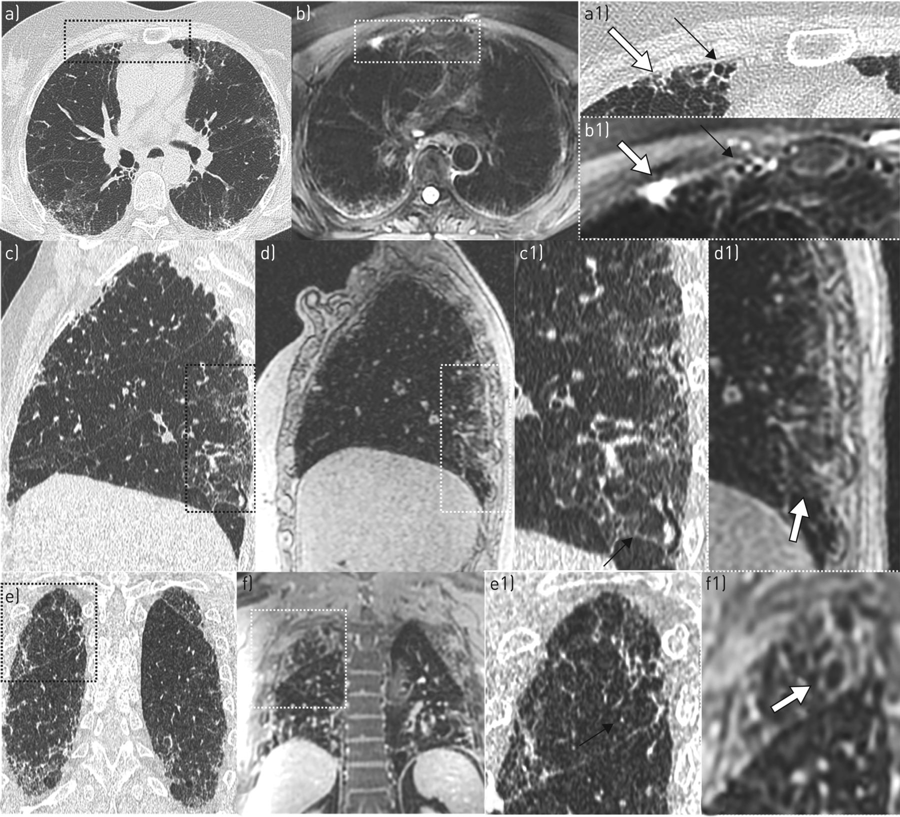
  • FIGURE 2
    • Download figure
    • Open in new tab
    • Download powerpoint
    FIGURE 2

    Comparison of radiological findings of interstitial lung disease with thin-slices multi-detector computed tomography (MDCT) and magnetic resonance imaging (MRI). Honeycombing at a) thin-slices MDCT and b) MRI two-dimensional axial PROPELLER T2-weighted sequence in a 78-year-old female with secondary usual interstitial pneumonia from collagen vascular disease. The magnified images of MDCT (a1) and MRI (b1) show honeycombing (black arrow) and the area of ground-glass opacity (white arrow). The high signal of ground-glass opacity might be due to water content and therefore indicates active inflammation. Ground-glass opacity at c) thin-slices MDCT and d) MRI sagittal reformat three-dimensional SPGR proton density-weighted sequence pre-contrast in a 62-year-old male with idiopathic pulmonary fibrosis. On the magnified images of MDCT (c1) and MRI (d1), the ground-glass opacity is harder to identify on MRI (white arrow) than on MDCT (black arrow). Reticulation at e) thin-slices MDCT and f) MRI coronal reformat three-dimensional SPGR after T1-weighted contrast-enhanced administration (at 20 min) in a 78-year-old female with secondary usual interstitial pneumonia from collagen vascular disease. In the magnified images of MDCT (e1) and MRI (f1) the different appearance of reticulation on MRI (white arrow) and MDCT (black arrow) is probably due to both a different breathing condition (MDCT inspiration versus MRI expiration) and slice thickness (MDCT 1 mm versus MRI 3 mm).
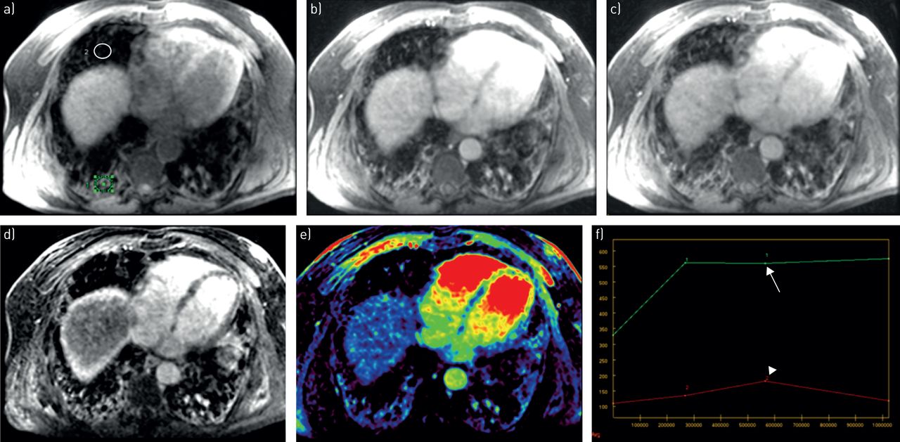
  • FIGURE 3
    • Download figure
    • Open in new tab
    • Download powerpoint
    FIGURE 3

    T1-contrast enhancement signal characteristics in the fibrotic and normal lung parenchyma pre- and post-contrast injection at ∼5, 10 and 20 min in a 73-year-old female with fibrotic nonspecific interstitial pneumonia. a) Chest magnetic resonance imaging axial reformat three-dimensional SPGR proton density-weighted pre-contrast. Axial reformats three-dimensional SPGR T1-weighted post-contrast at b) 5 min and c) 10 min. d) Contrast enhancement subtraction image (10 min image subtracted to pre-contrast). e) Axial maximum slope of increase of contrast enhancement. f) Contrast enhancement curves for fibrotic and normal lung at different time-points, Green region of interest in (a) and arrow in (f) indicate fibrotic lung tissue, while white region of interest in (a) and arrowhead in (f) indicate normal lung tissue. Note progressive contrast enhancement of fibrotic lung with peak at 10 min (arrow), while normal lung has a lower contrast enhancement with signal decay after the first 5 min (arrowhead).

  • FIGURE 4
    • Download figure
    • Open in new tab
    • Download powerpoint
    FIGURE 4

    Diffusion weighted imaging (DWI) at multiple b-values in a 72-year-old female with nonspecific interstitial pneumonia. a) Three-dimensional SPGR proton density-weighted (PD-w) image, b) two-dimensional fat suppressed PROPELLER T2-weighted image, c–e) DWI at b-values c) 200, d) 400 and e) 800 s·mm−2, and f) apparent diffusion coefficient (ADC) map. Note area of inflammation in proton density-weighted image (arrow in a), which has a high T2-weighted signal (arrow in b) and shows signal decay by increasing b-values in the DWI images (arrows in c–e). In the apparent diffusion coefficient maps, the same area in the right lower lobe is hyperintense possibly indicating either high water content or increased perfusion.
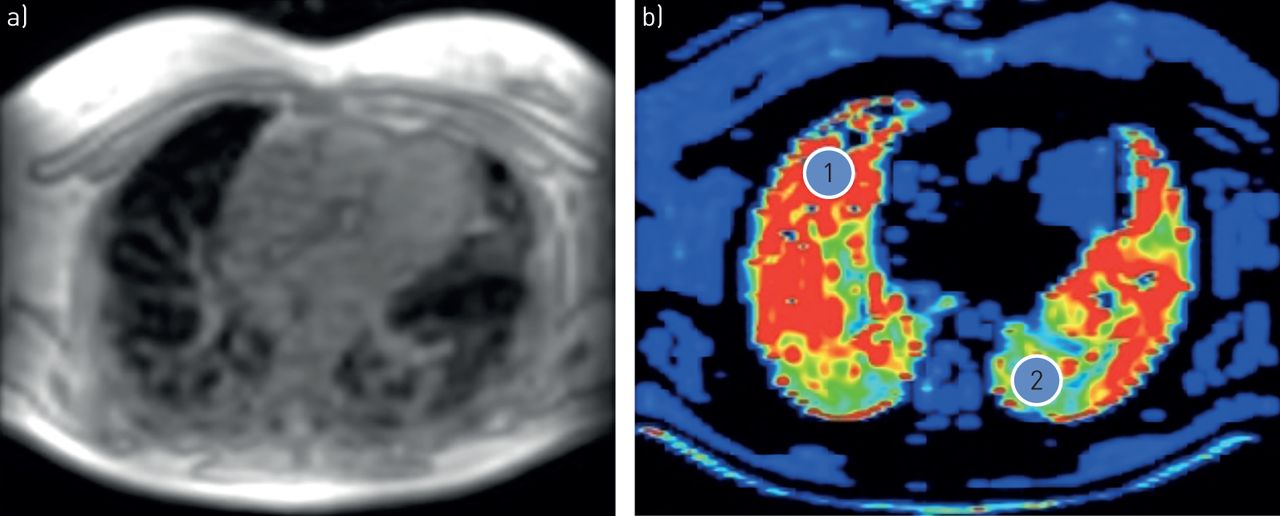
  • FIGURE 5
    • Download figure
    • Open in new tab
    • Download powerpoint
    FIGURE 5

    T2 mapping. a) Native axial multi-echo three-dimensional SPGR in a 72-year-old female with nonspecific interstitial pneumonia. b) Post-processed R2* map (R2*=1/T2*) axial with region of interest 1 in normal lung tissue (2395 Hz=0.42 ms) and region of interest 2 in fibrotic lung tissue (887 Hz=1.13 ms). Note how T2* is longer in fibrotic tissue, therefore R2 is smaller (low signal-to-noise ratio) compared to normal tissue. Post-processing performed with Advantage Window Server 2.0 (GE Healthcare, Milwaukee, WI, USA).

Supplementary Materials

  • Figures
  • Supplementary Material

    Please note: supplementary material is not edited by the Editorial Office, and is uploaded as it has been supplied by the author.

    supplementary tables supplementary tables

PreviousNext
Back to top
View this article with LENS
Vol 27 Issue 150 Table of Contents
European Respiratory Review: 27 (150)
  • Table of Contents
  • Index by author
Email

Thank you for your interest in spreading the word on European Respiratory Society .

NOTE: We only request your email address so that the person you are recommending the page to knows that you wanted them to see it, and that it is not junk mail. We do not capture any email address.

Enter multiple addresses on separate lines or separate them with commas.
The use of chest magnetic resonance imaging in interstitial lung disease: a systematic review
(Your Name) has sent you a message from European Respiratory Society
(Your Name) thought you would like to see the European Respiratory Society web site.
CAPTCHA
This question is for testing whether or not you are a human visitor and to prevent automated spam submissions.
Print
Alerts
Sign In to Email Alerts with your Email Address
Citation Tools
The use of chest magnetic resonance imaging in interstitial lung disease: a systematic review
Chiara Romei, Laura Turturici, Laura Tavanti, Jelle Miedema, Sara Fiorini, Massimo Marletta, Piotr Wielopolski, Harm Tiddens, Fabio Falaschi, Pierluigi Ciet
European Respiratory Review Dec 2018, 27 (150) 180062; DOI: 10.1183/16000617.0062-2018

Citation Manager Formats

  • BibTeX
  • Bookends
  • EasyBib
  • EndNote (tagged)
  • EndNote 8 (xml)
  • Medlars
  • Mendeley
  • Papers
  • RefWorks Tagged
  • Ref Manager
  • RIS
  • Zotero

Share
The use of chest magnetic resonance imaging in interstitial lung disease: a systematic review
Chiara Romei, Laura Turturici, Laura Tavanti, Jelle Miedema, Sara Fiorini, Massimo Marletta, Piotr Wielopolski, Harm Tiddens, Fabio Falaschi, Pierluigi Ciet
European Respiratory Review Dec 2018, 27 (150) 180062; DOI: 10.1183/16000617.0062-2018
del.icio.us logo Digg logo Reddit logo Technorati logo Twitter logo CiteULike logo Connotea logo Facebook logo Google logo Mendeley logo
Full Text (PDF)

Jump To

  • Article
    • Abstract
    • Abstract
    • Introduction
    • Methods
    • Results
    • Future directions
    • Supplementary material
    • Footnotes
    • References
  • Figures & Data
  • Info & Metrics
  • PDF

Subjects

  • Lung imaging
  • Interstitial and orphan lung disease
  • Tweet Widget
  • Facebook Like
  • Google Plus One

More in this TOC Section

  • AATD and chronic respiratory disorders
  • Subglottic secretion drainage for preventing ventilator-associated pneumonia
  • Current and future applications of liquid biopsy in NSCLC
Show more Review

Related Articles

Navigate

  • Home
  • Current issue
  • Archive

About the ERR

  • Journal information
  • Editorial board
  • Reviewers
  • Press
  • Permissions and reprints
  • Advertising
  • Sponsorship

The European Respiratory Society

  • Society home
  • myERS
  • Privacy policy
  • Accessibility

ERS publications

  • European Respiratory Journal
  • ERJ Open Research
  • European Respiratory Review
  • Breathe
  • ERS books online
  • ERS Bookshop

Help

  • Feedback

For authors

  • Instructions for authors
  • Submit a manuscript
  • ERS author centre

For readers

  • Alerts
  • Subjects
  • RSS

Subscriptions

  • Accessing the ERS publications

Contact us

European Respiratory Society
442 Glossop Road
Sheffield S10 2PX
United Kingdom
Tel: +44 114 2672860
Email: journals@ersnet.org

ISSN

Print ISSN: 0905-9180
Online ISSN: 1600-0617

Copyright © 2021 by the European Respiratory Society