
Main menu

  • Home
  • Current issue
  • Past issues
  • Authors/reviewers
    • Instructions for authors
    • Submit a manuscript
    • COVID-19 submission information
    • Institutional open access agreements
    • Peer reviewer login
  • Alerts
  • Subscriptions
  • ERS Publications
    • European Respiratory Journal
    • ERJ Open Research
    • European Respiratory Review
    • Breathe
    • ERS Books
    • ERS publications home

User menu

  • Log in
  • Subscribe
  • Contact Us
  • My Cart

Search

  • Advanced search
  • ERS Publications
    • European Respiratory Journal
    • ERJ Open Research
    • European Respiratory Review
    • Breathe
    • ERS Books
    • ERS publications home

Login

European Respiratory Society

Advanced Search

  • Home
  • Current issue
  • Past issues
  • Authors/reviewers
    • Instructions for authors
    • Submit a manuscript
    • COVID-19 submission information
    • Institutional open access agreements
    • Peer reviewer login
  • Alerts
  • Subscriptions

Unclassifiable interstitial lung disease: a pathologist's perspective

Kirk D. Jones
European Respiratory Review 2018 27: 170132; DOI: 10.1183/16000617.0132-2017
Kirk D. Jones
Dept of Pathology, University of California San Francisco, San Francisco, CA, USA
  • Find this author on Google Scholar
  • Find this author on PubMed
  • Search for this author on this site
  • For correspondence: kirk.jones@ucsf.edu
  • Article
  • Figures & Data
  • Info & Metrics
  • PDF
Loading

Figures

  • Tables
  • FIGURE 1
    • Download figure
    • Open in new tab
    • Download powerpoint
    FIGURE 1

    Bronchiolocentric fibrosis. a) This low power view shows fibrosis of peribronchiolar alveolar septa. Scale bar=500 µm. b) A higher power view shows peribronchiolar metaplasia with lining of the bronchiolocentric alveolar septa by ciliated columnar respiratory epithelium. Scale bar=200 µm.

  • FIGURE 2
    • Download figure
    • Open in new tab
    • Download powerpoint
    FIGURE 2

    Nonspecific interstitial pneumonia. a) This low power view shows diffuse alveolar septal thickening by moderate interstitial fibrosis. Scale bar=1 mm. b) A higher power view shows the architectural preservation of lung tissue. Scale bar=500 µm.
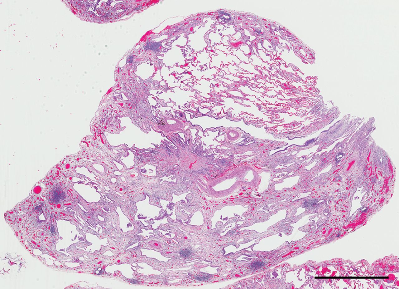
  • FIGURE 3
    • Download figure
    • Open in new tab
    • Download powerpoint
    FIGURE 3

    Usual interstitial pneumonia. a) This low power view shows the heterogeneous pattern of fibrosis with subpleural microscopic honeycombing and centrilobular lack of alveolar inflammation or fibrosis. Scale bar=2 mm. b) A high power view shows a fibroblast focus with typical layered appearance within a loose myxoid stroma. Scale bar=100 µm.

  • FIGURE 4
    • Download figure
    • Open in new tab
    • Download powerpoint
    FIGURE 4

    Unclassifiable interstitial fibrosis. This low power view shows patchy fibrosis with subpleural and bronchiolocentric accentuation as well as prominent lymphoid aggregates. Further clinical history for this 68-year-old woman suggested the likely multidisciplinary diagnosis was nitrofurantoin toxicity. Scale bar= 2 mm.

  • FIGURE 5
    • Download figure
    • Open in new tab
    • Download powerpoint
    FIGURE 5

    Unclassifiable interstitial fibrosis. This low power view shows patchy fibrosis with focal microscopic honeycombing, airspace enlargement, scattered lymphoid aggregates, and central nonspecific interstitial pneumonia fibrosis-like changes. Further clinical history for this 49-year-old woman with early greying of hair and a sibling with pulmonary fibrosis suggested the likely diagnosis was familial interstitial pulmonary fibrosis. Scale bar=2 mm.

Tables

  • Figures
  • TABLE 1

    Multidisciplinary diagnosis of patients pathologically unclassifiable by surgical lung biopsy

    Cases n
     Number of surgical lung biopsies performed: 849
     Number of non-diagnostic biopsies: 2
     Number with pathologic diagnosis of “fibrosing interstitial pneumonia: unclassifiable”: 41
    Final multidisciplinary diagnosis n
     Unclassifiable: 19
     Idiopathic pulmonary fibrosis: 8
     Chronic hypersensitivity pneumonia: 5
     Autoimmune connective tissue disease: 4
     Smoking-related interstitial lung disease: 2
     Infection (Mycobacterium avium): 1
     Sarcoidosis: 1
     Idiopathic nonspecific interstitial pneumonia: 1
PreviousNext
Back to top
View this article with LENS
Vol 27 Issue 147 Table of Contents
European Respiratory Review: 27 (147)
  • Table of Contents
  • Index by author
Email

Thank you for your interest in spreading the word on European Respiratory Society .

NOTE: We only request your email address so that the person you are recommending the page to knows that you wanted them to see it, and that it is not junk mail. We do not capture any email address.

Enter multiple addresses on separate lines or separate them with commas.
Unclassifiable interstitial lung disease: a pathologist's perspective
(Your Name) has sent you a message from European Respiratory Society
(Your Name) thought you would like to see the European Respiratory Society web site.
CAPTCHA
This question is for testing whether or not you are a human visitor and to prevent automated spam submissions.
Print
Citation Tools
Unclassifiable interstitial lung disease: a pathologist's perspective
Kirk D. Jones
European Respiratory Review Mar 2018, 27 (147) 170132; DOI: 10.1183/16000617.0132-2017

Citation Manager Formats

  • BibTeX
  • Bookends
  • EasyBib
  • EndNote (tagged)
  • EndNote 8 (xml)
  • Medlars
  • Mendeley
  • Papers
  • RefWorks Tagged
  • Ref Manager
  • RIS
  • Zotero

Share
Unclassifiable interstitial lung disease: a pathologist's perspective
Kirk D. Jones
European Respiratory Review Mar 2018, 27 (147) 170132; DOI: 10.1183/16000617.0132-2017
Reddit logo Technorati logo Twitter logo Connotea logo Facebook logo Mendeley logo
Full Text (PDF)

Jump To

  • Article
    • Abstract
    • Abstract
    • Introduction
    • Surgical lung biopsy: reducing pre-histological barriers to classification
    • Typical patterns of fibrosis
    • Physiological basis of the typical patterns of fibrosis
    • Histological sources of unclassifiable interstitial fibrosis
    • Post-biopsy evaluation: the MDD
    • Conclusions
    • Footnotes
    • References
  • Figures & Data
  • Info & Metrics
  • PDF

Subjects

  • Interstitial and orphan lung disease
  • Tweet Widget
  • Facebook Like
  • Google Plus One

More in this TOC Section

Series

  • Supplemental oxygen and noninvasive ventilation
  • Nonpharmacological management of psychological distress in COPD
  • Lung transplantation for COPD/pulmonary emphysema
Show more Series

Pathology for the Clinician

  • Lung ageing and COPD
  • Differential diagnosis of granulomatous lung disease
Show more Pathology for the Clinician

Related Articles

Navigate

  • Home
  • Current issue
  • Archive

About the ERR

  • Journal information
  • Editorial board
  • Press
  • Permissions and reprints
  • Advertising
  • Sponsorship

The European Respiratory Society

  • Society home
  • myERS
  • Privacy policy
  • Accessibility

ERS publications

  • European Respiratory Journal
  • ERJ Open Research
  • European Respiratory Review
  • Breathe
  • ERS books online
  • ERS Bookshop

Help

  • Feedback

For authors

  • Instructions for authors
  • Publication ethics and malpractice
  • Submit a manuscript

For readers

  • Alerts
  • Subjects
  • RSS

Subscriptions

  • Accessing the ERS publications

Contact us

European Respiratory Society
442 Glossop Road
Sheffield S10 2PX
United Kingdom
Tel: +44 114 2672860
Email: journals@ersnet.org

ISSN

Print ISSN: 0905-9180
Online ISSN: 1600-0617

Copyright © 2023 by the European Respiratory Society