
Main menu

  • Home
  • Current issue
  • Past issues
  • For authors
    • Instructions for authors
    • Submit a manuscript
    • ERS author centre
    • COVID-19 submission information
  • Alerts
  • Subscriptions
  • ERS Publications
    • European Respiratory Journal
    • ERJ Open Research
    • European Respiratory Review
    • Breathe
    • ERS Books
    • ERS publications home

User menu

  • Log in
  • Subscribe
  • Contact Us
  • My Cart

Search

  • Advanced search
  • ERS Publications
    • European Respiratory Journal
    • ERJ Open Research
    • European Respiratory Review
    • Breathe
    • ERS Books
    • ERS publications home

Login

European Respiratory Society

Advanced Search

  • Home
  • Current issue
  • Past issues
  • For authors
    • Instructions for authors
    • Submit a manuscript
    • ERS author centre
    • COVID-19 submission information
  • Alerts
  • Subscriptions

Management of antithrombotic agents in patients undergoing flexible bronchoscopy

Sami Abuqayyas, Shine Raju, John R. Bartholomew, Roulan Abu Hweij, Atul C. Mehta
European Respiratory Review 2017 26: 170001; DOI: 10.1183/16000617.0001-2017
Sami Abuqayyas
1Internal Medicine Institute, Cleveland Clinic, Cleveland, OH, USA
4Both authors contributed equally
  • Find this author on Google Scholar
  • Find this author on PubMed
  • Search for this author on this site
Shine Raju
2Respiratory Institute, Cleveland Clinic, Cleveland, OH, USA
4Both authors contributed equally
  • Find this author on Google Scholar
  • Find this author on PubMed
  • Search for this author on this site
John R. Bartholomew
3Vascular Medicine Institute, Cleveland Clinic, Cleveland, OH, USA
  • Find this author on Google Scholar
  • Find this author on PubMed
  • Search for this author on this site
Roulan Abu Hweij
1Internal Medicine Institute, Cleveland Clinic, Cleveland, OH, USA
  • Find this author on Google Scholar
  • Find this author on PubMed
  • Search for this author on this site
Atul C. Mehta
2Respiratory Institute, Cleveland Clinic, Cleveland, OH, USA
  • Find this author on Google Scholar
  • Find this author on PubMed
  • Search for this author on this site
  • For correspondence: mehtaa1@ccf.org
  • Article
  • Figures & Data
  • Info & Metrics
  • PDF
Loading

Abstract

Bleeding is one of the most feared complications of flexible bronchoscopy. Although infrequent, it can be catastrophic and result in fatal outcomes. Compared to other endoscopic procedures, the risk of morbidity and mortality from the bleeding is increased, as even a small amount of blood can fill the tracheobronchial tree and lead to respiratory failure. Patients using antithrombotic agents (ATAs) have higher bleeding risk. A thorough understanding of the different ATAs is critical to manage patients during the peri-procedural period. A decision to stop an ATA before bronchoscopy should take into account a variety of factors, including indication for its use and the type of procedure. This article serves as a detailed review on the different ATAs, their pharmacokinetics and the pre- and post-bronchoscopy management of patients receiving these medications.

Abstract

A thorough understanding of antithrombotic agents is critical to manage patients in the peri-bronchoscopic period http://ow.ly/zcqn30bRCjf

Introduction

A bronchoscopist is often involved in the management of antithrombotic agents (ATAs) in the peri-procedural period. Patients are prescribed these medications for a variety of indications, and the decision to withhold them prior to bronchoscopy can be challenging. The increasing number of novel oral anticoagulants (also known as direct oral anticoagulants or target-specific oral anticoagulants), with different pharmacokinetics and safety profiles, has made these decisions more complex. The recent American College of Chest Physicians (ACCP) guidelines emphasising the use of direct thrombin or factor Xa inhibitors over vitamin K antagonists for atrial fibrillation, deep vein thrombosis (DVT) and pulmonary embolism (PE) in non-cancer patients [1] will certainly result in their increasing use. With more patients being prescribed newer ATAs, it is imperative for a bronchoscopist to have a thorough knowledge of these drugs. During the peri-procedural management of ATAs, one has to weigh up several clinical factors (table 1): the invasiveness of the procedure, patient-related risk factors, the indication for antithrombotic therapy and the need for bridging. The pharmacokinetics and other properties of the individual ATAs, which are broadly classified into antiplatelet agents and anticoagulants, will also influence the management of antithrombotic therapy. All these factors will be discussed in this review.

View this table:
  • View inline
  • View popup
TABLE 1

Factors influencing antithrombotic therapy during the peri-bronchoscopic period

Invasiveness of the procedure and patient-related risk factors

The data for bleeding risks associated with different types of bronchoscopic procedure are scant. Bleeding rates vary by the type of bronchoscopic procedure performed [2–5]. Cordasco et al. [2] noted that the rate of bleeding was related to the type of biopsy performed, with transbronchial biopsy carrying a much higher risk than endobronchial biopsy. The authors reported an overall bleeding rate of 0.83% with flexible bronchoscopy and 1.9% if either brushing or biopsy was performed. In a prospective multicentre study involving 20 986 bronchoscopies, Facciolongo et al. [3] reported different rates of bleeding with various bronchoscopic procedures. Transbronchial biopsy was associated with the highest rates of bleeding complications, followed by therapeutic bronchoscopy (which entails the use of Nd:YAG laser, argon-plasma, electrocautery and stent positioning), endobronchial biopsy, transbronchial needle aspiration (TBNA), bronchoalveolar lavage and explorative bronchoscopy, which had the lowest rates of bleeding complications. Thus, transbronchial biopsy and therapeutic bronchoscopy are considered to be relatively high-risk procedures, while explorative bronchoscopy with or without bronchoalveolar lavage and endobronchial needle aspiration are low-risk interventions (table 2). In the latter category, although discontinuation of ATAs is recommended in most instances, exceptions can be made depending on the skill and experience of the bronchoscopist, the capabilities of the centre in managing life-threatening bleeding and when the situation presents itself as emergent. It must, however, be noted that the decision should be individualised, and bleeding can ensue with any procedure, even those with minimal risk. Hence, the best clinical and procedural skills should be practised. Additionally, patient-related risk factors can independently increase the risk of bleeding. These risk factors include uraemia, thrombocytopenia, HIV/AIDS, solid-organ transplant, haematological disorders and thrombocytosis [2, 6]. Evidence is currently lacking on the effect of bleeding risk when multiple risk factors present in combination. In these situations, coagulation parameters, platelet counts and blood urea levels should be routinely monitored, and therapeutic intervention should be attempted to reverse coagulopathy in the form of fresh frozen plasma (FFP), platelet transfusions and desmopressin, if necessary [2].

View this table:
  • View inline
  • View popup
TABLE 2

Invasiveness of bronchoscopic procedures based on relative bleeding rates

Indication for the use of ATAs

The bronchoscopist must consider the rationale behind the use of ATAs prior to their discontinuation. In some instances, the risks of thromboembolic events can outweigh the risk of bleeding. The most common indications for their use are acute coronary syndrome (ACS), non-valvular atrial fibrillation, metallic heart valves, percutaneous coronary intervention (PCI), PE and DVT.

In patients with non-valvular atrial fibrillation, the risk of stroke can be determined using CHADS₂ [7] and CHA₂DS₂-VASc [8] scores (table 3). Patients with valvular atrial fibrillation are at much higher risk of thromboembolic events [9]. For patients with mechanical heart valves, the risk for thromboembolic events is based on their type, number and location. Metallic valves in the mitral position have the highest risk for arterial thromboembolic events [9, 10]. The decision to perform bronchoscopy with transbronchial biopsy in patients with recent ACS or coronary stent insertion should take into account the time since the most recent ACS, the type of stent (bare metal stent (BMS) versus drug-eluting stent (DES)) and the time since its insertion. In fact, the strongest predictor of stent thrombosis among patients receiving more than one ATA is premature discontinuation of dual-antiplatelet therapy (DAPT) (within 6 weeks of BMS insertion and 6 months of DES insertion) [11].

View this table:
  • View inline
  • View popup
TABLE 3

Comparison of CHADS2 versus CHA2DS2-VASc scoring systems

According to current guidelines, DAPT should be continued in patients with stable ischaemic heart disease for at least 1 month after a BMS is implanted and for at least 6 months after a DES is implanted (class of recommendation: I); a longer duration of DAPT is recommended in these patients if they are not at high risk of bleeding and able to tolerate DAPT without bleeding complications (class of recommendation: IIb) [12].

Need for bridging

When a long-acting ATA is withheld during the peri-procedural period, short-acting antiplatelet agents such as glycoprotein IIb/IIIa inhibitors or anticoagulants such as unfractionated heparin and low-molecular-weight heparin (LMWH) products may be recommended during the interruption period (bridging). At present, the recommendations regarding the need for bridging in patients with atrial fibrillation are inconsistent. Moreover, data on the short-term risk of thromboembolic events during the peri-procedural interruption period vary between published studies. Raunso et al. [13] found a significant increase in short-term (90-day) risk of thromboembolic events or death when warfarin was interrupted in patients with atrial fibrillation, while Douketis et al. [14], in the “bridge trial”, noted that no bridging was non-inferior to bridging in the prevention of arterial thromboembolism in patients with atrial fibrillation. Douketis et al. [14] also reported that bridging was associated with three times the risk for major bleeding. We recommend that bridging should be reserved for patients with high venous and arterial thromboembolic risk [14–16]. Additionally, current evidence does not recommend bridging for low- and moderate-risk patients, and the decision should be individualised based on the patient's risk factors and procedure-related factors (table 4) [17]. The bridging protocol is described in table 5.

View this table:
  • View inline
  • View popup
TABLE 4

Risk for thromboembolic events during the interruption period of antithrombotics

View this table:
  • View inline
  • View popup
TABLE 5

Bridging for warfarin and P2Y12 receptor blockers

Antiplatelet drugs

Antiplatelet drugs are grouped into various subclasses based on their mechanism of action. These are described in detail in the following sections and summarised in table 6 and figure 1.

View this table:
  • View inline
  • View popup
TABLE 6

Management of antiplatelet agents in the peri-bronchoscopic period

FIGURE 1
  • Download figure
  • Open in new tab
  • Download powerpoint
FIGURE 1

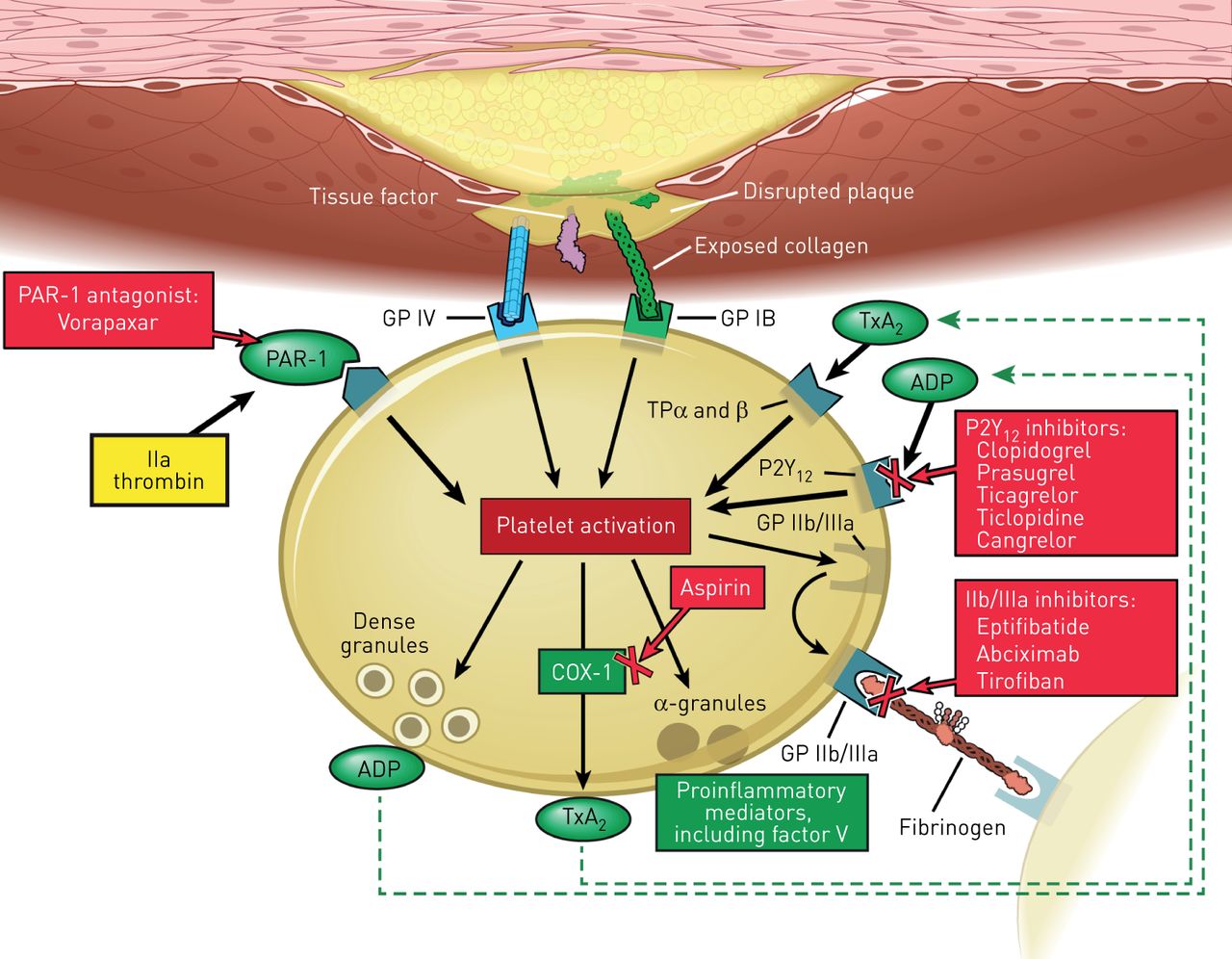
Mechanism and site of action of antiplatelet agents. COX-1: cyclooxygenase-1; GP: glycoprotein; PAR-1: protease-activated receptor 1; TP: thromboxane prostanoid; TxA2: thromboxane A2. Reprinted with permission, Cleveland Clinic © 2016–2017. All rights reserved.

Irreversible cyclooxygenase inhibitors

Aspirin (salicylate)

Aspirin is the most commonly used antiplatelet agent. Aspirin irreversibly blocks the enzyme cyclooxygenase-1 enzyme and interrupts the first step in the prostaglandin and thromboxane A2 synthesis pathway, thereby inhibiting platelet aggregation. Aspirin's maximal antiplatelet effect occurs within minutes and lasts through the life of the platelets (5–7 days) [18–20].

Current evidence does not suggest any increased risk of bleeding with singular use of aspirin. Zakaria and Abdel-Fattah [21] found no association between the use of aspirin and the increased bleeding risk after transbronchial biopsy. Herth et al. [22] concluded that the use of aspirin was not associated with increased risk of bleeding and the recommendation was to not stop aspirin prior to bronchoscopy.

Monitoring of the aspirin antiplatelet effect is generally not indicated. Additionally, blood testing for monitoring purposes has not been shown to correlate with clinical outcomes [23]. Due to its irreversible effect on platelets, bleeding complications can be controlled with platelet transfusions.

P2Y12 receptor blockers

Thienopyridines

Clopidogrel (Plavix)

Active metabolites of clopidogrel irreversibly bind to the platelet P2Y12 receptor, thereby inhibiting ADP-induced platelet activation and aggregation. The onset of action is dose dependent. Maximum platelet inhibition takes 5–10 days with a maintenance dose of 75 mg·day−1, as compared to 12–15 h after a loading dose of 300–600 mg [24–26]. Clopidogrel is commonly used as part of DAPT, in combination with aspirin in the management of ACS, recent myocardial infarction or stroke, or established peripheral arterial disease [27].

The decision to stop clopidogrel prior to the bronchoscopic procedure can be challenging, especially when there is an increased risk for thromboembolic events such as in-stent thrombosis and death. The 2012 ACCP guidelines recommend delaying surgical procedures for at least 6 weeks after BMS insertion and for at least 6 months after DES [17]. DAPT is associated with significant increase in bleeding, as evidenced by the CURE (Clopidogrel in Unstable Angina to Prevent Recurrent Events) trial [28]. The rate of spontaneous major bleeding over a 9-month follow-up period was 3.7% with DAPT versus 2.7% with aspirin monotherapy, and 5.1% versus 2.4% for spontaneous minor bleeding, respectively.

Ernst et al. [29] evaluated the safety of performing transbronchial biopsy in patients receiving clopidogrel. The study was stopped early because of the high bleeding rate in the clopidogrel-only group (89%; p<0.001). It also showed a 100% bleeding rate in the group receiving a combination of clopidogrel and aspirin (p<0.001). The authors suggested stopping clopidogrel 5–7 days prior to transbronchial biopsy. Stather et al. [30] evaluated the safety of endobronchial ultrasound-guided TBNA (EBUS-TBNA) in patients taking clopidogrel. They identified 12 cases with no significant bleeding and hence suggested that EBUS-TBNA can be performed safely in patients taking clopidogrel. However, due to the limitations of the retrospective nature of the study and the small study sample, they recommended continuing clopidogrel through the procedure only if the risk of short-term thrombosis outweighed the risk of bleeding.

Clopidogrel's antiplatelet effect is not routinely monitored and there are no standardised tests.

Prasugrel (Effient)

Prasugrel irreversibly inhibits platelet aggregation by blocking the P2Y12 receptor. In contrast to clopidogrel, prasugrel has more rapid absorption and onset of action, reaching its peak plasma concentration within 30 min. Its active metabolite has an average elimination half-life of 7 h and is mainly cleared by the kidneys [20, 31]. It is indicated for the reduction of thrombotic events in patients with ACS who are to be managed with PCI. Compared to clopidogrel, it has a significantly lower risk of stent thrombosis (1.1% versus 2.4%; p<0.001) but an increased risk of bleeding (major bleeding not related to coronary-artery bypass grafting 2.4% versus 1.8%; p=0.03) [32].

Recommendations for the peri-procedural management of prasugrel are identical to that for clopidogrel [20]. Due to its irreversible inhibitory effect on platelets, treatment of bleeding can be achieved by platelet transfusions.

Ticlopidine (Ticlid)

Ticlopidine is an oral irreversible inhibitor of ADP-induced platelet aggregation. It can reach its peak plasma concentration within 1–3 h after a single oral dose of 250 mg and has a half-life of 4–5 days after chronic usage [33]. Although studies on ticlopidine show benefit when used for stroke prevention and coronary stent occlusion, it is used rarely due to its potential serious side-effects, thrombocytopenia and neutropenia [34]. It can be continued throughout the peri-procedural period if the bronchoscopic procedure is considered a low risk and discontinuation of ticlopidine has a higher risk of thromboembolic events. It should be stopped 10–14 days prior to high-risk procedures in patients with low risk for thromboembolic events [35].

ADP receptor antagonists

Cangrelor (Kengreal)

Cangrelor is an intravenous rapidly acting reversible inhibitor of the P2Y12 receptor with a half-life of 3–6 min. Due to its reversible effect, normalisation of its antiplatelet effect occurs within 60 min of discontinuation. Its efficacy has been evaluated for use in the peri-procedural period during PCI [36].

Cangrelor can be stopped as early as 1–2 h prior to bronchoscopic procedures and can be used to bridge patients on the oral P2Y12 receptor blockers in whom prolonged discontinuation is needed prior to surgical intervention [37].

Ticagrelor (Brilinta)

Ticagrelor is a cyclopentyltriazolopyrimidine and an oral reversible P2Y12 ADP-receptor blocker. It is indicated for use in patients with ACS, it has a more potent antiplatelet activity than clopidogrel with a lower rate of stent thrombosis (1.3% versus 1.9%; p=0.009) and a nonsignificant difference in the rate of major bleeding [38, 39]. The drug has predictable pharmacokinetics with linear dose-related increases in plasma concentration, achieving its peak effect within 2–4 h after dosing [40, 41].

Similar to prasugrel, ticagrelor should be stopped 5–7 days prior to bronchoscopy and can be restarted within 24–48 h of the procedure [20, 25].

PAR-1 antagonists

Vorapaxar (Zontivity)

Vorapaxar is a potent orally active thrombin receptor (protease-activated receptor 1 (PAR-1)) antagonist. It is indicated for the reduction of thrombotic cardiovascular events in patients with a history of myocardial infarction or peripheral arterial disease [42]. It has a half-life of 5–13 days. After a single dose of vorapaxar, platelet function is not restored for 2–3 weeks [43]. Based on its prolonged half-life, it should be stopped at least 2 weeks prior to the bronchoscopic procedure.

Phosphodiesterase inhibitors: quinolone derivatives

Cilostazol (Pletal)

Cilostazol is a phosphodiesterase-3 inhibitor with vasodilatory, antiproliferative and reversible antiplatelet activity with a half-life of 11 h (longer in patients with renal impairment) [44]. It is approved for moderate to severe intermittent claudication for patients with peripheral arterial disease [45].

Having a relatively shorter half-life compared to the other antiplatelet drugs, it should be stopped 1–2 days prior to the bronchoscopic procedure.

Dipyridamole (Persantine)

Dipyridamole is phosphodiesterase inhibitor with vasodilatory and reversible antiplatelet effects. It produces sustained platelet inhibition via multiple mechanisms, including increasing cAMP inhibition of nucleotide phosphodiesterase and blockade of adenosine uptake, causing increase in the amount of adenosine at the platelet vascular interface. It can cause vasodilation at high doses [46]. It is used in conjunction with aspirin for the prevention of cerebral ischaemic events in patients with non-cardioembolic transient ischaemic attacks or strokes [47, 48].

The drug is excreted in bile and has a terminal half-life ranging between 10 h and 19 h [20, 25]. It should be stopped within 1–2 days prior to the bronchoscopic procedure.

Glycoprotein IIb/IIIa inhibitors

These intravenously administered antiplatelet agents include abciximab, eptifibatide and tirofiban. They inhibit platelet aggregation and thrombus formation and have proven of benefit in patients with ACS undergoing PCI [49–51]. Bleeding and thrombocytopenia are potential side-effects of these antiplatelet drugs [52, 53]. In addition, spontaneous alveolar haemorrhage has been reported as a rare side-effect (0.5–0.9%) [54–57]. Based on their pharmacokinetics and current guidelines, eptifibatide and tirofiban should be discontinued for at least 2–4 h before bronchoscopic procedures, whereas abciximab should be withheld for at least 12 h [58].

Abciximab (ReoPro)

Abciximab is a chimeric monoclonal antibody-binding fragment specific for platelet glycoprotein IIb/IIIa receptors. It has a rapid onset of action, with slow recovery of platelet function after discontinuation compared with the other glycoprotein IIb/IIIa inhibitors. It has a half-life of 10–30 min [25].

Eptifibatide (Integrilin)

Eptifibatide is a cyclic heptapeptide that binds to platelet glycoprotein IIb/IIIa receptors. It has a rapid onset of action and rapid reversal of platelet function with a half-life of 2.5 h, which can become prolonged in patients with renal disease [25, 59].

Tirofiban (Aggrastat)

Tirofiban is a non-peptide tyrosine derivative glycoprotein IIb/IIIa receptor inhibitor. Similar to eptifibatide, it has a rapid onset of action and rapid recovery of platelet function after discontinuation with a half-life of 2 h [60]. Dose adjustment is required in patients with impaired renal function [61].

Anticoagulant drugs

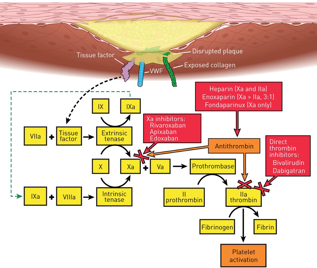
Anticoagulant drugs are likewise grouped into various subclasses based on their mechanism of action. These are described in detail in the following sections and summarised in table 7 and figure 2.

View this table:
  • View inline
  • View popup
TABLE 7

Management of anticoagulant agents in the peri-bronchoscopic period

FIGURE 2
  • Download figure
  • Open in new tab
  • Download powerpoint
FIGURE 2

Coagulation cascade and site of action of anticoagulant agents. VWF: von Willebrand factor. Reprinted with permission, Cleveland Clinic © 2016–2017. All rights reserved.

Oral anticoagulants

Vitamin K antagonists

Warfarin, acenocoumarol, phenindione and phenprocoumon constitute the vitamin K antagonists and act by inhibiting the production of vitamin K-dependent clotting factors II, VII, IX and X. In addition, they inhibit the carboxylation of the regulatory anticoagulant proteins C, S and Z [62, 63].

Warfarin (Coumadin)

Warfarin is the most commonly used oral anticoagulant. Drug–drug interactions and dietary restrictions are major drawbacks for its use. It is indicated for the primary and secondary prevention of venous thromboembolic diseases, for anticoagulation in atrial fibrillation and mechanical heart valves [64]. Warfarin should be stopped 5 days before bronchoscopic procedures and can be safely resumed within 12–24 h after bronchoscopy [65].

The prothrombin time and the international normalised ratio (INR) monitor its antithrombotic effects. In patients with impaired liver function, the use of the INR can be misleading. Checking the INR value 7 days before bronchoscopy is recommended to allow adequate time for planning of peri-bronchoscopic management. Rechecking the INR the day before surgery is suggested by multiple studies to allow correction with vitamin K supplementation and to avoid delaying the procedure [17, 66, 67].

The management of complications can be guided by the clinical scenario (urgent versus elective procedure). Multiple reversal strategies have been proposed, including the interruption of treatment, administration of vitamin K (oral or parenteral phytonadione, keeping in mind that the parenteral route should be reserved for patients in whom the risk is justified), FFP, prothrombin complex concentrate (PCC) or recombinant activated factor VII (rFVIIa) [68–70].

In situations where life-threatening bleeding has occurred and an immediate reversal of the anticoagulant effect of warfarin is needed, PCC has been shown in a randomised clinical trial to be non-inferior when compared with the traditional use of FFP to achieve haemostasis, and was associated with more rapid INR reduction [71]. Moreover, the use of FFP has its own drawbacks when compared with PCC; for example, FFP entails the use of larger volumes to achieve haemostasis, which may compromise the respiratory status and put certain patients at risk for transfusion-associated circulatory overload.

New oral anticoagulants

Due to the major drawbacks of vitamin K antagonists, more emphasis is being put on the use of newer oral anticoagulants such as the direct thrombin inhibitors and factor Xa inhibitors. These anticoagulants have been shown to be more effective in managing certain thromboembolic events, with fewer side-effects [72]. The use of these drugs over vitamin K antagonists for lower-extremity DVT or PE in non-cancer patients has been recommended by the recent ACCP guidelines [1].

Direct thrombin inhibitors

Dabigatran (Pradaxa)

Dabigatran is an oral, selective, reversible direct thrombin inhibitor. It is metabolised by the liver and eliminated by renal excretion. Its indications include the treatment and prevention of venous thromboembolism (VTE) and the prevention of stroke and arterial embolism in patients with non-valvular atrial fibrillation [73, 74]. Dose adjustment is needed in patients with a creatinine clearance of <30 mL·min−1 or in patients on selective glycoprotein IIb/IIIa inhibitors. Its use should be avoided in patients with advanced liver disease.

The half-life of dabigatran in plasma is 15 h and its time to peak effect is 2 h. Current recommendations are to stop dabigatran 1–2 days prior to procedures in patients with creatinine clearance >50 mL·min−1 and 3–5 days with creatinine clearance <50 mL·min−1 (longer if major bleeding is expected). Dabigatran can be safely restarted within 24 h after bronchoscopy if haemostasis is achieved. No monitoring is required [75].

An advantage of dabigatran is the availability of an antidote, idarucizumab, which can completely and rapidly reverse the anticoagulant effect of dabigatran; this received approval from the US Food and Drug Administration (FDA) in 2015 [76]. Dialysis is an another option for the management of dabigatran-induced bleeding [77].

Factor Xa inhibitors

Apixaban (Eliquis)

Apixaban is an oral, selective, reversible, direct factor Xa inhibitor. It is indicated for reducing the risk of stroke and systemic embolism in non-valvular atrial fibrillation, the treatment and prevention of DVT and PE, as well as prophylaxis of DVT following hip or knee replacement surgery. Apixaban undergoes hepatic metabolism with partial renal excretion. Dosage adjustment is recommended for renal disease as well as in patients aged ≥80 years, with body weight ≤60 kg or serum creatinine ≥1.5 mg·dL−1. It should be avoided in advanced liver disease. Apixaban has an approximate plasma half-life of 12 h, and reaches its peak effect at 3 h.

Edoxaban (Savaysa)

Edoxaban is a once-daily administered, rapidly acting direct factor Xa inhibitor. It has a similar safety and adverse reaction profile to other medications in this group. Its half-life is 12 h and peak time to onset is 2 h. It is indicated for the treatment of DVT and PE following 5–10 days of initial therapy with a parenteral anticoagulant [78]. It is also indicated for patients with non-valvular atrial fibrillation [79]. Dose adjustments are needed if a patient's creatinine clearance is 15–50 mL·min−1, if the patient weighs <60 kg and with the use of selective glycoprotein IIb/IIIa inhibitors. It is also recommended that edoxaban should not be used in patients with a creatinine clearance >95 mL·min−1 because of the increased risk of ischaemic stroke.

Rivaroxaban (Xarelto)

Rivaroxaban is an oral, selective and competitive active-site-directed, reversible factor Xa inhibitor; it has a predictable dose-dependent effect without the need for laboratory monitoring. It undergoes hepatic metabolism and partial renal excretion. It should be used cautiously in renal and liver disease patients.

Controlling bleeding

Along with the usual bronchoscopic manoeuvres to control bleeding, current measures include the use of activated and non-activated PCCs and rFVIIa [80–82]. In addition, andexanet alfa is a new antidote for factor Xa inhibitors [83]. In a multicentre, prospective study evaluating 67 patients who developed acute major bleeding after 18 h of using factor Xa inhibitors (apixaban, rivaroxaban, edoxaban or enoxaparin), it was shown that andexanet alfa can rapidly reverse their anticoagulant effect and achieve effective haemostasis in 79% of patients after 12 h of administration [84]. However, it should be noted that andexanet alfa has yet to be approved for use in the USA by the FDA.

Factor Xa inhibitors should be stopped 1–2 days before bronchoscopy (longer in patients prone to bleeding complications) and can be restarted within 24 h of bronchoscopy if haemostasis is achieved. However, the decision to restart these anticoagulants should be individualised based on the nature of the procedure performed and the risk of thromboembolic events.

Parenteral anticoagulants

Unfractionated heparin

Heparin is the most commonly used parenteral anticoagulant. It is indicated for primary and secondary prevention of VTE, ACS, mechanical heart valves, atrial fibrillation and for bridging from and to long-acting oral anticoagulants. Heparin binds to antithrombin, leading to conformational structural changes converting antithrombin from a slow thrombin inhibitor to a rapid inhibitor. In addition, it inhibits activated coagulation factors IX, X, XI, XII and plasmin, as well as the conversion of fibrinogen to fibrin [85, 86].

Unfractionated heparin has a half-life of 60–90 min and should be stopped 4–6 h prior to the bronchoscopic procedure when used intravenously. Discontinuing the drug for a longer period is suggested if major bleeding is expected [87, 88]. Heparin can be restarted 12–24 h post-bronchoscopy in intermediate- to high-risk procedures and sooner (4–6 h, preferably without a bolus) if the procedure was low-risk and intra-procedural haemostasis was achieved. Heparin's anticoagulant response varies among patients, so the need to monitor is of great significance. The activated partial thromboplastin time (APTT) or an anti-Xa assay is used to monitor the effect and adjust the dose of heparin [89, 90]. In addition to general measures for bleeding control, intravenous protamine sulfate at 1 mg per 100 circulating heparin units can rapidly reverse its anticoagulant effects [91, 92].

Low-molecular-weight heparin

Enoxaparin (Lovenox), dalteparin and tinzaparin are LMWH products available in the USA. They have smaller molecular weights than heparin, have a reduced inhibitory effect on thrombin, and have more selectivity to bind antithrombin than other plasma proteins [93].

LMWH products have multiple indications, including prophylaxis and treatment for VTE, especially in cancer patients, ACS and for bridging to and from the new oral anticoagulants and vitamin K antagonists [1, 94–96]. They have a more predictable dose–response relationship, longer half-life, and less incidence of heparin-induced thrombocytopenia (HIT) and bone loss compared to unfractionated heparin [97, 98]. The half-life of enoxaparin is 3–5 h, which can be prolonged in patients with renal impairment due to its renal elimination [99]. Dose adjustment is necessary for patients with a creatinine clearance <30 mL·min−1.

Due to their predictable effect, routine laboratory monitoring is not indicated (exceptions are obesity, pregnancy, renal insufficiency and paediatric age group). If necessary, an anti-Xa assay is the recommended test [100]. Enoxaparin should be stopped 24 h before bronchoscopy if the patient is on a dose of 1 mg·kg−1 every 12 h, and should be stopped 36 h before if the patient is on a schedule of 1.5 mg every 24 h. It can be restarted within 24–72 h post-bronchoscopy after homeostasis is achieved [88].

The inability of protamine sulfate to fully bind LMWH explains its incomplete neutralisation of the anticoagulant effect. Nevertheless, its use as an antidote is justifiable if bleeding is severe. Hirsh et al. [87] have suggested administering protamine sulfate at a dose of 1 mg per mg of LMWH within 8 h of the last dose of LMWH, and a second dose of 0.5 mg per mg of LMWH if bleeding continues.

Fondaparinux (Arixtra)

Fondaparinux, an indirect factor Xa inhibitor, is a synthetic pentasaccharide that binds antithrombin in the plasma, potentiating its effect on factor Xa. Fondaparinux is administered subcutaneously once daily and has a half-life of 17 h. Owing to its predictable anticoagulant effect, drug monitoring is not required. It is excreted unchanged in the urine, hindering its use in patients with renal disease [87, 101, 102]. Fondaparinux is used for the prevention of VTE in hip fracture, hip and knee replacement and abdominal surgery, and for treatment of VTE, HIT (off-label use) and ACS [103–108].

Due to its renal elimination, peri-procedural management of fondaparinux should take into account the creatinine clearance. For creatinine clearance >50 mL·min−1 it should be stopped 3 days before and restarted 24 h after bronchoscopy once haemostasis is achieved. It is recommended that it should be stopped for longer in patients with creatinine clearance <50 mL·min−1.

Unlike heparin, fondaparinux does not interact with protamine sulfate. In cases where uncontrolled bleeding occurs, PCCs or rFVIIa can be used in an attempt to reverse its anticoagulant effect [109, 110].

Direct thrombin inhibitors

Argatroban

Argatroban is an intravenously administered competitive reversible direct thrombin inhibitor. It is metabolised by the liver and can therefore be used safely in patients with renal failure [111]. It is approved for prophylaxis or treatment in patients with HIT and for patients with or at risk for HIT who undergo PCI [112, 113].

Argatroban has a plasma half-life of 45 min. In patients with normal liver function, it should be stopped 3 h before and restarted 24 h post-bronchoscopy [114]. The APTT is the recommended test for monitoring its anticoagulant effect [115].

Desirudin (Iprivask)

Desirudin is a recombinant hirudin derivative with irreversible direct thrombin inhibitor action [116]. Compared to argatroban, its use is not recommended in patients with renal failure, as it is cleared primarily by the kidneys, but it can be used in patients with impaired liver function [117]. Desirudin is used for VTE prophylaxis in patients undergoing total hip replacement [118]. Hirudin use for ACS has been studied, but not yet approved [119]. It has a plasma half-life of 2–3 h after intravenous administration [120]. Based on this, it should be stopped at least 6–8 h before bronchoscopy if administered intravenously and longer if subcutaneously. The APTT correlates well with the plasma level of desirudin and should be used for monitoring its anticoagulant effect [121].

Bivalirudin (Angiomax)

Bivalirudin is an analogue of hirudin, directly and reversibly inhibiting thrombin. The half-life is 25 min after intravenous injection, longer in patients with impaired renal clearance as it is partially cleared by the kidneys [122].

It has been approved by the FDA to be used as an alternative to heparin in patients undergoing PCI, especially in patients with a history of HIT [123]. It is also approved in patients who are undergoing urgent cardiac surgery and acute HIT. Because of its short half-life it can be stopped 1.5 h before the procedure in patients with creatinine clearance >30 mL·min−1 and can be restarted within 24 h after bronchoscopy.

There is no specific antidote for direct thrombin inhibitors. rFVIIa can be used to control excessive bleeding [110].

Conclusion

Peri-procedural management of ATAs can be a challenge. An individualised approach should be adopted prior to each bronchoscopic procedure. The risks and benefits should be thoroughly evaluated, and patient safety should always take precedence. A sound understanding of the different ATAs and their individual properties can help guide and simplify the decision-making. Should a bleeding complication occur, the bronchoscopist should be competent at managing complications and make use of the available drug-specific reversal agents.

Acknowledgements

All authors contributed to the content of this manuscript and to approval of the final version to be submitted.

Footnotes

  • Conflict of interest: None declared.

  • Provenance: Submitted article, peer reviewed.

  • Received January 3, 2017.
  • Accepted April 9, 2017.
  • Copyright ©ERS 2017.

ERR articles are open access and distributed under the terms of the Creative Commons Attribution Non-Commercial Licence 4.0.

References

  1. ↵
    1. Kearon C,
    2. Akl EA,
    3. Ornelas J, et al.
    Antithrombotic therapy for VTE disease: CHEST Guideline and Expert Panel Report. Chest 2016; 149: 315–352.
    OpenUrlCrossRefPubMed
  2. ↵
    1. Cordasco E,
    2. Mehta A,
    3. Ahmad M
    . Bronchoscopically induced bleeding. A summary of nine years’ Cleveland clinic experience and review of the literature. Chest 1991; 100: 1141–1147.
    OpenUrlCrossRefPubMed
  3. ↵
    1. Facciolongo N,
    2. Patelli M,
    3. Gasparini S, et al.
    Incidence of complications in bronchoscopy. Multicentre prospective study of 20,986 bronchoscopies. Monaldi Arch Chest Dis 2009; 71: 8–14.
    OpenUrlPubMed
    1. Hanson R,
    2. Zavala D,
    3. Rhodes M, et al.
    Transbronchial biopsy via flexible fiberoptic bronchoscope: results in 164 patients. Am Rev Respir Dis 1976; 114: 67–72.
    OpenUrlPubMed
  4. ↵
    1. Pincus PS,
    2. Kallenbach JM,
    3. Hurwitz MD, et al.
    Transbronchial biopsy during mechanical ventilation. Crit Care Med 1987; 15: 1136–1139.
    OpenUrlCrossRefPubMed
  5. ↵
    1. Panchabhai TS,
    2. Bandyopadhyay D,
    3. Farver C, et al.
    Fatal post-bronchoscopy bleeding in a patient with essential thrombocytosis. Ann Am Thorac Soc 2014; 11: 1668–1670.
    OpenUrl
  6. ↵
    1. Gage BF,
    2. Waterman AD,
    3. Shannon W, et al.
    Validation of clinical classification schemes for predicting stroke: results from the National Registry of Atrial Fibrillation. JAMA 2001; 285: 2864–2870.
    OpenUrlCrossRefPubMed
  7. ↵
    1. Lip GY,
    2. Nieuwlaat R,
    3. Pisters R, et al.
    Refining clinical risk stratification for predicting stroke and thromboembolism in atrial fibrillation using a novel risk factor-based approach: the Euro Heart Survey on Atrial Fibrillation. Chest 2010; 137: 263–272.
    OpenUrlCrossRefPubMed
  8. ↵
    1. Bonow R,
    2. Carabello B,
    3. de Leon A, et al.
    ACC/AHA guidelines for the management of patients with valvular heart disease: a report of the American College of Cardiology/American Heart Association Task Force on Practice Guidelines (Committee on Management of Patients With Valvular Heart Disease). J Am Coll Cardiol 1998; 32: 1486–1582.
    OpenUrlFREE Full Text
  9. ↵
    1. Bonow RO,
    2. Carabello BA,
    3. Chatterjee K, et al.
    ACC/AHA 2006 guidelines for the management of patients with valvular heart disease: a report of the American College of Cardiology/American Heart Association Task Force on Practice Guidelines (Writing Committee to Revise the 1998 Guidelines for the Management of Patients with Valvular Heart Disease) developed in collaboration with the Society of Cardiovascular Anesthesiologists endorsed by the Society for Cardiovascular Angiography and Interventions and the Society of Thoracic Surgeons. J Am Coll Cardiol 2006; 48: e1–e148.
    OpenUrlFREE Full Text
  10. ↵
    1. Holmes DR,
    2. Dehmer GJ,
    3. Kaul S, et al.
    ACCF/AHA clopidogrel clinical alert: approaches to the FDA “boxed warning”: a report of the American College of Cardiology Foundation Task Force on clinical expert consensus documents and the American Heart Association endorsed by the Society for Cardiovascular Angiography and Interventions and the Society of Thoracic Surgeons. J Am Coll Cardiol 2010; 56: 321–341.
    OpenUrlFREE Full Text
  11. ↵
    1. Mauri L,
    2. Smith SC
    . Focused update on duration of dual antiplatelet therapy for patients with coronary artery disease. JAMA Cardiol 2016; 1: 733–734.
    OpenUrl
  12. ↵
    1. Raunso J,
    2. Selmer C,
    3. Olesen JB, et al.
    Increased short-term risk of thrombo-embolism or death after interruption of warfarin treatment in patients with atrial fibrillation. Eur Heart J 2012; 33: 1886–1892.
    OpenUrlCrossRefPubMed
  13. ↵
    1. Douketis JD,
    2. Spyropoulos AC,
    3. Kaatz S, et al.
    Perioperative bridging anticoagulation in patients with atrial fibrillation. N Engl J Med 2015; 373: 823–833.
    OpenUrlCrossRefPubMed
    1. Siegal D,
    2. Yudin J,
    3. Kaatz S, et al.
    Periprocedural heparin bridging in patients receiving vitamin K antagonists: systematic review and meta-analysis of bleeding and thromboembolic rates. Circulation 2012; 126: 1630–1639.
    OpenUrlAbstract/FREE Full Text
  14. ↵
    1. Birnie DH,
    2. Healey JS,
    3. Wells GA, et al.
    Pacemaker or defibrillator surgery without interruption of anticoagulation. N Engl J Med 2013; 368: 2084–2093.
    OpenUrlCrossRefPubMed
  15. ↵
    1. Douketis JD,
    2. Spyropoulos AC,
    3. Spencer FA, et al.
    Perioperative management of antithrombotic therapy: antithrombotic therapy and prevention of thrombosis: American College of Chest Physicians evidence-based clinical practice guidelines. Chest 2012; 141: Suppl. 2, e326S–e350S.
    OpenUrlCrossRefPubMed
  16. ↵
    1. Fuster V,
    2. Chesebro JH
    . Antithrombotic therapy: role of platelet-inhibitor drugs. I. Current concepts of thrombogenesis: role of platelets. Mayo Clin Proc 1981; 56: 102–112.
    OpenUrlPubMed
    1. FitzGerald GA,
    2. Oates JA,
    3. Hawiger J, et al.
    Endogenous biosynthesis of prostacyclin and thromboxane and platelet function during chronic administration of aspirin in man. J Clin Invest 1983; 71: 676–688.
    OpenUrlCrossRefPubMed
  17. ↵
    1. Hall R,
    2. Mazer CD
    . Antiplatelet drugs: a review of their pharmacology and management in the perioperative period. Anesth Analg 2011; 112: 292–318.
    OpenUrlCrossRefPubMed
  18. ↵
    1. Zakaria MW,
    2. Abdel-Fattah I
    . Bleeding complications after transbronchial biopsy in patients using aspirin. J Egyptian Soc Cardio-Thoracic Surg 2013; 21: 149–152.
    OpenUrl
  19. ↵
    1. Herth FJ,
    2. Becker HD,
    3. Ernst A
    . Aspirin does not increase bleeding complications after transbronchial biopsy. Chest 2002; 122: 1461–1464.
    OpenUrlCrossRefPubMed
  20. ↵
    1. Watson HG,
    2. Greaves M
    . Can we predict bleeding? Semin Thromb Hemost 2008; 34: 97–103.
    OpenUrlPubMed
  21. ↵
    1. Savcic M,
    2. Hauert J,
    3. Bachmann F, et al.
    Clopidogrel loading dose regimens: kinetic profile of pharmacodynamic response in healthy subjects. Semin Thromb Hemost 1999; 25: Suppl. 2, 15–19.
    OpenUrlPubMed
  22. ↵
    1. Eikelboom JW,
    2. Hirsh J,
    3. Spencer FA, et al.
    Antiplatelet drugs: antithrombotic therapy and prevention of thrombosis, 9th ed: American College of Chest Physicians evidence-based clinical practice guidelines. Chest 2012; 141: Suppl. 2, e89S–e119S.
    OpenUrlCrossRefPubMed
  23. ↵
    1. Hochholzer W,
    2. Trenk D,
    3. Frundi D, et al.
    Time dependence of platelet inhibition after a 600-mg loading dose of clopidogrel in a large, unselected cohort of candidates for percutaneous coronary intervention. Circulation 2005; 111: 2560–2564.
    OpenUrlAbstract/FREE Full Text
  24. ↵
    1. O'Gara PT,
    2. Kushner FG,
    3. Ascheim DD, et al.
    2013 ACCF/AHA guideline for the management of ST-elevation myocardial infarction: a report of the American College of Cardiology Foundation/American Heart Association Task Force on Practice Guidelines. J Am Coll Cardiol 2013; 61: e78–e140.
    OpenUrlFREE Full Text
  25. ↵
    1. Yusuf S,
    2. Fox K,
    3. Tognoni G, et al.
    Effects of clopidogrel in addition to aspirin in patients with acute coronary syndromes without ST-segment elevation. N Engl J Med 2001; 345: 494–502.
    OpenUrlCrossRefPubMed
  26. ↵
    1. Ernst A,
    2. Eberhardt R,
    3. Wahidi M, et al.
    Effect of routine clopidogrel use on bleeding complications after transbronchial biopsy in humans. Chest 2006; 129: 734–737.
    OpenUrlCrossRefPubMed
  27. ↵
    1. Stather DR,
    2. MacEachern P,
    3. Chee A, et al.
    Safety of endobronchial ultrasound-guided transbronchial needle aspiration for patients taking clopidogrel: a report of 12 consecutive cases. Respiration 2012; 83: 330–334.
    OpenUrlCrossRefPubMed
  28. ↵
    1. Brandt JT,
    2. Payne CD,
    3. Wiviott SD, et al.
    A comparison of prasugrel and clopidogrel loading doses on platelet function: magnitude of platelet inhibition is related to active metabolite formation. Am Heart J 2007; 153: 66.e9–66.e16.
    OpenUrlCrossRefPubMed
  29. ↵
    1. Wiviott SD,
    2. Braunwald E,
    3. McCabe CH, et al.
    Prasugrel versus clopidogrel in patients with acute coronary syndromes. N Engl J Med 2007; 357: 2001–2015.
    OpenUrlCrossRefPubMed
  30. ↵
    1. Savi P,
    2. Herbert JM
    . Clopidogrel and ticlopidine: P2Y12 adenosine diphosphate-receptor antagonists for the prevention of atherothrombosis. Semin Thromb Hemost 2005; 31: 174–183.
    OpenUrlCrossRefPubMed
  31. ↵
    1. Hass WK,
    2. Easton JD,
    3. Adams HP Jr., et al.
    A randomized trial comparing ticlopidine hydrochloride with aspirin for the prevention of stroke in high-risk patients. Ticlopidine Aspirin Stroke Study Group. N Engl J Med 1989; 321: 501–507.
    OpenUrlCrossRefPubMed
  32. ↵
    1. Boustiere C,
    2. Veitch A,
    3. Vanbiervliet G, et al.
    Endoscopy and antiplatelet agents. European Society of Gastrointestinal Endoscopy (ESGE) guideline. Endoscopy 2011; 43: 445–461.
    OpenUrlCrossRefPubMed
  33. ↵
    1. Bhatt DL,
    2. Lincoff AM,
    3. Gibson CM, et al.
    Intravenous platelet blockade with cangrelor during PCI. N Engl J Med 2009; 361: 2330–2341.
    OpenUrlCrossRefPubMed
  34. ↵
    1. Angiolillo DJ,
    2. Firstenberg MS,
    3. Price MJ, et al.
    Bridging antiplatelet therapy with cangrelor in patients undergoing cardiac surgery: a randomized controlled trial. JAMA 2012; 307: 265–274.
    OpenUrlCrossRefPubMed
  35. ↵
    1. Schomig A
    . Ticagrelor – is there need for a new player in the antiplatelet-therapy field? N Engl J Med 2009; 361: 1108–1111.
    OpenUrlCrossRefPubMed
  36. ↵
    1. Wallentin L,
    2. Becker RC,
    3. Budaj A, et al.
    Ticagrelor versus clopidogrel in patients with acute coronary syndromes. N Engl J Med 2009; 361: 1045–1057.
    OpenUrlCrossRefPubMed
  37. ↵
    1. Husted S,
    2. Emanuelsson H,
    3. Heptinstall S, et al.
    Pharmacodynamics, pharmacokinetics, and safety of the oral reversible P2Y12 antagonist AZD6140 with aspirin in patients with atherosclerosis: a double-blind comparison to clopidogrel with aspirin. Eur Heart J 2006; 27: 1038–1047.
    OpenUrlCrossRefPubMed
  38. ↵
    1. Teng R,
    2. Butler K
    . Pharmacokinetics, pharmacodynamics, tolerability and safety of single ascending doses of ticagrelor, a reversibly binding oral P2Y12 receptor antagonist, in healthy subjects. Eur J Clin Pharmacol 2010; 66: 487–496.
    OpenUrlCrossRefPubMed
  39. ↵
    1. Morrow DA,
    2. Braunwald E,
    3. Bonaca MP, et al.
    Vorapaxar in the secondary prevention of atherothrombotic events. N Engl J Med 2012; 366: 1404–1413.
    OpenUrlCrossRefPubMed
  40. ↵
    1. Kosoglou T,
    2. Reyderman L,
    3. Tiessen RG, et al.
    Pharmacodynamics and pharmacokinetics of the novel PAR-1 antagonist vorapaxar (formerly SCH 530348) in healthy subjects. Eur J Clin Pharmacol 2012; 68: 249–258.
    OpenUrlCrossRefPubMed
  41. ↵
    1. Thompson PD,
    2. Zimet R,
    3. Forbes WP, et al.
    Meta-analysis of results from eight randomized, placebo-controlled trials on the effect of cilostazol on patients with intermittent claudication. Am J Cardiol 2002; 90: 1314–1319.
    OpenUrlCrossRefPubMed
  42. ↵
    1. Money SR,
    2. Herd JA,
    3. Isaacsohn JL, et al.
    Effect of cilostazol on walking distances in patients with intermittent claudication caused by peripheral vascular disease. J Vasc Surg 1998; 27: 267–275.
    OpenUrlCrossRefPubMed
  43. ↵
    1. Schaper W
    . Dipyridamole, an underestimated vascular protective drug. Cardiovasc Drugs Ther 2005; 19: 357–363.
    OpenUrlCrossRefPubMed
  44. ↵
    1. Diener H,
    2. Cunha L,
    3. Forbes C, et al.
    European Stroke Prevention Study. 2. Dipyridamole and acetylsalicylic acid in the secondary prevention of stroke. J Neurol Sci 1996; 143: 1–13.
    OpenUrlCrossRefPubMed
  45. ↵
    1. Hirsh J,
    2. Guyatt G,
    3. Albers GW, et al.
    Executive summary: American College of Chest Physicians evidence-based clinical practice guidelines. Chest 2008; 133: Suppl. 6, 71S–109S.
    OpenUrlCrossRefPubMed
  46. ↵
    EPILOG Investigators. Platelet glycoprotein IIb/IIIa receptor blockade and low-dose heparin during percutaneous coronary revascularization. N Engl J Med 1997; 336: 1689–1696.
    OpenUrlCrossRefPubMed
    1. Simoons M,
    2. Rutsch W,
    3. Vahanian A, et al.
    Randomised placebo-controlled trial of abciximab before and during coronary intervention in refractory unstable angina: the CAPTURE study. Lancet 1997; 349: 1429–1435.
    OpenUrlCrossRefPubMed
  47. ↵
    EPISTENT Investigators. Randomised placebo-controlled and balloon-angioplasty-controlled trial to assess safety of coronary stenting with use of platelet glycoprotein-IIb/IIIa blockade. Lancet 1998; 352: 87–92.
    OpenUrlCrossRefPubMed
  48. ↵
    1. Bougie DW,
    2. Wilker PR,
    3. Wuitschick ED, et al.
    Acute thrombocytopenia after treatment with tirofiban or eptifibatide is associated with antibodies specific for ligand-occupied GPIIb/IIIa. Blood 2002; 100: 2071–2076.
    OpenUrlAbstract/FREE Full Text
  49. ↵
    1. Merlini PA,
    2. Rossi M,
    3. Menozzi A, et al.
    Thrombocytopenia caused by abciximab or tirofiban and its association with clinical outcome in patients undergoing coronary stenting. Circulation 2004; 109: 2203–2206.
    OpenUrlAbstract/FREE Full Text
  50. ↵
    1. Kalra S,
    2. Bell MR,
    3. Rihal CS
    . Alveolar hemorrhage as a complication of treatment with abciximab. Chest 2001; 120: 126–131.
    OpenUrlCrossRefPubMed
    1. Ali A,
    2. Hashem M,
    3. Rosman HS, et al.
    Use of platelet glycoprotein IIb/IIIa inhibitors and spontaneous pulmonary hemorrhage. J Invasive Cardiol 2003; 15: 186–188.
    OpenUrlPubMed
    1. Orford JL,
    2. Fasseas P,
    3. Holmes DR, et al.
    Alveolar hemorrhage associated with periprocedural eptifibatide administration. J Invasive Cardiol 2004; 16: 341–342.
    OpenUrlPubMed
  51. ↵
    1. Guo J,
    2. Xu M,
    3. Xi Y
    . Tirofiban-induced diffuse alveolar hemorrhage: after primary angioplasty. Tex Heart Inst J 2012; 39: 99–103.
    OpenUrlPubMed
  52. ↵
    1. Hillis LD,
    2. Smith PK,
    3. Anderson JL, et al.
    2011 ACCF/AHA Guideline for Coronary Artery Bypass Graft Surgery: executive summary: a report of the American College of Cardiology Foundation/American Heart Association Task Force on Practice Guidelines. Circulation 2011; 124: 2610–2642.
    OpenUrlFREE Full Text
  53. ↵
    1. Reddan DN,
    2. O'Shea JC,
    3. Sarembock IJ, et al.
    Treatment effects of eptifibatide in planned coronary stent implantation in patients with chronic kidney disease (ESPRIT Trial). Am J Cardiol 2003; 91: 17–21.
    OpenUrlPubMed
  54. ↵
    1. Hartman GD,
    2. Egbertson MS,
    3. Halczenko W, et al.
    Non-peptide fibrinogen receptor antagonists. 1. Discovery and design of exosite inhibitors. J Med Chem 1992; 35: 4640–4642.
    OpenUrlCrossRefPubMed
  55. ↵
    1. Vickers S,
    2. Theoharides AD,
    3. Arison B, et al.
    In vitro and in vivo studies on the metabolism of tirofiban. Drug Metab Dispos 1999; 27: 1360–1366.
    OpenUrlAbstract/FREE Full Text
  56. ↵
    1. Hirsh J,
    2. Fuster V,
    3. Ansell J, et al.
    American Heart Association/American College of Cardiology Foundation guide to warfarin therapy. J Am Coll Cardiol 2003; 41: 1633–1652.
    OpenUrlFREE Full Text
  57. ↵
    1. Ansell J,
    2. Hirsh J,
    3. Hylek E, et al.
    Pharmacology and management of the vitamin K antagonists: American College of Chest Physicians evidence-based clinical practice guidelines. Chest 2008; 133: Suppl. 6, 160S–198S.
    OpenUrlCrossRefPubMed
  58. ↵
    Thrombosis prevention trial: randomised trial of low-intensity oral anticoagulation with warfarin and low-dose aspirin in the primary prevention of ischaemic heart disease in men at increased risk. The Medical Research Council's General Practice Research Framework. Lancet 1998; 351: 233–241.
    OpenUrlCrossRefPubMed
  59. ↵
    1. Douketis JD,
    2. Johnson JA,
    3. Turpie AG
    . Low-molecular-weight heparin as bridging anticoagulation during interruption of warfarin: assessment of a standardized periprocedural anticoagulation regimen. Arch Intern Med 2004; 164: 1319–1326.
    OpenUrlCrossRefPubMed
  60. ↵
    1. Douketis JD
    . Perioperative anticoagulation management in patients who are receiving oral anticoagulant therapy: a practical guide for clinicians. Thromb Res 2002; 108: 3–13.
    OpenUrlCrossRefPubMed
  61. ↵
    1. Woods K,
    2. Douketis JD,
    3. Kathirgamanathan K, et al.
    Low-dose oral vitamin K to normalize the international normalized ratio prior to surgery in patients who require temporary interruption of warfarin. J Thromb Thrombolysis 2007; 24: 93–97.
    OpenUrlCrossRefPubMed
  62. ↵
    1. Deveras RAE,
    2. Kessler CM
    . Reversal of warfarin-induced excessive anticoagulation with recombinant human factor VIIa concentrate. Ann Intern Med 2002; 137: 884–888.
    OpenUrlCrossRefPubMed
    1. Demeyere R,
    2. Gillardin S,
    3. Arnout J, et al.
    Comparison of fresh frozen plasma and prothrombin complex concentrate for the reversal of oral anticoagulants in patients undergoing cardiopulmonary bypass surgery: a randomized study. Vox Sang 2010; 99: 251–260.
    OpenUrlCrossRefPubMed
  63. ↵
    1. Leissinger CA,
    2. Blatt PM,
    3. Hoots WK, et al.
    Role of prothrombin complex concentrates in reversing warfarin anticoagulation: a review of the literature. Am J Hematol 2008; 83: 137–143.
    OpenUrlCrossRefPubMed
  64. ↵
    1. Sarode R,
    2. Milling TJ Jr.,
    3. Refaai MA, et al.
    Efficacy and safety of a 4-?factor prothrombin complex concentrate in patients on vitamin K antagonists presenting with major bleeding: a randomized, plasma-controlled, phase IIIb study. Circulation 2013; 128: 1234–1243.
    OpenUrlAbstract/FREE Full Text
  65. ↵
    1. Scaglione F
    . New oral anticoagulants: comparative pharmacology with vitamin K antagonists. Clin Pharmacokinet 2013; 52: 69–82.
    OpenUrlCrossRefPubMed
  66. ↵
    1. Connolly SJ,
    2. Ezekowitz MD,
    3. Yusuf S, et al.
    Dabigatran versus warfarin in patients with atrial fibrillation. N Engl J Med 2009; 361: 1139–1151.
    OpenUrlCrossRefPubMed
  67. ↵
    1. Schulman S,
    2. Kearon C,
    3. Kakkar AK, et al.
    Extended use of dabigatran, warfarin, or placebo in venous thromboembolism. N Engl J Med 2013; 368: 709–718.
    OpenUrlCrossRefPubMed
  68. ↵
    1. Stangier J,
    2. Clemens A
    . Pharmacology, pharmacokinetics, and pharmacodynamics of dabigatran etexilate, an oral direct thrombin inhibitor. Clin Appl Thromb Hemost 2009; 15: Suppl. 1, 9S–16S.
    OpenUrlCrossRefPubMed
  69. ↵
    1. Pollack CV Jr.,
    2. Reilly PA,
    3. Eikelboom J, et al.
    Idarucizumab for dabigatran reversal. N Engl J Med 2015; 373: 511–520.
    OpenUrlCrossRefPubMed
  70. ↵
    1. Singh T,
    2. Maw TT,
    3. Henry BL, et al.
    Extracorporeal therapy for dabigatran removal in the treatment of acute bleeding: a single center experience. Clin J Am Soc Nephrol 2013; 8: 1533–1539.
    OpenUrlAbstract/FREE Full Text
  71. ↵
    Hokusai-VTE Investigators. Edoxaban versus warfarin for the treatment of symptomatic venous thromboembolism. N Engl J Med 2013; 369: 1406–1415.
    OpenUrlCrossRefPubMed
  72. ↵
    1. Giugliano RP,
    2. Ruff CT,
    3. Braunwald E, et al.
    Edoxaban versus warfarin in patients with atrial fibrillation. N Engl J Med 2013; 369: 2093–2104.
    OpenUrlCrossRefPubMed
  73. ↵
    1. Akwaa F,
    2. Spyropoulos AC
    . Treatment of bleeding complications when using oral anticoagulants for prevention of strokes. Curr Treat Options Cardiovasc Med 2013; 15: 288–298.
    OpenUrl
    1. Majeed A,
    2. Schulman S
    . Bleeding and antidotes in new oral anticoagulants. Best Pract Res Clin Haematol 2013; 26: 191–202.
    OpenUrlCrossRef
  74. ↵
    1. Nitzki-George D,
    2. Wozniak I,
    3. Caprini JA
    . Current state of knowledge on oral anticoagulant reversal using procoagulant factors. Ann Pharmacother 2013; 47: 841–855.
    OpenUrlCrossRefPubMed
  75. ↵
    1. Siegal DM,
    2. Curnutte JT,
    3. Connolly SJ, et al.
    Andexanet alfa for the reversal of factor Xa inhibitor activity. N Engl J Med 2015; 373: 2413–2424.
    OpenUrlCrossRefPubMed
  76. ↵
    1. Connolly SJ,
    2. Milling TJ Jr.,
    3. Eikelboom JW, et al.
    Andexanet alfa for acute major bleeding associated with factor Xa inhibitors. N Engl J Med 2016; 375: 1131–1141.
    OpenUrlCrossRefPubMed
  77. ↵
    1. Beguin S,
    2. Lindhout T,
    3. Hemker HC
    . The mode of action of heparin in plasma. Thromb Haemost 1988; 60: 457–462.
    OpenUrlPubMed
  78. ↵
    1. Colman RW,
    2. Hirsh J,
    3. Marder VJ, et al., eds
    1. Rosenberg RD,
    2. Bauer KA
    . The heparin–antithrombin system: a natural anticoagulant mechanisn. In: Colman RW, Hirsh J, Marder VJ, et al., eds. Hemostasis and Thrombosis: Basic Principles and Clinical Practice. 3rd Edn. Philadelphia, J.B. Lippincott, 1994; pp. 837–860.
  79. ↵
    1. Hirsh J,
    2. Bauer KA,
    3. Donati MB, et al.
    Parenteral anticoagulants: American College of Chest Physicians evidence-based clinical practice guidelines. Chest 2008; 133: Suppl. 6, 141S–159S.
    OpenUrlCrossRefPubMed
  80. ↵
    1. Baron TH,
    2. Kamath PS,
    3. McBane RD
    . Management of antithrombotic therapy in patients undergoing invasive procedures. N Engl J Med 2013; 368: 2113–2124.
    OpenUrlCrossRefPubMed
  81. ↵
    1. Basu D,
    2. Gallus A,
    3. Hirsh J, et al.
    A prospective study of the value of monitoring heparin treatment with the activated partial thromboplastin time. N Engl J Med 1972; 287: 324–327.
    OpenUrlCrossRefPubMed
  82. ↵
    1. Koerber JM,
    2. Smythe MA,
    3. Begle RL, et al.
    Correlation of activated clotting time and activated partial thromboplastin time to plasma heparin concentration. Pharmacotherapy 1999; 19: 922–931.
    OpenUrlCrossRefPubMed
  83. ↵
    1. Levy JH,
    2. Cormack JG,
    3. Morales A
    . Heparin neutralization by recombinant platelet factor 4 and protamine. Anesth Analg 1995; 81: 35–37.
    OpenUrlCrossRefPubMed
  84. ↵
    1. Warkentin TE,
    2. Crowther MA
    . Reversing anticoagulants both old and new. Can J Anaesth 2002; 49: S11–S25.
    OpenUrlPubMed
  85. ↵
    1. Hirsh J,
    2. Levine MN
    . Low molecular weight heparin. Blood 1992; 79: 1–17.
    OpenUrlFREE Full Text
  86. ↵
    1. Nishimura RA,
    2. Otto CM,
    3. Bonow RO, et al.
    2014 AHA/ACC guideline for the management of patients with valvular heart disease: a report of the American College of Cardiology/American Heart Association Task Force on Practice Guidelines. J Am Coll Cardiol 2014; 63: e57–e185.
    OpenUrlFREE Full Text
    1. Hull RD,
    2. Raskob GE,
    3. Pineo GF, et al.
    Subcutaneous low-molecular-weight heparin compared with continuous intravenous heparin in the treatment of proximal-vein thrombosis. N Engl J Med 1992; 326: 975–982.
    OpenUrlCrossRefPubMed
  87. ↵
    1. Wong GC,
    2. Giugliano RP,
    3. Antman EM
    . Use of low-molecular-weight heparins in the management of acute coronary artery syndromes and percutaneous coronary intervention. JAMA 2003; 289: 331–342.
    OpenUrlCrossRefPubMed
  88. ↵
    1. Warkentin TE,
    2. Levine MN,
    3. Hirsh J, et al.
    Heparin-induced thrombocytopenia in patients treated with low-molecular-weight heparin or unfractionated heparin. N Engl J Med 1995; 332: 1330–1336.
    OpenUrlCrossRefPubMed
  89. ↵
    1. Bhandari M,
    2. Hirsh J,
    3. Weitz JI, et al.
    The effects of standard and low molecular weight heparin on bone nodule formation in vitro. Thromb Haemost 1998; 80: 413–417.
    OpenUrlPubMed
  90. ↵
    1. Nutescu EA,
    2. Spinler SA,
    3. Wittkowsky A, et al.
    Low-molecular-weight heparins in renal impairment and obesity: available evidence and clinical practice recommendations across medical and surgical settings. Ann Pharmacother 2009; 43: 1064–1083.
    OpenUrlCrossRefPubMed
  91. ↵
    1. Laposata M,
    2. Green D,
    3. Van Cott E, et al.
    The clinical use and laboratory monitoring of low-molecular-weight heparin, danaparoid, hirudin and related compounds, and argatroban. Arch Pathol Lab Med 1998; 122: 799–807.
    OpenUrlPubMed
  92. ↵
    1. Bauer KA
    . Fondaparinux sodium: a selective inhibitor of factor Xa. Am J Health Syst Pharm 2001; 58: Suppl 2, S14–S17.
    OpenUrlAbstract/FREE Full Text
  93. ↵
    1. Boneu B,
    2. Necciari J,
    3. Cariou R, et al.
    Pharmacokinetics and tolerance of the natural pentasaccharide (SR90107/Org31540) with high affinity to antithrombin III in man. Thromb Haemost 1995; 74: 1468–1473.
    OpenUrlPubMed
  94. ↵
    1. Cohen A,
    2. Gallus A,
    3. Lassen M, et al.
    Fondaparinux vs. placebo for the prevention of venous thromboembolism in acutely ill medical patients (ARTEMIS). J Thromb Haemost 2003; 1: Suppl 1, P2046.
    OpenUrlCrossRef
    1. Bauer KA,
    2. Eriksson BI,
    3. Lassen MR, et al.
    Fondaparinux compared with enoxaparin for the prevention of venous thromboembolism after elective major knee surgery. N Engl J Med 2001; 345: 1305–1310.
    OpenUrlCrossRefPubMed
    1. Büller HR,
    2. Davidson BL,
    3. Decousus H, et al.
    Subcutaneous fondaparinux versus intravenous unfractionated heparin in the initial treatment of pulmonary embolism. N Engl J Med 2003; 349: 1695–1702.
    OpenUrlCrossRefPubMed
    1. Büller HR,
    2. Davidson BL,
    3. Decousus H, et al.
    Fondaparinux or enoxaparin for the initial treatment of symptomatic deep venous thrombosis: a randomized trial. Ann Intern Med 2004; 40: 867–873.
    OpenUrl
    1. Harenberg J,
    2. Jorg I,
    3. Fenyvesi T
    . Treatment of heparin-induced thrombocytopenia with fondaparinux. Haematologica 2004; 89: 1017–1018.
    OpenUrlAbstract/FREE Full Text
  95. ↵
    1. Yusuf S,
    2. Mehta SR,
    3. Chrolavicius S, et al.
    Effects of fondaparinux on mortality and reinfarction in patients with acute ST-segment elevation myocardial infarction: the OASIS-6 randomized trial. JAMA 2006; 295: 1519–1530.
    OpenUrlCrossRefPubMed
  96. ↵
    1. Bijsterveld NR,
    2. Moons AH,
    3. Boekholdt SM, et al.
    Ability of recombinant factor VIIa to reverse the anticoagulant effect of the pentasaccharide fondaparinux in healthy volunteers. Circulation 2002; 106: 2550–2554.
    OpenUrlAbstract/FREE Full Text
  97. ↵
    1. Young G,
    2. Yonekawa KE,
    3. Nakagawa PA, et al.
    Recombinant activated factor VII effectively reverses the anticoagulant effects of heparin, enoxaparin, fondaparinux, argatroban, and bivalirudin ex vivo as measured using thromboelastography. Blood Coagul Fibrinolysis 2007; 18: 547–553.
    OpenUrlCrossRefPubMed
  98. ↵
    1. Swan SK,
    2. Hursting MJ
    . The pharmacokinetics and pharmacodynamics of argatroban: effects of age, gender, and hepatic or renal dysfunction. Pharmacotherapy 2000; 20: 318–329.
    OpenUrlCrossRefPubMed
  99. ↵
    1. Lewis BE,
    2. Wallis DE,
    3. Berkowitz SD, et al.
    Argatroban anticoagulant therapy in patients with heparin-induced thrombocytopenia. Circulation 2001; 103: 1838–1843.
    OpenUrlAbstract/FREE Full Text
  100. ↵
    1. Lewis BE,
    2. Matthai WH,
    3. Cohen M, et al.
    Argatroban anticoagulation during percutaneous coronary intervention in patients with heparin-induced thrombocytopenia. Catheter Cardiovasc Interv 2002; 57: 177–184.
    OpenUrlCrossRefPubMed
  101. ↵
    1. Douketis JD
    . Perioperative management of patients receiving anticoagulant or antiplatelet therapy: a clinician-oriented and practical approach. Hosp Pract 2011; 39: 41–53.
    OpenUrlPubMed
  102. ↵
    1. Walenga JM,
    2. Fasanella AR,
    3. Iqbal O, et al.
    Coagulation laboratory testing in patients treated with argatroban. Semin Thromb Hemost 1999; 25: Suppl. 1, 61–66.
    OpenUrl
  103. ↵
    1. Markwardt F,
    2. Fink G,
    3. Kaiser B, et al.
    Pharmacological survey of recombinant hirudin. Pharmazie 1988; 43: 202–207.
    OpenUrlPubMed
  104. ↵
    1. Lefèvre G,
    2. Duval M,
    3. Gauron S, et al.
    Effect of renal impairment on the pharmacokinetics and pharmacodynamics of desirudin. Clin Pharmacol Ther 1997; 62: 50–59.
    OpenUrlCrossRefPubMed
  105. ↵
    1. Eriksson BI,
    2. Wille-Jørgensen P,
    3. Kälebo P, et al.
    A comparison of recombinant hirudin with a low-molecular-weight heparin to prevent thromboembolic complications after total hip replacement. N Engl J Med 1997; 337: 1329–1335.
    OpenUrlCrossRefPubMed
  106. ↵
    1. Antman EM
    . Hirudin in acute myocardial infarction. Thrombolysis and Thrombin Inhibition in Myocardial Infarction (TIMI) 9B trial. Circulation 1996; 94: 911–921.
    OpenUrlAbstract/FREE Full Text
  107. ↵
    1. Matheson AJ,
    2. Goa KL
    . Desirudin: a review of its use in the management of thrombotic disorders. Drugs 2000; 60: 679–700.
    OpenUrlCrossRefPubMed
  108. ↵
    1. Zoldhelyi P,
    2. Webster MW,
    3. Fuster V, et al.
    Recombinant hirudin in patients with chronic, stable coronary artery disease. Safety, half-life, and effect on coagulation parameters. Circulation 1993; 88: 2015–2022.
    OpenUrlAbstract/FREE Full Text
  109. ↵
    1. Robson R,
    2. White H,
    3. Aylward P, et al.
    Bivalirudin pharmacokinetics and pharmacodynamics: effect of renal function, dose, and gender. Clin Pharmacol Ther 2002; 71: 433–439.
    OpenUrlCrossRefPubMed
  110. ↵
    1. Mahaffey KW,
    2. Lewis BE,
    3. Wildermann NM, et al.
    The anticoagulant therapy with bivalirudin to assist in the performance of percutaneous coronary intervention in patients with heparin-induced thrombocytopenia (ATBAT) study: main results. J Invasive Cardiol 2003; 15: 611–616.
    OpenUrlPubMed
PreviousNext
Back to top
View this article with LENS
Vol 26 Issue 145 Table of Contents
European Respiratory Review: 26 (145)
  • Table of Contents
  • Table of Contents (PDF)
  • Index by author
Email

Thank you for your interest in spreading the word on European Respiratory Society .

NOTE: We only request your email address so that the person you are recommending the page to knows that you wanted them to see it, and that it is not junk mail. We do not capture any email address.

Enter multiple addresses on separate lines or separate them with commas.
Management of antithrombotic agents in patients undergoing flexible bronchoscopy
(Your Name) has sent you a message from European Respiratory Society
(Your Name) thought you would like to see the European Respiratory Society web site.
CAPTCHA
This question is for testing whether or not you are a human visitor and to prevent automated spam submissions.
Print
Alerts
Sign In to Email Alerts with your Email Address
Citation Tools
Management of antithrombotic agents in patients undergoing flexible bronchoscopy
Sami Abuqayyas, Shine Raju, John R. Bartholomew, Roulan Abu Hweij, Atul C. Mehta
European Respiratory Review Sep 2017, 26 (145) 170001; DOI: 10.1183/16000617.0001-2017

Citation Manager Formats

  • BibTeX
  • Bookends
  • EasyBib
  • EndNote (tagged)
  • EndNote 8 (xml)
  • Medlars
  • Mendeley
  • Papers
  • RefWorks Tagged
  • Ref Manager
  • RIS
  • Zotero

Share
Management of antithrombotic agents in patients undergoing flexible bronchoscopy
Sami Abuqayyas, Shine Raju, John R. Bartholomew, Roulan Abu Hweij, Atul C. Mehta
European Respiratory Review Sep 2017, 26 (145) 170001; DOI: 10.1183/16000617.0001-2017
del.icio.us logo Digg logo Reddit logo Technorati logo Twitter logo CiteULike logo Connotea logo Facebook logo Google logo Mendeley logo
Full Text (PDF)

Jump To

  • Article
    • Abstract
    • Abstract
    • Introduction
    • Invasiveness of the procedure and patient-related risk factors
    • Indication for the use of ATAs
    • Need for bridging
    • Antiplatelet drugs
    • Anticoagulant drugs
    • Conclusion
    • Acknowledgements
    • Footnotes
    • References
  • Figures & Data
  • Info & Metrics
  • PDF

Subjects

  • Respiratory clinical practice
  • Pulmonary pharmacology and therapeutics
  • Tweet Widget
  • Facebook Like
  • Google Plus One

More in this TOC Section

  • Natural variability in the disease course of SSc-ILD
  • Current evidence for COVID-19 therapies
  • Asthma-COPD overlap: current understanding and experimental models
Show more Reviews

Related Articles

Navigate

  • Home
  • Current issue
  • Archive

About the ERR

  • Journal information
  • Editorial board
  • Reviewers
  • Press
  • Permissions and reprints
  • Advertising
  • Sponsorship

The European Respiratory Society

  • Society home
  • myERS
  • Privacy policy
  • Accessibility

ERS publications

  • European Respiratory Journal
  • ERJ Open Research
  • European Respiratory Review
  • Breathe
  • ERS books online
  • ERS Bookshop

Help

  • Feedback

For authors

  • Instructions for authors
  • Submit a manuscript
  • ERS author centre

For readers

  • Alerts
  • Subjects
  • RSS

Subscriptions

  • Accessing the ERS publications

Contact us

European Respiratory Society
442 Glossop Road
Sheffield S10 2PX
United Kingdom
Tel: +44 114 2672860
Email: journals@ersnet.org

ISSN

Print ISSN: 0905-9180
Online ISSN: 1600-0617

Copyright © 2021 by the European Respiratory Society