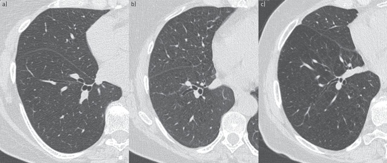
Main menu

  • Home
  • Current issue
  • Past issues
  • Authors/reviewers
    • Instructions for authors
    • Submit a manuscript
    • COVID-19 submission information
    • Institutional open access agreements
    • Peer reviewer login
  • Alerts
  • Subscriptions
  • ERS Publications
    • European Respiratory Journal
    • ERJ Open Research
    • European Respiratory Review
    • Breathe
    • ERS Books
    • ERS publications home

User menu

  • Log in
  • Subscribe
  • Contact Us
  • My Cart

Search

  • Advanced search
  • ERS Publications
    • European Respiratory Journal
    • ERJ Open Research
    • European Respiratory Review
    • Breathe
    • ERS Books
    • ERS publications home

Login

European Respiratory Society

Advanced Search

  • Home
  • Current issue
  • Past issues
  • Authors/reviewers
    • Instructions for authors
    • Submit a manuscript
    • COVID-19 submission information
    • Institutional open access agreements
    • Peer reviewer login
  • Alerts
  • Subscriptions

Lung imaging

Sebastian Ley
European Respiratory Review 2015 24: 240-245; DOI: 10.1183/16000617.00011214
Sebastian Ley
1Dept of Diagnostic and Interventional Radiology, Chirurgische Klinik Dr. Rinecker, Munich, Germany
2 Dept of Clinical Radiology, Ludwig Maximilians University, Munich, Germany
  • Find this author on Google Scholar
  • Find this author on PubMed
  • Search for this author on this site
  • For correspondence: ley@gmx.net
  • Article
  • Figures & Data
  • Info & Metrics
  • PDF
Loading

Figures

  • FIGURE 1
    • Download figure
    • Open in new tab
    • Download powerpoint
    FIGURE 1

    Axial inspiratory computed tomography (CT) scans at the level of the right inferior pulmonary vein, from three 54-year-old females, showing typical patterns. a) A healthy never-smoker. b) A smoker (note the subtle uniform diffuse increased opacity, which makes vessels more visible, and mild interlobular septal thickening, especially in middle lobe). c) A chronic obstructive pulmonary disease patient (note the diffusely distributed centrilobular emphysema, more in central part of the scans, with radiolucency and an irregular vascular pattern). Reproduced from [5] with permission from the publisher.

  • FIGURE 2
    • Download figure
    • Open in new tab
    • Download powerpoint
    FIGURE 2

    a) For segments in the middle and lower lobes centrilobular emphysema was found to be the predominant type on computed tomography. b) The corresponding colour-coded map of maximum peak enhancement of magnetic resonance perfusion showed heterogeneous perfusion with defects as the predominant pattern. Reproduced from [8] with permission from the publisher.

  • FIGURE 3
    • Download figure
    • Open in new tab
    • Download powerpoint
    FIGURE 3

    Images from a 62-year-old male patient presenting with a heterogeneous mass in the superior lobe of the right lung. a) Fusion of axial fat-saturated T1-weighted and diffusion-weighted sequences. Magnetic resonance imaging (MRI) demonstrated that only the anteromedial portion (arrow) of the mass showed high signal intensity on the diffusion-weighted MRI sequence, suggesting that the rest of the mass may be composed of atelectasis, consolidation or necrosis (*). b) Computed tomography guided biopsy was directed to this area, and histopathological examination yielded a diagnosis of lung adenocarcinoma. Reproduced from [16] with permission from the publisher.

PreviousNext
Back to top
View this article with LENS
Vol 24 Issue 136 Table of Contents
  • Table of Contents
  • Table of Contents (PDF)
  • Index by author
Email

Thank you for your interest in spreading the word on European Respiratory Society .

NOTE: We only request your email address so that the person you are recommending the page to knows that you wanted them to see it, and that it is not junk mail. We do not capture any email address.

Enter multiple addresses on separate lines or separate them with commas.
Lung imaging
(Your Name) has sent you a message from European Respiratory Society
(Your Name) thought you would like to see the European Respiratory Society web site.
CAPTCHA
This question is for testing whether or not you are a human visitor and to prevent automated spam submissions.
Print
Citation Tools
Lung imaging
Sebastian Ley
European Respiratory Review Jun 2015, 24 (136) 240-245; DOI: 10.1183/16000617.00011214

Citation Manager Formats

  • BibTeX
  • Bookends
  • EasyBib
  • EndNote (tagged)
  • EndNote 8 (xml)
  • Medlars
  • Mendeley
  • Papers
  • RefWorks Tagged
  • Ref Manager
  • RIS
  • Zotero

Share
Lung imaging
Sebastian Ley
European Respiratory Review Jun 2015, 24 (136) 240-245; DOI: 10.1183/16000617.00011214
del.icio.us logo Digg logo Reddit logo Technorati logo Twitter logo CiteULike logo Connotea logo Facebook logo Google logo Mendeley logo
Full Text (PDF)

Jump To

  • Article
    • Abstract
    • Abstract
    • Introduction
    • Functional lung imaging
    • Imaging in oncology
    • Conclusion
    • Footnotes
    • References
  • Figures & Data
  • Info & Metrics
  • PDF

Subjects

  • Lung imaging
  • Tweet Widget
  • Facebook Like
  • Google Plus One

More in this TOC Section

  • Diffuse parenchymal lung disease
  • Acute respiratory distress syndrome
  • Short-course treatment for MDR-TB: the STREAM trials
Show more Clinical Year in Review

Related Articles

Navigate

  • Home
  • Current issue
  • Archive

About the ERR

  • Journal information
  • Editorial board
  • Reviewers
  • Press
  • Permissions and reprints
  • Advertising
  • Sponsorship

The European Respiratory Society

  • Society home
  • myERS
  • Privacy policy
  • Accessibility

ERS publications

  • European Respiratory Journal
  • ERJ Open Research
  • European Respiratory Review
  • Breathe
  • ERS books online
  • ERS Bookshop

Help

  • Feedback

For authors

  • Instructions for authors
  • Publication ethics and malpractice
  • Submit a manuscript

For readers

  • Alerts
  • Subjects
  • RSS

Subscriptions

  • Accessing the ERS publications

Contact us

European Respiratory Society
442 Glossop Road
Sheffield S10 2PX
United Kingdom
Tel: +44 114 2672860
Email: journals@ersnet.org

ISSN

Print ISSN: 0905-9180
Online ISSN: 1600-0617

Copyright © 2023 by the European Respiratory Society