
Main menu

  • Home
  • Current issue
  • Past issues
  • Authors/reviewers
    • Instructions for authors
    • Submit a manuscript
    • Institutional open access agreements
    • Peer reviewer login
  • Alerts
  • Subscriptions
  • ERS Publications
    • European Respiratory Journal
    • ERJ Open Research
    • European Respiratory Review
    • Breathe
    • ERS Books
    • ERS publications home

User menu

  • Log in
  • Subscribe
  • Contact Us
  • My Cart

Search

  • Advanced search
  • ERS Publications
    • European Respiratory Journal
    • ERJ Open Research
    • European Respiratory Review
    • Breathe
    • ERS Books
    • ERS publications home

Login

European Respiratory Society

Advanced Search

  • Home
  • Current issue
  • Past issues
  • Authors/reviewers
    • Instructions for authors
    • Submit a manuscript
    • Institutional open access agreements
    • Peer reviewer login
  • Alerts
  • Subscriptions

Management of idiopathic pulmonary fibrosis: selected case reports

Michael Kreuter, Peter Kardos, Victor Hoffstein
European Respiratory Review 2014 23: 239-248; DOI: 10.1183/09059180.00002014
Michael Kreuter
1Dept of Pneumology and Respiratory Critical Care Medicine, Thoraxklinik, University of Heidelberg, Heidelberg, Germany. 2Group Practice and Respiratory, Allergology and Sleep Unit, Maingau Hospital, Frankfurt, Germany. 3Division of Respirology, St. Michael’s Hospital, Toronto, Canada
  • Find this author on Google Scholar
  • Find this author on PubMed
  • Search for this author on this site
  • For correspondence: kreuter@uni-heidelberg.de
Peter Kardos
1Dept of Pneumology and Respiratory Critical Care Medicine, Thoraxklinik, University of Heidelberg, Heidelberg, Germany. 2Group Practice and Respiratory, Allergology and Sleep Unit, Maingau Hospital, Frankfurt, Germany. 3Division of Respirology, St. Michael’s Hospital, Toronto, Canada
  • Find this author on Google Scholar
  • Find this author on PubMed
  • Search for this author on this site
Victor Hoffstein
1Dept of Pneumology and Respiratory Critical Care Medicine, Thoraxklinik, University of Heidelberg, Heidelberg, Germany. 2Group Practice and Respiratory, Allergology and Sleep Unit, Maingau Hospital, Frankfurt, Germany. 3Division of Respirology, St. Michael’s Hospital, Toronto, Canada
  • Find this author on Google Scholar
  • Find this author on PubMed
  • Search for this author on this site
  • Article
  • Figures & Data
  • Info & Metrics
  • PDF
Loading

Figures

  • Tables
  • Additional Files
  • Figure 1.
    • Download figure
    • Open in new tab
    • Download powerpoint
    Figure 1.

    Chest high-resolution computed tomography scans. Multi-slice computed tomography demonstrates a) apical emphysema and b) honeycombing and reticulation with subpleural, basal predominance. Extensive ground-glass opacities can be seen in both panels. Image courtesy of Claus P. Heussel (Thoraxklinik, University of Heidelberg, Heidelberg, Germany).
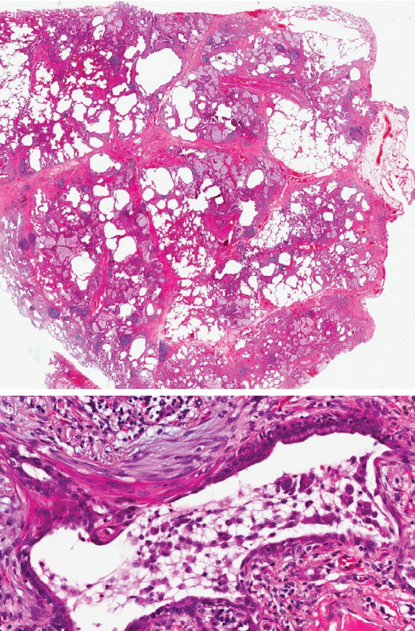
  • Figure 2.
    • Download figure
    • Open in new tab
    • Download powerpoint
    Figure 2.

    a) Low magnification histopathological biopsy showing typical features of usual interstitial pneumonia pattern with a heterogeneous appearance and areas of fibrosis with scarring and honeycomb change. Areas with less affected parenchyma also show emphysematous changes. b) Higher magnification of the histopathological biopsy reveals diffuse pulmonary involvement of numerous macrophage accumulations within most of the distal airspaces, consistent with a desquamative interstitial pneumonia pattern. Image courtesy of Philipp A. Schnabel (Institute of Pathology, University of Heidelberg, Heidelberg, Germany).
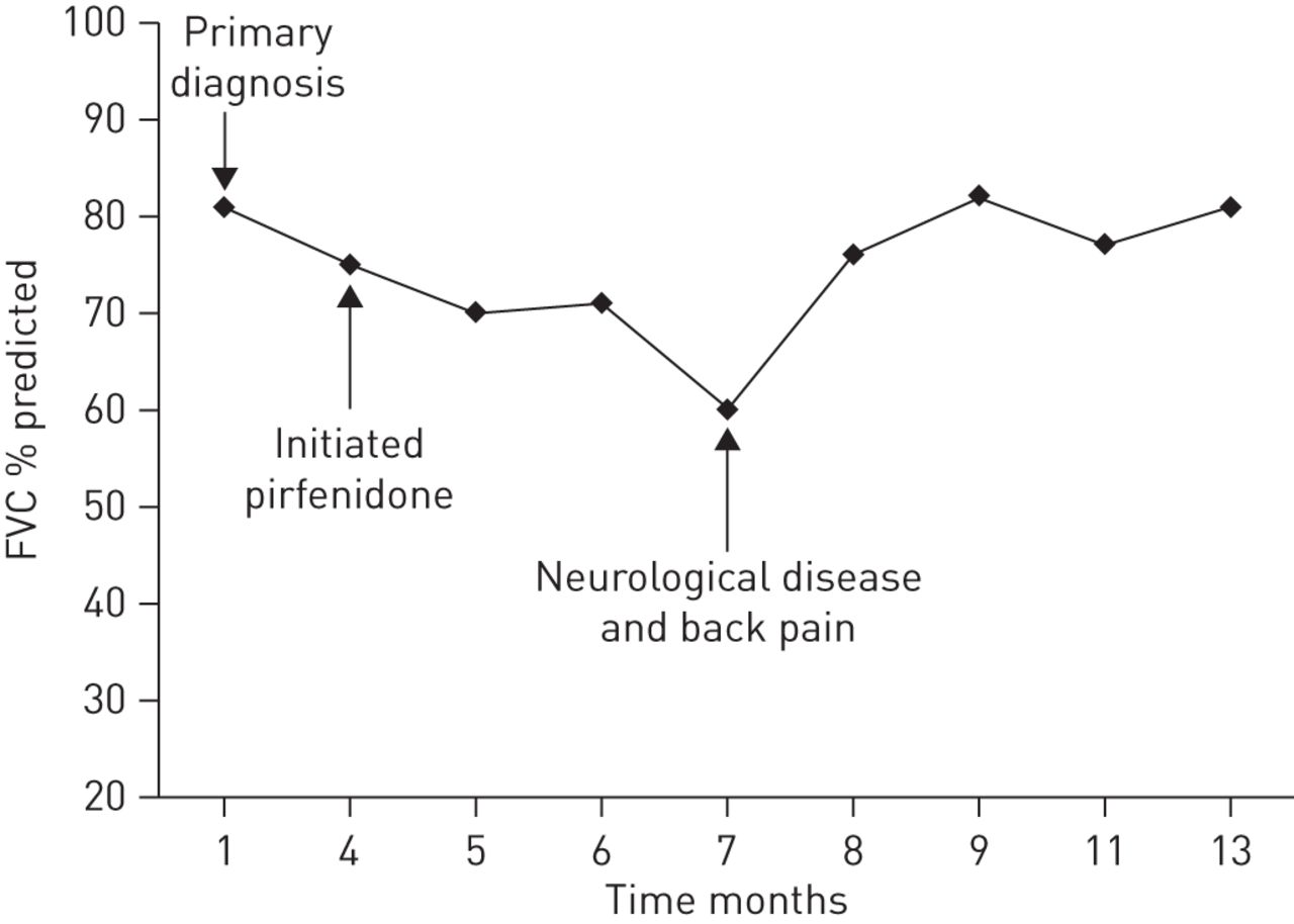
  • Figure 3.
    • Download figure
    • Open in new tab
    • Download powerpoint
    Figure 3.

    Forced vital capacity (FVC) over the course of the disease, some disease stabilisation can be seen following initiation of pirfenidone therapy. However, there was a significant decline in FVC due to neurological disease. After treatment for this disease the patient’s FVC increased again.
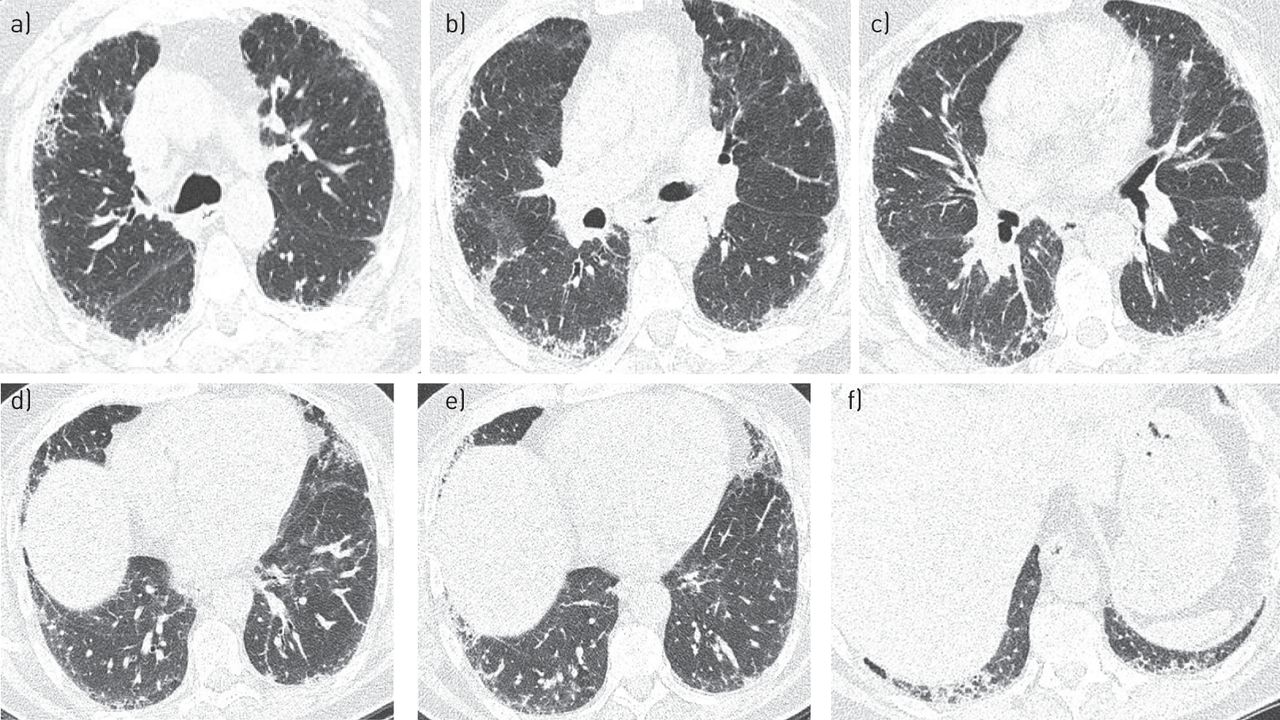
  • Figure 4.
    • Download figure
    • Open in new tab
    • Download powerpoint
    Figure 4.

    Chest high-resolution computed tomography scans performed on a) August 16, 2011 and b) September 4, 2012.

  • Figure 5.
    • Download figure
    • Open in new tab
    • Download powerpoint
    Figure 5.

    Initial high-resolution computed tomography scans performed in November 2011 showing mild reticulation and sub-pleural honeycombing. Representative sections of the lungs from a) the upper to f) the lower zones.

  • Figure 6.
    • Download figure
    • Open in new tab
    • Download powerpoint
    Figure 6.

    Progression in the patient’s pulmonary function tests from 2010 to 2013. a) Forced vital capacity (FVC); b) total lung capacity (TLC); c) diffusing capacity of the lung for carbon monoxide (DLCO); d) 6-min walking distance (6MWD); and e) oxygen saturation (arrow indicates patient was receiving supplemental oxygen). Start: oxygen saturation at the beginning of the 6MWD; end: oxygen saturation at the end of the 6MWD.
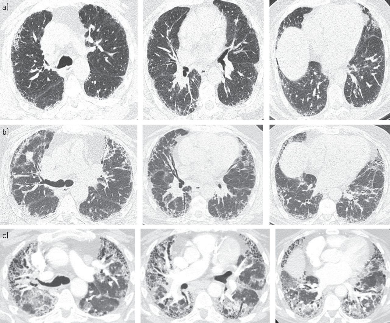
  • Figure 7.
    • Download figure
    • Open in new tab
    • Download powerpoint
    Figure 7.

    Patient computed tomography scans performed in a) 2010, b) 2012 and c) 2013. Representative sections of the lungs from the upper region to the lower region (left to right).

Tables

  • Figures
  • Additional Files
  • Table 1. Pulmonary function tests
    ParameterAbsolute% Predicted
    FVC L2.581
    FEV1 L2.182
    FEV1/FVC %84106
    RV L2.2124
    TLC L4.794
    DLCO-SB36
    DLCO–VA54
    • FVC: forced vital capacity; FEV1: forced expiratory volume in 1 s; RV: residual volume; TLC: total lung capacity; DLCO: diffusing capacity of the lung for carbon monoxide; SB: single breath; VA: alveolar volume.

  • Table 2. Pulmonary function tests performed between 2000 and 2012#
    Lung functionDate of test
    Feb 1, 2000May 29, 2006Oct 31, 2011April 21, 2012Sept 15, 2012Oct 27, 2012
    VC % predicted10010180888888
    FVC % predicted10110380949194
    FEV1 % predicted959872939192
    FEV1/VC % predicted717165767467
    DLCO % predicted10076324251.2
    • VC: vital capacity; FVC: forced vital capacity; FEV1: forced expiratory volume in 1 s; DLCO: diffusing capacity of the lung for carbon monoxide. #: the full dose of pirfenidone was started in November 2011.

  • Table 3. Initial pulmonary function tests performed in November 2011
    PredictedPatient
    NormalRangeMeasured% Predicted
    Lung volumes
        TLCpleth L6.44>5.074.61#72
        VC L4.13>3.302.97#72
        RV L2.30<2.991.6471
        RV/TLC35.8<46.935.6100
        FRC L2.2
    Spirometry
        FVC L4.13>3.302.86#69
        FEV1 L3.19>2.512.30#72
        FEV1/FVC %77.1>67.980.4
        FEF25–75 L·s−12.8>1.52.175
        FEFmax L·s−17.8>4.411.0142
        FIFmax L·s−13.7
        FEF50/FIF500.7
        MVV L·min−1124>9187#71
    Diffusing capacity
        DLCO-SB L·min−1·mmHg−125.3>17.315.4#61
        DLCO adjusted for Hb16.0 g·dL−114.8#59
        VA L6.23>5.073.82#61
    Oximetry
        Oxygen saturation %96≥9396
    Heart rate
        Pulse beats·min−170
    • TLCpleth: total lung capacity (TLC) measured by plethysmography; VC: vital capacity; RV: residual volume; FRC: functional residual capacity; FVC: functional vital capacity; FEV1: forced expiratory volume in 1 s; FEF25–75: forced expiratory flow at 25–75% of FVC; FEFmax: maximal forced expiratory flow; FIFmax: maximum forced inspiratory flow; FEF50: forced expiratory flow at 50% of FVC; FIF50: forced inspiratory flow at 50% of FVC; MVV: maximum voluntary ventilation; DLCO: diffusing capacity of the lung for carbon monoxide; SB: single breath; Hb: haemoglobin; VA: alveolar volume. #: outside normal limits.

Additional Files

  • Figures
  • Tables
  • Disclosures

    Files in this Data Supplement:

    • P. Kardos
    • M. Kreuter
PreviousNext
Back to top
View this article with LENS
Vol 23 Issue 132 Table of Contents
  • Table of Contents
  • Table of Contents (PDF)
  • Index by author
Email

Thank you for your interest in spreading the word on European Respiratory Society .

NOTE: We only request your email address so that the person you are recommending the page to knows that you wanted them to see it, and that it is not junk mail. We do not capture any email address.

Enter multiple addresses on separate lines or separate them with commas.
Management of idiopathic pulmonary fibrosis: selected case reports
(Your Name) has sent you a message from European Respiratory Society
(Your Name) thought you would like to see the European Respiratory Society web site.
CAPTCHA
This question is for testing whether or not you are a human visitor and to prevent automated spam submissions.
Print
Citation Tools
Management of idiopathic pulmonary fibrosis: selected case reports
Michael Kreuter, Peter Kardos, Victor Hoffstein
European Respiratory Review Jun 2014, 23 (132) 239-248; DOI: 10.1183/09059180.00002014

Citation Manager Formats

  • BibTeX
  • Bookends
  • EasyBib
  • EndNote (tagged)
  • EndNote 8 (xml)
  • Medlars
  • Mendeley
  • Papers
  • RefWorks Tagged
  • Ref Manager
  • RIS
  • Zotero

Share
Management of idiopathic pulmonary fibrosis: selected case reports
Michael Kreuter, Peter Kardos, Victor Hoffstein
European Respiratory Review Jun 2014, 23 (132) 239-248; DOI: 10.1183/09059180.00002014
del.icio.us logo Digg logo Reddit logo Technorati logo Twitter logo CiteULike logo Connotea logo Facebook logo Google logo Mendeley logo
Full Text (PDF)

Jump To

  • Article
    • Abstract
    • Introduction
    • Case 1
    • Case 2
    • Case 3
    • Acknowledgments
    • Footnotes
    • References
  • Figures & Data
  • Info & Metrics
  • PDF

Subjects

  • Interstitial and orphan lung disease
  • Tweet Widget
  • Facebook Like
  • Google Plus One

More in this TOC Section

  • Challenges in managing pulmonary hypertension
  • CTD-PAH-: management of severe haemodynamic impairment
Show more Case-Based Review

Related Articles

Navigate

  • Home
  • Current issue
  • Archive

About the ERR

  • Journal information
  • Editorial board
  • Press
  • Permissions and reprints
  • Advertising
  • Sponsorship

The European Respiratory Society

  • Society home
  • myERS
  • Privacy policy
  • Accessibility

ERS publications

  • European Respiratory Journal
  • ERJ Open Research
  • European Respiratory Review
  • Breathe
  • ERS books online
  • ERS Bookshop

Help

  • Feedback

For authors

  • Instructions for authors
  • Publication ethics and malpractice
  • Submit a manuscript

For readers

  • Alerts
  • Subjects
  • RSS

Subscriptions

  • Accessing the ERS publications

Contact us

European Respiratory Society
442 Glossop Road
Sheffield S10 2PX
United Kingdom
Tel: +44 114 2672860
Email: journals@ersnet.org

ISSN

Print ISSN: 0905-9180
Online ISSN: 1600-0617

Copyright © 2023 by the European Respiratory Society