
Main menu

  • Home
  • Current issue
  • Past issues
  • Authors/reviewers
    • Instructions for authors
    • Submit a manuscript
    • Institutional open access agreements
    • Peer reviewer login
  • Alerts
  • Subscriptions
  • ERS Publications
    • European Respiratory Journal
    • ERJ Open Research
    • European Respiratory Review
    • Breathe
    • ERS Books
    • ERS publications home

User menu

  • Log in
  • Subscribe
  • Contact Us
  • My Cart

Search

  • Advanced search
  • ERS Publications
    • European Respiratory Journal
    • ERJ Open Research
    • European Respiratory Review
    • Breathe
    • ERS Books
    • ERS publications home

Login

European Respiratory Society

Advanced Search

  • Home
  • Current issue
  • Past issues
  • Authors/reviewers
    • Instructions for authors
    • Submit a manuscript
    • Institutional open access agreements
    • Peer reviewer login
  • Alerts
  • Subscriptions

Endobronchial ultrasound in the management of nonsmall cell lung cancer

Christophe Dooms, Inge Muylle, Jonas Yserbyt, Vincent Ninane
European Respiratory Review 2013 22: 169-177; DOI: 10.1183/09059180.00001113
Christophe Dooms
*Respiratory Division, University Hospitals Leuven, Leuven, Dept of Clinical and Experimental Medicine, KU LeuvenLeuven, #Dept of Clinical and Experimental Medicine, KU Leuven, Leuven, and ¶Chest Service, Saint-Pierre University Hospital, Brussels, Belgium.
  • Find this author on Google Scholar
  • Find this author on PubMed
  • Search for this author on this site
Inge Muylle
*Respiratory Division, University Hospitals Leuven, Leuven, Dept of Clinical and Experimental Medicine, KU LeuvenLeuven, #Dept of Clinical and Experimental Medicine, KU Leuven, Leuven, and ¶Chest Service, Saint-Pierre University Hospital, Brussels, Belgium.
  • Find this author on Google Scholar
  • Find this author on PubMed
  • Search for this author on this site
Jonas Yserbyt
*Respiratory Division, University Hospitals Leuven, Leuven, Dept of Clinical and Experimental Medicine, KU LeuvenLeuven, #Dept of Clinical and Experimental Medicine, KU Leuven, Leuven, and ¶Chest Service, Saint-Pierre University Hospital, Brussels, Belgium.
  • Find this author on Google Scholar
  • Find this author on PubMed
  • Search for this author on this site
Vincent Ninane
*Respiratory Division, University Hospitals Leuven, Leuven, Dept of Clinical and Experimental Medicine, KU LeuvenLeuven, #Dept of Clinical and Experimental Medicine, KU Leuven, Leuven, and ¶Chest Service, Saint-Pierre University Hospital, Brussels, Belgium.
  • Find this author on Google Scholar
  • Find this author on PubMed
  • Search for this author on this site
  • For correspondence: Vincent_ninane@stpierre-bru.be
  • Article
  • Figures & Data
  • Info & Metrics
  • PDF
Loading

Figures

  • Tables
  • Additional Files
  • Figure 1.
    • Download figure
    • Open in new tab
    • Download powerpoint
    Figure 1.

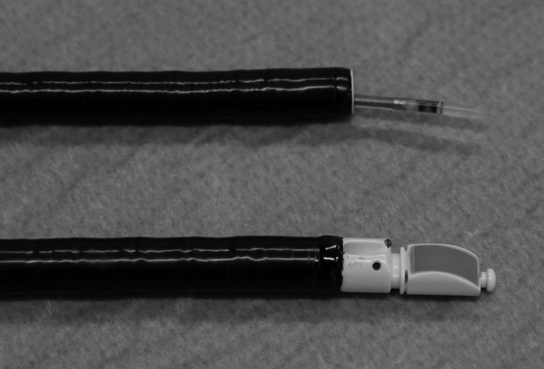
    A convex ultrasound transducer is located at the tip of a dedicated bronchoscope for peribronchial linear scanning (bottom). A radial ultrasound transducer miniprobe is introduced through the biopsy channel of a standard bronchoscope for endobronchial scanning (top).

  • Figure 2.
    • Download figure
    • Open in new tab
    • Download powerpoint
    Figure 2.

    The radial endobronchial ultrasound miniprobe can visualise a) normal lung parenchyma (“snow-storm” pattern) or b) solid peripheral pulmonary lesion.

  • Figure 3.
    • Download figure
    • Open in new tab
    • Download powerpoint
    Figure 3.

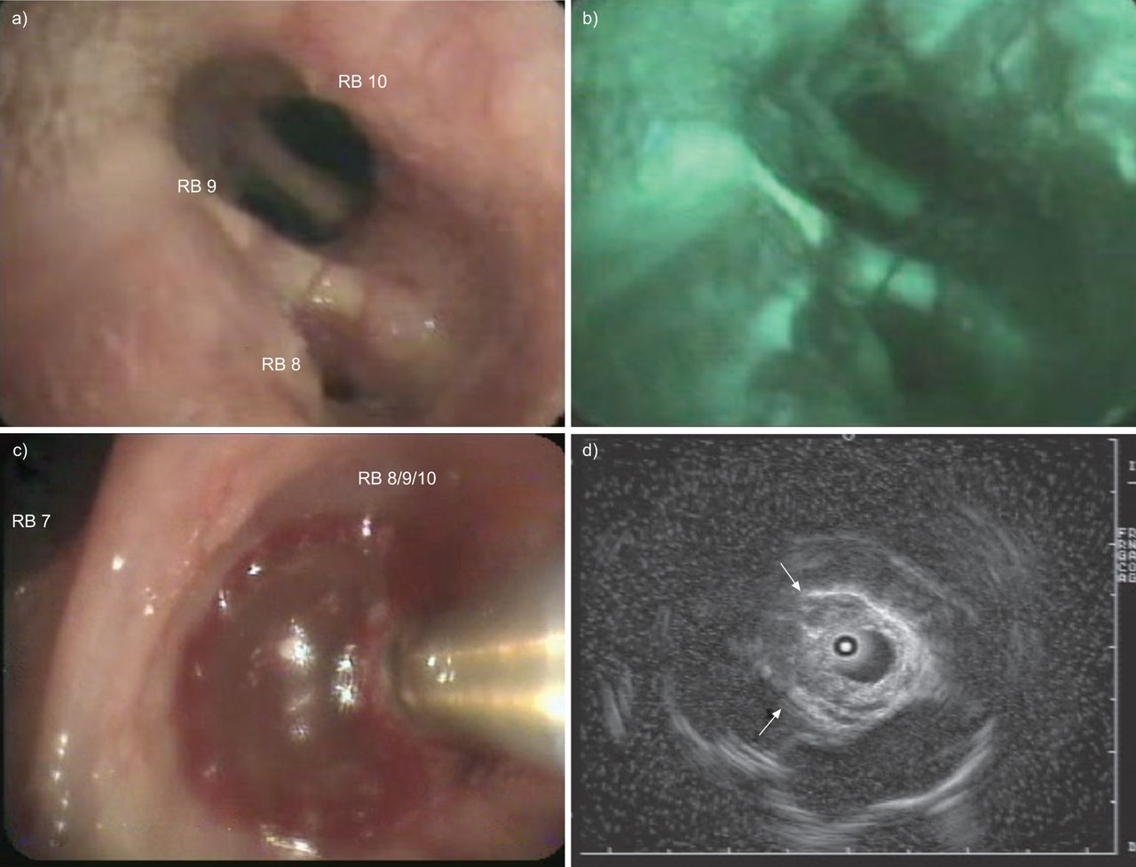
    a) White light and b) autofluorescence bronchoscopy of an early stage radio-occult squamous cell carcinoma, which has been proven invasive by radial endobronchial ultrasound miniprobe with filled balloon sheath (c and d). The ultrasound image (d) illustrates interruption of white “cartilage” line, as indicated by the arrows. RB: right bronchus.

  • Figure 4.
    • Download figure
    • Open in new tab
    • Download powerpoint
    Figure 4.

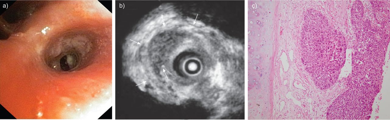
    a) White light bronchoscopy of an early stage radio-occult squamous cell carcinoma, which has been proven micro-invasive by radial endobronchial ultrasound miniprobe with filled balloon sheath (b), and on histopathology (c). The arrows in b) indicate the intact white “cartilage” line on the ultrasound image.

  • Figure 5.
    • Download figure
    • Open in new tab
    • Download powerpoint
    Figure 5.

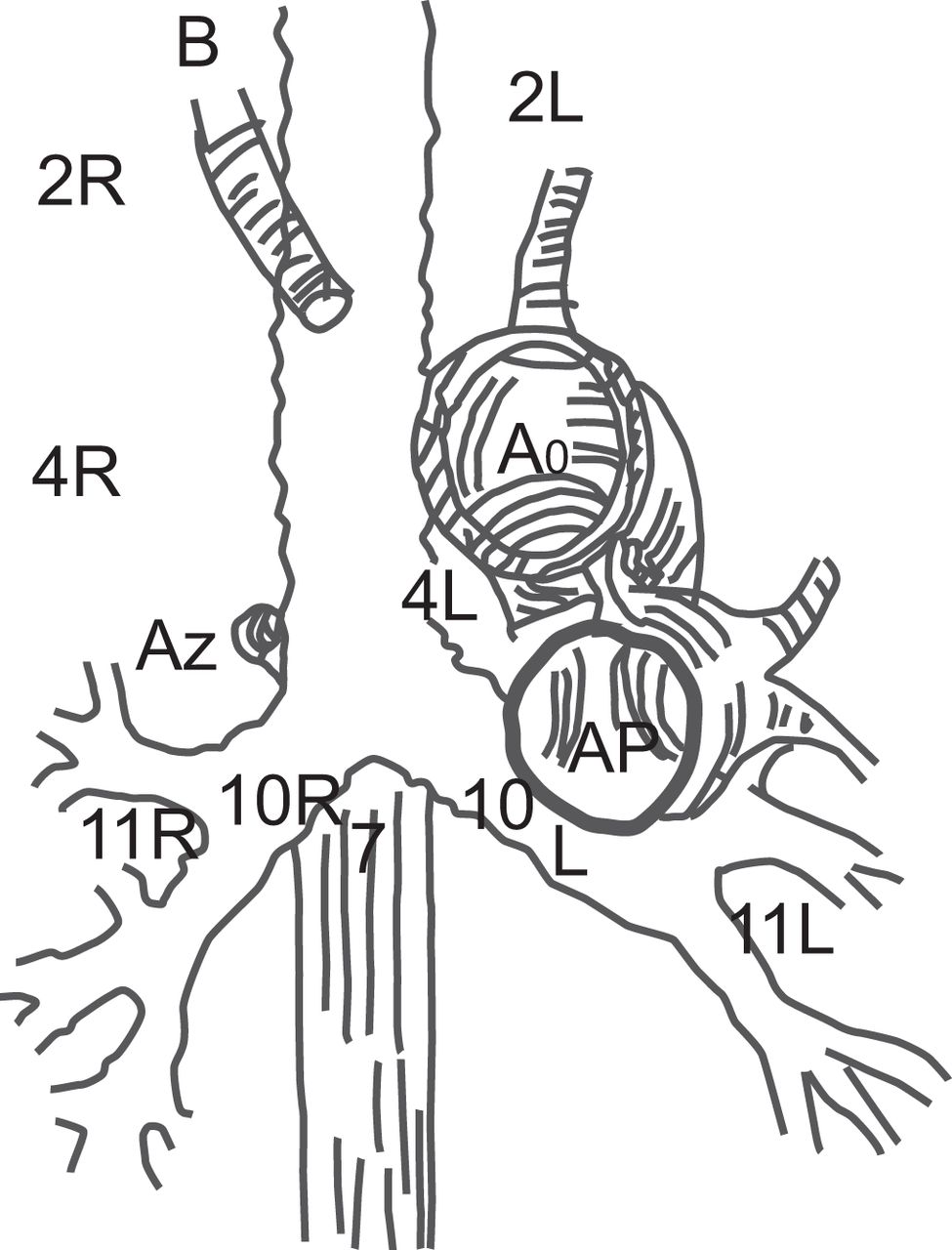
    Schematic representation of the mediastinum and nodal stations that are accessible to EBUS. Ao: aorta; AP: pulmonary artery; Az: azygos vein; B: brachiocephalic artery; L: left; R: right. For precise borders of the nodal stations see [36].

Tables

  • Figures
  • Additional Files
  • Table 1. Accessibility to nodal stations with different sampling techniques
    MediastinoscopyEBUS-TBNAEUS-FNA
    Supraclavicular zone
        1±–-–
    Superior mediastinal nodes
        2R++–
        2L+++
        4R++–
        4L+++
        3a–––
        3p_±#±#
    Aortic nodes
        5–––#
        6–––#
    Inferior mediastinal nodes
        7+++
        8––+
        9––+
    N1 nodes
        10 Hilar±+±#
        11 Interlobar–+–
        12 Lobar–+–
    • EBUS-TBNA: endobronchial ultrasound-transbronchial needle aspiration; EUS-FNA: endoscopic ultrasound-fine needle aspiration. +: accessible; –: inaccessible; ±: may be accessible. #: casuistic reports that it is feasible.

Additional Files

  • Figures
  • Tables
  • Disclosures

    Files in this Data Supplement:

    • V. Ninane
PreviousNext
Back to top
View this article with LENS
Vol 22 Issue 128 Table of Contents
  • Table of Contents
  • Table of Contents (PDF)
  • Cover (PDF)
  • Index by author
Email

Thank you for your interest in spreading the word on European Respiratory Society .

NOTE: We only request your email address so that the person you are recommending the page to knows that you wanted them to see it, and that it is not junk mail. We do not capture any email address.

Enter multiple addresses on separate lines or separate them with commas.
Endobronchial ultrasound in the management of nonsmall cell lung cancer
(Your Name) has sent you a message from European Respiratory Society
(Your Name) thought you would like to see the European Respiratory Society web site.
CAPTCHA
This question is for testing whether or not you are a human visitor and to prevent automated spam submissions.
Print
Citation Tools
Endobronchial ultrasound in the management of nonsmall cell lung cancer
Christophe Dooms, Inge Muylle, Jonas Yserbyt, Vincent Ninane
European Respiratory Review Jun 2013, 22 (128) 169-177; DOI: 10.1183/09059180.00001113

Citation Manager Formats

  • BibTeX
  • Bookends
  • EasyBib
  • EndNote (tagged)
  • EndNote 8 (xml)
  • Medlars
  • Mendeley
  • Papers
  • RefWorks Tagged
  • Ref Manager
  • RIS
  • Zotero

Share
Endobronchial ultrasound in the management of nonsmall cell lung cancer
Christophe Dooms, Inge Muylle, Jonas Yserbyt, Vincent Ninane
European Respiratory Review Jun 2013, 22 (128) 169-177; DOI: 10.1183/09059180.00001113
del.icio.us logo Digg logo Reddit logo Technorati logo Twitter logo CiteULike logo Connotea logo Facebook logo Google logo Mendeley logo
Full Text (PDF)

Jump To

  • Article
    • Abstract
    • CONVEX AND RADIAL PROBE EBUS: TECHNICAL AND PRACTICAL PATHOLOGICAL ASPECTS
    • RADIAL PROBE EBUS FOR PRIMARY TUMOUR STAGING (T-FACTOR)
    • CONVEX PROBE EBUS FOR MEDIASTINAL NODAL STAGING (N-FACTOR)
    • RADIAL PROBE EBUS FOR DIAGNOSIS OF PERIPHERAL PULMONARY LESIONS
    • CONVEX EBUS PROBE FOR THE DIAGNOSIS OF CENTRAL PARENCHYMAL LUNG CANCERS
    • CONCLUSIONS
    • Footnotes
    • REFERENCES
  • Figures & Data
  • Info & Metrics
  • PDF

Subjects

  • Lung cancer
  • Respiratory clinical practice
  • Tweet Widget
  • Facebook Like
  • Google Plus One

More in this TOC Section

  • The oesophageal balloon for respiratory monitoring in ventilated patients
  • QoL in patients with chronic respiratory failure on HMV
  • Awake prone positioning in acute hypoxaemic respiratory failure
Show more Series

Related Articles

Navigate

  • Home
  • Current issue
  • Archive

About the ERR

  • Journal information
  • Editorial board
  • Press
  • Permissions and reprints
  • Advertising
  • Sponsorship

The European Respiratory Society

  • Society home
  • myERS
  • Privacy policy
  • Accessibility

ERS publications

  • European Respiratory Journal
  • ERJ Open Research
  • European Respiratory Review
  • Breathe
  • ERS books online
  • ERS Bookshop

Help

  • Feedback

For authors

  • Instructions for authors
  • Publication ethics and malpractice
  • Submit a manuscript

For readers

  • Alerts
  • Subjects
  • RSS

Subscriptions

  • Accessing the ERS publications

Contact us

European Respiratory Society
442 Glossop Road
Sheffield S10 2PX
United Kingdom
Tel: +44 114 2672860
Email: journals@ersnet.org

ISSN

Print ISSN: 0905-9180
Online ISSN: 1600-0617

Copyright © 2023 by the European Respiratory Society