
Main menu

  • Home
  • Current issue
  • Past issues
  • Authors/reviewers
    • Instructions for authors
    • Submit a manuscript
    • COVID-19 submission information
    • Institutional open access agreements
    • Peer reviewer login
  • Alerts
  • Subscriptions
  • ERS Publications
    • European Respiratory Journal
    • ERJ Open Research
    • European Respiratory Review
    • Breathe
    • ERS Books
    • ERS publications home

User menu

  • Log in
  • Subscribe
  • Contact Us
  • My Cart

Search

  • Advanced search
  • ERS Publications
    • European Respiratory Journal
    • ERJ Open Research
    • European Respiratory Review
    • Breathe
    • ERS Books
    • ERS publications home

Login

European Respiratory Society

Advanced Search

  • Home
  • Current issue
  • Past issues
  • Authors/reviewers
    • Instructions for authors
    • Submit a manuscript
    • COVID-19 submission information
    • Institutional open access agreements
    • Peer reviewer login
  • Alerts
  • Subscriptions

Why you do or do not need thoracoscopy

J.P. Janssen
European Respiratory Review 2010 19: 213-216; DOI: 10.1183/09059180.00005410
J.P. Janssen
  • Find this author on Google Scholar
  • Find this author on PubMed
  • Search for this author on this site
  • For correspondence: j.janssen@cwz.nl
  • Article
  • Figures & Data
  • Info & Metrics
  • PDF
Loading

Abstract

Thoracoscopy is an old but still very valuable technique for the evaluation of pleural pathology and, especially for the further investigation of the aetiology of pleural fluid. It remains of great importance, since it is able to not only provide an exact diagnosis, but also can have therapeutic potential. In this review, the differential diagnostic aspects of transudate versus exudate are further elaborated, and the role of thoracoscopy is compared to closed pleural biopsy and image guided biopsy.

  • Medical
  • pleuroscopy
  • thoracoscopy

Thoracoscopy is not new; this year we will celebrate the Centennial of Thoracoscopy, as the first thoracoscopic procedure was performed by the Swedish internist H.C. Jacobaeus in 1910. Today, thoracoscopy is still a valuable tool in the diagnosis and treatment of pleural diseases. In this review, we will discuss the diagnostic and therapeutic aspects of thoracoscopy in patients with pleural effusion. Thoracoscopy has a place in the diagnosis and treatment of other diseases, such as pneumothorax, palmary hyperhidrosis and empyema. These diseases will not be discussed here.

DIAGNOSTIC ASPECTS OF THORACOSCOPY

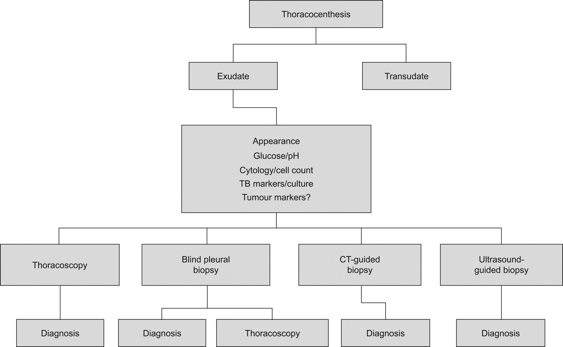
The flow chart of the work-up of pleural effusion is presented in figure 1. Thoracocenthesis is the first procedure to be performed in a pleural effusion of >1 cm diameter in lateral decubitus position [1]. The most important step in narrowing the differential diagnosis is to distinguish a transudate from an exudate. To do this, Light's original citeria (ratio of pleural fluid/serum protein >0.5, ratio of pleural fluid/serum lactate dehydrogenase (LDH) >0.6 or pleural fluid LDH more than two-thirds of the upper limit of normal serum value) are still appropriate [1].

Figure 1.
  • Download figure
  • Open in new tab
  • Download powerpoint
Figure 1.

The diagnostic work-up of pleural effusion. CT: computed tomography.

If the protein level is >35 g·L−1, the effusion is most likely an exudate [2]. In borderline exudates (protein level >25 but <35 g·L−1) Light's criteria may misclassify transudates in up to 20% of cases, especially in patients with congestive heart failure who have been on diuretics. To discriminate true transudates from pseudo-exudates, measurement of the pleural protein gradient or the pleural fluid albumin gradient can be applied: if serum protein level minus pleural protein level is >3.1 g·dL−1, or serum albumin level minus pleural albumin level is >1.2 g·dL−1, it is a transudate [1]. In recent studies, measurement of pro-B-type natriuretic peptide (pro-BNP) in pleural fluid and serum appeared to be promising in the diagnosis of transudates in patients with chronic heart failure. In case of possible misclassification by the use of Light's criteria, measurement of pro-BNP level may appear to be a better tool to differentiate a transudate from a pseudo-exudate [3–6].

In the case of an exudate, and benign or non-conclusive cytology, infection, pulmonary embolism and abdominal disease should be considered. If these diseases have been ruled out or are very unlikely, the exudate is caused by malignant disease, tuberculosis or the exudate is idiopathic.

In the past, it has been stated that a malignant pleural effusion can also occur as a transudate [7]. This is most likely due to imperfect application of the diagnostic rules, or comorbid conditions like hypoalbuminaemia, cirrhosis with ascites or chronic heart failure.

ANALYSIS OF AN EXUDATE

In the case of a proven exudate with non-conclusive cytology after (repeated) thoracocenthesis, an additional procedure to obtain pleural histology tissue is the next step. This can be done with a minimal invasive procedure in four ways: closed pleural biopsy (CPB; Abrams biopsy), thoracoscopy, ultrasound (US)-guided biopsy, and computed tomography (CT)-guided biopsy. A comparison of these techniques is summarised in table 1.

View this table:
  • View inline
  • View popup
Table 1 Comparison of techniques to obtain pleural biopsy

CPB (ABRAMS BIOPSY)

CPB is an old technique. In patients with pleural effusion, a blind biopsy of the parietal pleura can be obtained. In malignant pleural disease, the additional diagnostic yield of CPB after thoracocenthesis is limited to 7% [8].

The diagnostic yield of CPB is better in areas with high incidence of tuberculosis (TB), as has been demonstrated by Diacon et al. [9]. In their study, the diagnostic yield of the combination of TB culture and histology reached 79%, with a sensitivity of 93%, if combined with serum adenosine deaminase level and lymphocytosis of the pleural fluid (lymphocytes/neutrophils >0.75). In a prospective study in the UK, Maskell et al. [10] compared CPB with CT-guided pleural biopsy. The results for the CT group were: sensitivity 87%, specificity 100% and negative predictive value 80%. In the CPB group the results were: sensitivity 47%, specificity 100% and negative predictive value 44%.

In conclusion, CPB should no longer be used in a setting where image-guided pleural biopsies can be obtained. Use of CPB is only indicated in areas with high incidence of TB and limited medical resources [9].

CT- VERSUS US-GUIDED PLEURAL BIOPSY

There are no comparative studies of the diagnostic yield of CT- and US-guided biopsy; the diagnostic sensitivity of both techniques is high (>83%) [11]. The characteristics of each procedure are summarised in table 1.

THORACOSCOPY VERSUS IMAGE-GUIDED PROCEDURES

The diagnostic yield of thoracoscopy is high; it is reported to be >90% in the majority of studies (table 2). Although it is a more invasive procedure compared with image-guided pleural biopsy, the big advantage of thoracoscopy is the possibility to perform a therapeutic intervention in the same session as the diagnostic biopsy of the pleura.

View this table:
  • View inline
  • View popup
Table 2 The diagnostic yield of thoracoscopy

The possible therapeutic procedures during thoracoscopy are: 1) removal of (septated) pleural effusions; 2) talc poudrage (under visual control if preferred); and 3) drain positioning under visual control.

Thoracoscopy is the preferred procedure if no clear target lesion is visible on the CT scan, and in patients with large or recurrent effusions, in whom drainage and pleurodesis is indicated. Image-guided pleural biopsy may be the procedure of choice in elderly, unfit patients with small pleural effusions. The diagnostic work-up of a pleural effusion is summarised in figure 1.

HOW TO DEAL WITH UNDIAGNOSED PLEURAL EFFUSION

After a complete work-up of pleural effusion including thoracoscopy, a specific diagnosis may not be available in a considerable amount of cases (table 2).

This condition is described as “idiopathic pleuritis”, “unspecific pleuritis”, “nonspecific pleuritis” or “pleuritis with indeterminate cause”. This specific problem has been retrospectively addressed by four studies, three of which included thoracoscopy in the work-up, and one thoracotomy [19, 22–24]. The results of these studies are summarised in table 3.

View this table:
  • View inline
  • View popup
Table 3 Long-term follow-up of nonspecific pleuritis (NSP) after a diagnostic thoracoscopy or thoracotomy; eventually, malignant pleuritis was found in 5–25%

In our study of 208 patients with a nonspecific pleuritis after thoracoscopy, 85% followed a benign course during follow-up [23]. A “wait and see” approach is therefore justified in the majority of these patients. An extended video-assisted thoracoscopic surgery procedure is indicated in case of a difficult thoracoscopy with adhesions and layers of fibrin, especially if there is a clinical suspicion of mesothelioma. Otherwise, the patients should be monitored with regular chest radiography. In case of a persistent or increasing pleural effusion, thoracocenthesis should be repeated. If the analysis of the pleural fluid is showing a lymphocytosis or increased LDH, the thoracoscopy should be repeated [23].

CONCLUSIONS

Thoracoscopy is an old, safe and simple procedure. Any pulmonologist who deals with pleural disease nowadays should be able to perform thoracoscopy. The diagnostic yield of pleural effusions under CT or US guidance is only slightly inferior to thoracoscopy (85% versus >90%). Thoracoscopy is the preferred procedure if no clear target lesion is visible on the CT scan. Also, thoracoscopy is preferred if removal of pleural effusion and/or pleurodesis is indicated, because during thoracoscopy the diagnostic and therapeutic procedure can be performed in the same session.

Footnotes

  • Provenance

    Publication of this peer-reviewed article was supported by GlaxoSmithKline, Belgium (principal sponsor, European Respiratory Review issue 117).

  • Statement of Interest

    None declared.

  • Received May 25, 2010.
  • Accepted May 26, 2010.
  • ©ERS 2010

References

  1. ↵
    1. Light RW,
    2. et al
    1. Light RW
    Approach to the patient. In: Light RW, et al. Pleural Diseases. 5th Edn. Philadephia, Lippincott Williams and Wilkins, 2007; pp. 109–119
  2. ↵
    1. Maskell NA,
    2. Butland RJA
    BTS guidelines for the investigation of a unilateral pleural effusion in adults. Thorax 2003; 58: ii8–ii17.
    OpenUrlAbstract/FREE Full Text
  3. ↵
    1. Porcel JM,
    2. Chorda J,
    3. Cao G,
    4. et al
    . Comparing serum and pleural fluid pro-brain natriuretic peptide (NT-proBNP) levels with pleural-to-serum albumin gradient for the identification of cardiac effusions misclassified by Light's criteria. Respirology 2007; 12: 654–659.
    OpenUrlCrossRefPubMed
    1. Kolditz M,
    2. Halank M,
    3. Schiemanck CS,
    4. et al
    . High diagnostic accuracy of NT-proBNP for cardiac origin of pleural effusions. Eur Respir J 2006; 28: 144–150.
    OpenUrlAbstract/FREE Full Text
    1. Han C H,
    2. Choi J E,
    3. Chung J H
    Clinical utility of pleural fluid NT-proBNP in patients with pleural effusions. Intern Med 2008; 47: 1669–1674.
    OpenUrlCrossRefPubMed
  4. ↵
    1. Seyhan EC,
    2. Altin S,
    3. Cetinkaya E,
    4. et al.
    The importance of pleural fluid and serum NT-proBNP levels in differentiating pleural effusion due to heart failure from other causes of effusion. Intern Med 2009; 48: 287–293.
    OpenUrlCrossRefPubMed
  5. ↵
    1. Ashchi M,
    2. Golish J,
    3. Eng P,
    4. et al
    . Transudative malignant pleural effusions; prevalence and mechanisms. South Med J 1998; 91: 23–32.
    OpenUrlCrossRefPubMed
  6. ↵
    1. Prakash UBS
    Comparison of needle biopsy with cytologic analysis for the evaluation of pleural effusion: analysis of 414 cases. Mayo Clin Proc 1985; 60: 158–164.
    OpenUrlCrossRefPubMed
  7. ↵
    1. Diacon AH,
    2. Van de Wal BW,
    3. Wyser C,
    4. et al
    . Diagnostic tools in tuberculous pleurisy: a direct comparative study. Eur Respir J 2003; 22: 589–591.
    OpenUrlAbstract/FREE Full Text
  8. ↵
    1. Maskell NA,
    2. Gleeson FV,
    3. Davies RJ
    Standard pleural biopsy versus CT-guided cutting-needle biopsy for diagnosis of malignant disease in pleural effusions: a randomised controlled trial. Lancet 2003; 361: 1326–1330.
    OpenUrlCrossRefPubMed
  9. ↵
    1. Rahman NM,
    2. Gleeson FV
    Image-guided pleural biopsy. Curr Opin Pulm Med 2008; 14: 331–336.
    OpenUrlCrossRefPubMed
  10. ↵
    1. Boutin C,
    2. Viallat JR,
    3. Cargnino P,
    4. et al
    . Thoracoscopy in malignant pleural effusions. Am Rev Respir Dis 1981; 124: 588–592.
    OpenUrlPubMed
  11. ↵
    1. Loddenkemper R
    Thoracoscopy: results in non cancerous and idiopathic pleural effusions. Poumon Coeur 1981; 37: 261–264.
    OpenUrlPubMed
  12. ↵
    1. Page RD,
    2. Jeffrey RR,
    3. Donnelly RJ
    Thoracoscopy: a review of 121 consecutive surgical procedures. Ann Thorac Surg 1989; 48: 66–68.
    OpenUrlCrossRefPubMed
  13. ↵
    1. Menzies R,
    2. Charbonneau M
    Thoracoscopy for the diagnosis of pleural disease. Ann Intern Med 1991; 114: 271–276.
    OpenUrlCrossRefPubMed
  14. ↵
    1. Hucker J,
    2. Bhatnagar NK,
    3. Al-Jilaihawi AN,
    4. et al
    . Thoracoscopy in the diagnosis and management of recurrent pleural effusions. Ann Thorac Surg 1991; 52: 1145–1147.
    OpenUrlCrossRefPubMed
  15. ↵
    1. Kendall SWH,
    2. Bryan AJ,
    3. Large SR,
    4. et al
    . Pleural effusions: is thoracoscopy a reliable investigation? A retrospective review. Respir Med 1992; 86: 437–440.
    OpenUrlPubMed
  16. ↵
    1. Ohri SK,
    2. Oswal SK,
    3. Townsend ER,
    4. et al
    . Early and late outcome after diagnostic thoracoscopy and talc pleurodesis. Ann Thorac Surg 1992; 53: 1038–1041.
    OpenUrlCrossRefPubMed
  17. ↵
    1. Ferrer JS,
    2. Munoz XG,
    3. Orriols RM,
    4. et al
    . Evolution of idiopathic effusion. A prospective long term follow-up study. Chest 1996; 109: 1508–1513.
    OpenUrlCrossRefPubMed
  18. ↵
    1. Hansen M,
    2. Faurschou P,
    3. Clementsen P
    Medical thoracoscopy, results and complications in 146 patients: a retrospective study. Respir Med 1998; 92: 228–232.
    OpenUrlCrossRefPubMed
  19. ↵
    1. Blanc FX,
    2. Atassi K,
    3. Bignon J,
    4. et al
    . Diagnostic value of medical thoracoscopy in pleural disease. A 6-year retrospective study. Chest 2002; 121: 1677–1683.
    OpenUrlCrossRefPubMed
  20. ↵
    1. Ryan CJ,
    2. Rodgers RF,
    3. Unni KK,
    4. et al
    . The outcome of patients with pleural effusion of indeterminate cause at thoracotomy. Mayo Clin Proc 1981; 56: 145–149.
    OpenUrlPubMed
  21. ↵
    1. Janssen JP,
    2. Ramlal S,
    3. Mravunac M
    The long-term follow-up of exudative pleural effusion after nondiagnostic thoracoscopy. J Bronchol 2004; 11: 169–174.
    OpenUrlCrossRef
  22. ↵
    1. Venekamp LN,
    2. Velkeniers B,
    3. Noppen M
    Does “idiopathic pleuritis” exist? Natural history of non-specific pleuritis diagnosed after thoracoscopy. Respiration 2005; 72: 74–78.
    OpenUrlCrossRefPubMed
View Abstract
PreviousNext
Back to top
View this article with LENS
Vol 19 Issue 117 Table of Contents
  • Table of Contents
  • Table of Contents (PDF)
  • Index by author
Email

Thank you for your interest in spreading the word on European Respiratory Society .

NOTE: We only request your email address so that the person you are recommending the page to knows that you wanted them to see it, and that it is not junk mail. We do not capture any email address.

Enter multiple addresses on separate lines or separate them with commas.
Why you do or do not need thoracoscopy
(Your Name) has sent you a message from European Respiratory Society
(Your Name) thought you would like to see the European Respiratory Society web site.
CAPTCHA
This question is for testing whether or not you are a human visitor and to prevent automated spam submissions.
Print
Citation Tools
Why you do or do not need thoracoscopy
J.P. Janssen
European Respiratory Review Sep 2010, 19 (117) 213-216; DOI: 10.1183/09059180.00005410

Citation Manager Formats

  • BibTeX
  • Bookends
  • EasyBib
  • EndNote (tagged)
  • EndNote 8 (xml)
  • Medlars
  • Mendeley
  • Papers
  • RefWorks Tagged
  • Ref Manager
  • RIS
  • Zotero

Share
Why you do or do not need thoracoscopy
J.P. Janssen
European Respiratory Review Sep 2010, 19 (117) 213-216; DOI: 10.1183/09059180.00005410
del.icio.us logo Digg logo Reddit logo Technorati logo Twitter logo CiteULike logo Connotea logo Facebook logo Google logo Mendeley logo
Full Text (PDF)

Jump To

  • Article
    • Abstract
    • DIAGNOSTIC ASPECTS OF THORACOSCOPY
    • ANALYSIS OF AN EXUDATE
    • CPB (ABRAMS BIOPSY)
    • CT- VERSUS US-GUIDED PLEURAL BIOPSY
    • THORACOSCOPY VERSUS IMAGE-GUIDED PROCEDURES
    • HOW TO DEAL WITH UNDIAGNOSED PLEURAL EFFUSION
    • CONCLUSIONS
    • Footnotes
    • References
  • Figures & Data
  • Info & Metrics
  • PDF

Subjects

  • Respiratory clinical practice
  • Tweet Widget
  • Facebook Like
  • Google Plus One

More in this TOC Section

  • Spontaneous pneumothorax: epidemiology, pathophysiology and cause
  • Empyema thoracis: new insights into an old disease
Show more Review: Endoscopy

Related Articles

Navigate

  • Home
  • Current issue
  • Archive

About the ERR

  • Journal information
  • Editorial board
  • Reviewers
  • Press
  • Permissions and reprints
  • Advertising
  • Sponsorship

The European Respiratory Society

  • Society home
  • myERS
  • Privacy policy
  • Accessibility

ERS publications

  • European Respiratory Journal
  • ERJ Open Research
  • European Respiratory Review
  • Breathe
  • ERS books online
  • ERS Bookshop

Help

  • Feedback

For authors

  • Instructions for authors
  • Publication ethics and malpractice
  • Submit a manuscript

For readers

  • Alerts
  • Subjects
  • RSS

Subscriptions

  • Accessing the ERS publications

Contact us

European Respiratory Society
442 Glossop Road
Sheffield S10 2PX
United Kingdom
Tel: +44 114 2672860
Email: journals@ersnet.org

ISSN

Print ISSN: 0905-9180
Online ISSN: 1600-0617

Copyright © 2022 by the European Respiratory Society