Article Text

Statistics from Altmetric.com
INTRODUCTION
Need for recommendations on managing passengers with lung disease planning air travel
Air travel is now a common mode of travel for millions, with a single UK airline carrying over 33 million passengers annually. It is estimated that over one billion passengers travel by air worldwide each year, and for the majority this is without hazard.
Despite current uncertainties about the future of the airline industry, it seems likely that air travel will continue to offer a convenient form of transport for many. In the longer term passenger numbers may therefore increase further. Given the rising age of western populations, the age of air travellers is also likely to increase, with greater propensity for medical impairment. Over 25 years ago it was already estimated that 5% of commercial airline passengers were ambulatory patients with some illness including chronic obstructive pulmonary disease (COPD).1
There are still no established methods for quantifying the risk of in-flight medical problems. However, a North American service offering expert assistance by radio link for in-flight medical emergencies logged 8500 calls in 2000, of which 11% were respiratory in nature.2 Physicians should therefore be aware of the potential effects of the flight environment in passengers with lung disease. One million residents of Denver, Colorado live at 5280 ft (1609 m) and coaches crossing high Alpine passes reach 10 000 ft (3048 m), indicating that moderate hypoxaemia is not generally hazardous. Nevertheless, greater awareness of the risks of air travel will enable physicians to encourage patients to fly safely wherever possible and increase the safety of fellow air passengers.
The aircraft crew are subject to regular medical examination but passengers are not. For potential passengers with lung disease it would clearly be valuable for their physician to have recommendations for assessing the fitness of their patients for flying. A recent national survey of respiratory physicians indicated that many would welcome advice.3 Sources of available information include British and European,4–6 North American,7 and Canadian8 COPD guidelines, aviation medicine textbooks,9 supplements to the journal Aviation, Space & Environmental Medicine10–12 and other publications on air travel.13 However, these references may not always be readily accessible to physicians and do not all provide consistent, practical, or comprehensive coverage. In particular, there is disparity between European and North American guidelines, uncertainty about assessment methods, and failure to consider other respiratory causes of hypoxaemia such as pulmonary fibrosis.
To meet the need for consistent, practical, and comprehensive advice, the British Thoracic Society (BTS) Standards of Care Committee set up a Working Party to formulate national recommendations for managing patients with lung disease planning air travel. There is currently insufficient evidence to produce formal guidelines. The following recommendations are derived from literature reviews and aim to provide practical advice for respiratory physicians. They apply to commercial flights only and exclude emergency aeromedical evacuation situations.
Purpose of recommendations
-
To enhance safety for passengers with lung disease travelling by air and reduce the number of in-flight medical incidents due to respiratory disease.
-
To increase recognition among healthcare professionals that patients with respiratory disease may require clinical assessment and advice before air travel.
-
To provide an authoritative up to date literature review of available evidence.
-
To provide consistent, practical, and comprehensive advice for healthcare professionals managing such patients.
-
To formulate key research questions to provoke further investigation. This should produce a strengthened, high quality evidence base from which clearer evidence-based guidelines can be developed.
-
To promote the development of methods for monitoring the size of the problem.
Methods of production
The Working Party defined the target and purpose of the recommendations. Independent literature searches were performed by Working Party members from which a draft document was produced summarising current evidence and containing recommendations regarding (1) the flight environment, (2) physiological effects of exposure to altitude, (3) clinical assessment, (4) respiratory disorders presenting a possible risk for potential air travellers, and (5) oxygen supplementation. The document was reviewed by the Working Party and redrafted. It was then circulated to the BTS Standards of Care Committee and reviewers listed in Appendix 1 before being made available to BTS members on the members only section of the BTS website. A final draft was produced incorporating feedback after discussion and further review by the BTS Standards of Care Committee. The strength of evidence was agreed and the recommendations graded according to the Scottish Intercollegiate Guideline Network (SIGN) criteria shown in Appendix 2.
SUMMARY OF KEY POINTS AND RECOMMENDATIONS WITH AHCPR GRADING
The flight environment and effects of altitude
Modern aircraft are pressurised to cabin altitudes up to 2438 m (8000 ft) although this maximum may be breached in emergencies. Cabin altitudes in Concorde are lower at around 1829 m (6000 ft). At 2438 m (8000 ft) the partial pressure of oxygen will have dropped to the equivalent of breathing 15.1% oxygen at sea level. In a healthy passenger the arterial oxygen tension (Pao2) at 2438 m (8000 ft) will be influenced by age and minute ventilation, but will fall to 7.0–8.5 kPa (53–64 mm Hg, Spo2 85–91%). There is thus concern that altitude exposure may exacerbate hypoxaemia in patients with lung disease, and particular caution seems justified in those who are hypoxaemic at sea level. The physiological compensations for acute hypoxaemia at rest are mild to moderate hyperventilation (lowering of arterial carbon dioxide tension (Paco2) moderates the hyperventilation) and a moderate tachycardia.
Pre-flight assessment for adults
The following groups should be assessed:
severe COPD or asthma; [B]
severe restrictive disease (including chest wall and respiratory muscle disease), especially with hypoxaemia and/or hypercapnia; [C]
patients with cystic fibrosis; [C]
history of air travel intolerance with respiratory symptoms (dyspnoea, chest pain, confusion or syncope); [C]
co-morbidity with other conditions worsened by hypoxaemia (cerebrovascular disease, coronary artery disease, heart failure); [C]
pulmonary tuberculosis; [C]
within 6 weeks of hospital discharge for acute respiratory illness; [C]
recent pneumothorax; [B]
risk of or previous venous thromboembolism; [B]
pre-existing requirement for oxygen or ventilator support. [C]
The following assessment is recommended:
history and examination with particular reference to cardiorespiratory disease, dyspnoea, and previous flying experience; [C]
spirometric tests (in non-tuberculous patients only); [C]
measurement of Spo2 by pulse oximetry. Readings should be taken from a warm ear or finger after sufficient delay for the oximeter to display a stable reading. Blood gas tensions are preferred if hypercapnia is known or suspected. [C]
In those who are screened who have resting sea level oximetry between 92% and 95% with additional risk factors (table 1, 293), hypoxic challenge testing is recommended (table 2, 293). [C]
Notes
-
The following groups should not fly:
patients with infectious tuberculosis must not travel by public air transportation until rendered non-infectious. Three smear negative sputum examinations on separate days in a person on effective antituberculous treatment indicates an extremely low potential for transmission, and a negative culture result virtually precludes potential for transmission; [B]
those with a current closed pneumothorax should avoid commercial air travel. [C]
-
Patients who have undergone major thoracic surgery should ideally delay flying for 6 weeks after an uncomplicated procedure. [C] Patients should only fly if essential, and formal medical assessment is required before departure. In practice, some airlines are prepared to accept patients within 2 weeks of major thoracic surgery. The relative risk of these two approaches is not known, but careful medical assessment is required beforehand, whichever is adopted.
-
Lung cancer per se is not a contraindication to flying. However, associated respiratory diseases should be considered in their own right. [C]
-
Additional precautions for all passengers:
excess alcohol should be avoided before and during the flight, particularly in those with obstructive sleep apnoea and those at risk of venous thromboembolism; [C]
individuals not receiving oxygen should remain mobile during the flight; [C]
exercise without supplemental oxygen may worsen hypoxaemia; it may be prudent for the most compromised to use oxygen while walking on the plane and to let a flight attendant know how long they expect to be away from their seat; [C]
the risk of thromboembolic disease should initiate prophylactic measures as detailed in the following summary; [B]
patients should carry preventative and relieving inhalers in their hand luggage; [C]
portable nebulisers may be used at the discretion of the cabin crew, but there is good evidence that spacers are as effective as nebulisers in treating asthma; [A]
patients should check with their local or hospital pharmacists whether any medicine may be adversely affected by the extreme temperature in the hold baggage compartment; [C]
dry cell battery powered continuous positive airway pressure (CPAP) machines may be required by patients with obstructive sleep apnoea on long haul flights, but they must be switched off before landing; [C]
ventilator dependent patients should inform the airline of their requirements at the time of reservation, and a doctor's letter is required outlining the medical diagnosis, necessary equipment, recent blood gas results, and ventilator settings. A medical attendant is likely to be needed. Arrangements must be made for proceeding through air terminals before and after the flight. [C]
-
Logistics of air travel with oxygen: supplementary in-flight oxygen is usually prescribed at a rate of 2 l/min and should be given by nasal cannulae. In-flight oxygen need not be switched on until the plane is at cruising altitude, and may be switched off at the start of descent. For patients on oxygen at sea level, the rate should only be increased while at cruising altitude. [B]
-
In complex circumstances patients can be referred for testing in a hypobaric chamber. Centres are listed in Appendix 3.
Even with in-flight oxygen, travel cannot be guaranteed to be safe. Air travel is almost always possible with appropriate medical support, but the logistics and economic costs may outweigh the benefits in individual cases.
Pre-flight assessment for children
-
It is prudent to wait for 1 week after birth before allowing infants to fly to ensure the infant is healthy. [C]
-
If the infant has had any neonatal respiratory problems, the proposed journey should be discussed with a paediatrician and a hypoxic challenge test considered. [B]
-
For oxygen dependent children including ex-premature infants with chronic lung disease (bronchopulmonary dysplasia) where flying is imperative, oxygen requirements should be titrated in a body box [B] as follows:
The infant, receiving oxygen via nasal cannulae, is placed in the body box in the company of a parent or carer, and Spo2 monitored. The air in the body box is then diluted to 15% oxygen with nitrogen. Any fall in Spo2 can be restored to the original value by titration of the flow of oxygen through the nasal cannulae. This flow of oxygen should then be supplied during the flight.
Disease specific recommendations
Asthma
-
Assessment is recommended as described above.
-
Preventative and relieving inhalers should be carried in the hand luggage.
-
Portable nebulisers may be used at the discretion of cabin crew. They may be connected to the aircraft electrical supply on some but not all airlines. Some airlines can provide nebulisers for in-flight use and patients should check with the carrier when booking. Spacers are as effective as nebulisers.
COPD
-
Assessment is recommended as described above.
-
Passengers should travel on a non-smoking flight.
-
Preventative and relieving inhalers should be carried in the hand luggage.
-
Portable nebulisers may be used at the discretion of cabin crew. They may be connected to the aircraft electrical supply on some but not all airlines. Some airlines can provide nebulisers for in-flight use and patients should check with the carrier when booking. Spacers are as effective as nebulisers.
-
Patients prescribed in-flight oxygen should receive oxygen while visiting high altitude destinations (see Appendix 4).
-
Many airports can provide wheelchairs for transport to and from the aircraft.
Cystic fibrosis
-
Assessment by the cystic fibrosis physician is recommended as described above.
-
Medications should be divided between hand and hold baggage to allow for delays and stopovers.
-
Portable nebulisers may be used at the discretion of cabin crew and can be connected to the aircraft electrical supply on some but not all airlines. Some airlines can provide nebulisers for in-flight use and patients should check with the carrier when booking. Spacers are as effective as nebulisers.
-
Passengers should undertake physiotherapy during stopovers.
-
In-flight nebulised antibiotics and DNase should not be necessary.
-
Passengers should check with their pharmacist whether any medicine may be adversely affected by extreme temperatures in the hold baggage compartment.
-
Many airports can provide wheelchairs for transport to and from the aircraft.
Infections
-
Assessment is recommended as described above.
-
Aircraft boarding should be denied to those known to have infectious tuberculosis.
-
Patients with infectious tuberculosis must not travel by public air transportation until rendered non-infectious. WHO guidelines state that three smear negative sputum examinations on separate days in a person on effective antituberculous treatment indicate an extremely low potential for transmission, and a negative sputum culture result virtually precludes potential for transmission.14 This may be over-cautious. While this remains the policy for HIV positive patients, HIV negative patients who have completed 2 weeks of effective antituberculous treatment are, in practice, generally considered non-infectious.15
Fibrosing alveolitis
-
Assessment is recommended as described above.
Neuromuscular disease and kyphoscoliosis
-
Assessment is recommended as described above.
Ventilator dependent patients
For all patients:
The airline must be consulted before reservation.
A doctor's letter is required outlining the medical diagnosis, necessary equipment, recent blood gas results, and ventilator settings. It should state that the ventilator must travel in the cabin as extra hand luggage.
Long haul flights are best avoided.
A dual 110/240 volt function is recommended so that the ventilator is compatible with the voltage at the intended destination.
A dry cell battery pack is essential for back-up and for proceeding through air terminals before and after the flight.
For patients on permanent (24 hour) ventilation:
Ventilator dependent patients need a medical escort.
An electrical supply can be provided on the flight if arranged in advance.
Wet acid batteries are prohibited.
The medical escort must be competent to change the tube, operate suction, and ambubag the patient for emergency ventilation if electrical power fails.
A spare tracheostomy tube and battery powered suction must be taken.
Owing to reduced barometric pressure at altitude, patients with a tracheostomy should have the air in the cuff of their tube replaced with an equal volume of saline before boarding.
Obstructive sleep apnoea (OSA)
-
Assessment is recommended as described above.
-
The airline must be consulted before reservation.
-
A doctor's letter is required outlining the medical diagnosis and necessary equipment. It should state that the CPAP machine should travel in the cabin as extra hand luggage.
-
Long haul flights are best avoided.
-
A dual 110/240 volt function is recommended so that the CPAP machine is compatible with the voltage at the intended destination.
-
Dry cell battery powered CPAP can be used during the flight but must be switched off before landing.
-
Patients should avoid alcohol immediately before and during the flight.
-
Patients with mild snoring and hypersomnolence are unlikely to require CPAP during the flight.
-
Patients with significant desaturation intending to sleep during the flight should consider using their CPAP machine.
-
Patients with significant desaturation should use CPAP during sleep while visiting high altitude destinations (see Appendix 4).
Previous pneumothorax
-
Patients with a current closed pneumothorax should not travel on commercial flights.
-
Patients may be able to fly 6 weeks after a definitive surgical intervention and resolution of the pneumothorax. Careful medical assessment is required beforehand.
-
Patients who have not had surgery must have had a chest radiograph confirming resolution, and at least 6 weeks must have elapsed following resolution before travel.
-
Although recurrence is unlikely during the flight, the consequences at altitude may be significant given the absence of prompt medical care. This is particularly true for those with additional co-existing lung disease. Passengers may wish to consider alternative forms of transport within 1 year of the initial event.
Venous thromboembolic disease (VTE)
-
All passengers should avoid excess alcohol and caffeine containing drinks, and preferably remain mobile or exercise their legs during the flight.
-
Passengers at slightly increased risk of VTE include those aged over 40, those who are obese or who have extensive varicose veins, polycythaemia, and those who have undergone minor surgery in the previous 72 hours. In addition to the above precautions they should avoid alcohol and caffeine containing drinks, take only short periods of sleep unless they can attain their normal sleeping position, and avoid sleeping pills. Physicians may wish to recommend support tights or non-elasticated long socks.
-
Passengers at moderately increased risk of VTE include those with a family history of VTE, recent myocardial infarction, pregnancy or oestrogen therapy (including hormone replacement therapy and some types of oral contraception), postnatal patients within 2 weeks of delivery, and those with lower limb paralysis, recent lower limb trauma or recent surgery. In addition to the above precautions, physicians may wish to recommend pre-flight aspirin and graduating compression stockings.
-
Passengers at high risk of VTE include those with previous VTE, thrombophilia, those who have undergone within the previous 6 weeks, those with a history of previous stroke, or current known malignancy. If flying cannot be avoided or delayed, as an alternative to low dose aspirin it may be prudent to recommend either low molecular weight heparin or formal anticoagulation with an international normalised ratio (INR) in the therapeutic range (2–3) before departure. Depending on the length of stay abroad, passengers may need to remain anticoagulated until the homeward journey.
Thoracic surgery
-
Assessment is recommended as described above.
-
Air travel should be delayed for at least 2 weeks after uncomplicated chest surgery, and confirmation of resolution of any pneumothorax or collected air by chest radiography is recommended. Careful medical assessment is required before travel.
Logistics of travel with oxygen
For all patients
-
The need for oxygen should be disclosed when the patient books with the airline.
-
The airline medical department will issue a MEDIF form (see Appendix 5) or their own medical form. This requires completion by both the patient and the GP or hospital specialist and requests information about the patient's condition and oxygen requirements. The airline's Medical Officer then evaluates the patient's needs.
-
The need for oxygen on the ground and while changing flights must be considered.
-
The airline should be consulted in advance if the patient wishes to use humidification equipment.
-
Airlines do not provide oxygen for use at the airport. Some airports restrict oxygen use in the airport because of the risk of explosion.
-
In-flight oxygen flow is usually limited to 2 l/min or 4 l/min.
-
Patients cannot use their own cylinder or concentrator but may be able to take these items with them as baggage if empty. They should check with the airline first. Charges may be made for this service, in addition to a charge for in-flight oxygen.
-
Patients are advised to check charges with several airlines before reservation as considerable variation exists in fees and services.
For totally oxygen dependent patients
-
Special arrangements must be made with the airline and airport authorities. Transport to the aircraft by ambulance is possible, and some airports have a specially designated medical unit.
-
Patients should have a supply of all their usual medication, a copy of their medical form, and be accompanied.
-
A direct flight is preferable. If connecting flights are unavoidable, separate arrangements must be made for oxygen while on the ground during stopovers. The main oxygen distributors have their own international distribution network and can supply oxygen at intended destinations if active in those areas.
-
Patients normally using long term oxygen therapy (LTOT) should ensure that they have LTOT throughout their stay. In case of difficulty, the major UK lung charities may be able to advise.
-
Attention should be drawn to the need to make prior arrangements for the return as well as outward journey.
Results of initial assessment
Results of hypoxic challenge test (15% Fio2 for 20 minutes) with AHCPR grading (Appendix 2)
The search engines used were Medline (English language) 1966–99 and the Cochrane Library database. The word titles were: accidents, altitude, anoxia, aeroplane, aerospace medicine, asthma, aircraft, aircraft emergencies, air travel, aviation, bronchiectasis, bronchitis, COPD, cross infection, cystic fibrosis, decompression chamber, emergencies, emphysema, fibrosing alveolitis, fitness for air travel, fitness to fly, hypoxia inhalation simulation test, hypoxia inhalation test, infection, lung diseases (restrictive), Mycobacterium tuberculosis, opportunistic infections, passenger, pneumothorax, rehabilitation, pulmonary fibrosis, respiratory failure, respiratory tract disease, respiratory tract infections, thoracic surgery, travel, traveller, venous thromboembolism, walking test.
A summary of the recommendations for general practitioners is available on the Thorax website (www.thoraxjnl.com) and the website of the British Thoracic Society (www.brit-thoracic.org.uk).
BACKGROUND LITERATURE REVIEW
The flight environment
To understand how the flight environment influences physiology and occasionally pathology, it is useful to consider the physical properties of the atmosphere and changes that occur on ascent to altitude. The atmosphere consists of several concentric “shells” around the Earth. The innermost shell is the troposphere, which extends from ground level to 9144 m (30 000 ft) at the poles and 18 288 m (60 000 ft) at the Equator. Conventional aircraft operate in this region. It is characterised by a relatively constant decline in temperature with increasing altitude at a rate of 1.98°C/305 m (1000 ft) ascent. Air is compressed by gravity. Atmospheric pressure is therefore greatest at sea level and declines logarithmically with ascent (fig 1). Small changes in height at low altitude thus cause a much greater pressure change than the same change in height at high altitude. A conversion chart from feet to metres is shown in the table in Appendix 6.

Relationship between atmospheric pressure (mm Hg) and altitude (feet).
The troposphere has a constant composition containing 21% oxygen, 78% nitrogen, and 1% other gases (including argon and carbon dioxide, the latter being present at a concentration of 0.03%). It is the fall in the partial pressure of oxygen as total pressure declines on ascent that can give rise to hypobaric hypoxia, not a change in its percentage in air. The changes in pressure and temperature have other physical effects as described by the gas laws. Boyle's law predicts that, as pressure falls on ascent, there will be an inversely proportional increase in gas volumes. This affects body parts where gases are trapped, including the middle and inner ear, sinuses, and intestines. The same effect occurs in the lungs, although gas in free communication with ambient air equilibrates easily. Gas trapped in bullae or a closed pneumothorax is unlikely to equilibrate as rapidly, if at all. The volume of a gas is also related to temperature, but the temperature of gases trapped in the body stays constant at 37°C.
Cabin pressurisation in modern aircraft ensures that the effective altitude to which occupants are exposed is much lower than that at which the aircraft is flying. Commercial aircraft are not pressurised to sea level but to a relatively modest intermediate cabin altitude. This allows the aircraft to fly at much higher altitudes, which is fuel efficient for jet engines and more comfortable since it avoids much turbulence. Aircraft cabin altitude can thus approach 2438 m (8000 ft) while the aircraft is flying at 11 582 m (38 000 ft). A pressure differential therefore exists across the cabin wall, commonly of up to 9 pounds per square inch (psi). International aviation regulations16 stipulate that, at a plane's maximum cruising altitude, the cabin pressure should not exceed 2438 m (8000 ft). This may be exceeded in emergencies. One study of in-flight cabin altitude on 204 scheduled commercial aircraft flights has revealed significant variations in cabin altitude.17
In the event of failure of the cabin pressurisation system at high altitude, all occupants would require supplemental oxygen to prevent an unacceptable degree of hypoxaemia. Commercial aircraft are thus equipped with an emergency oxygen system for passengers, demonstrated before each flight in accordance with civil aviation regulations. However, some passengers with impaired respiratory function may be unusually susceptible to the effects of ascent even to normal cabin altitudes. It is these problems which are addressed here. These recommendations apply only to larger commercial aircraft. They do not apply to small private or unpressurised aircraft operating under general aviation regulations.18
Physiological effects of exposure to altitude
Breathing air at 2438 m (8000 ft) and 1524 m (5000 ft) is equivalent to breathing 15.1% and 17.1% oxygen at sea level. In healthy subjects exposed to these conditions, their Pao2 will be influenced by their age and minute ventilation, but the Pao2 is likely to fall to 7.0–8.5 kPa (53–64 mm Hg, Spo2 85–91%).19,20 However, healthy passengers do not generally experience symptoms. Conversion data to kPa and mm Hg are shown in Appendix 7.
Clinical pre-flight assessment
There are currently three procedures used to assess whether patients are fit to fly: (1) the 50 metre walk, (2) predicting hypoxaemia from equations, and (3) the hypoxic challenge test.
The 50 metre walk
The ability to walk 50 metres without distress is traditionally favoured by airline medical departments because of its simplicity, but it is often the only subject of enquiry and is not verified. There is no evidence validating this test. Although this may seem a crude assessment, the ability to increase minute ventilation and cardiac output in response to an exercise load is a good test of cardiorespiratory reserve. It is also a common sense approach to simulating the stress of the additional hypoxaemia patients will experience at rest during a flight. Respiratory physicians have experience of the value of walk tests in other contexts, including the six or 12 minute walk and the shuttle walk test.21–23 Such tests are increasingly being used as part of the assessment of patients for lung volume reduction surgery and lung transplantation.
The walk test used should be that in use in the laboratory where the assessment is being performed. Failure to complete the task (in terms of distance or time) or moderate to severe respiratory distress (recorded on a visual analogue scale) will alert the physician and the patient to the possible need for in-flight oxygen. Walk tests are obviously not suitable for those with lower limb arthritis or neuromuscular weakness.
Predicting hypoxaemia from equations
Some centres use one of several equations to predict Pao2 or Spo2 from measurements at sea level (see Appendix 8).24–28 The equations have been derived almost exclusively from patients with COPD who have had measurements of Pao2 in a hypobaric chamber, or before and during exposure to simulated altitude while breathing 15% inspired oxygen from a reservoir bag. Measurements of forced expiratory volume in 1 second (FEV1) may improve the accuracy of predicted values.25,26 One weakness is that the 90% confidence limits are ±1 kPa (±2–4% Spo2). However, the predictions are reliable enough to establish upper and lower thresholds for “no in-flight oxygen required” (Spo2 >95%) or “in-flight oxygen needed” (Spo2 <92%) (table 1). Flight duration and cabin conditions are not reproduced.
Hypoxic challenge test
The ideal test, which is to expose a subject to hypoxia in a hypobaric chamber, is not widely available. The hypoxic challenge test described by Gong et al is therefore often used.27 It assumes that breathing hypoxic gas mixtures at sea level (normobaric hypoxia) equates to the hypobaric hypoxia of altitude.29 The maximum cabin altitude of 2438 m (8000 ft) can be simulated at sea level with a gas mixture containing 15% oxygen in nitrogen. Subjects are usually asked to breathe the hypoxic gas mixture for 20 minutes or until equilibration. Saturation is monitored throughout, and blood gas tensions measured before and on completion.
Fifteen percent oxygen can be administered in several ways. Oxygen and nitrogen can be mixed in the appropriate proportions in a Douglas bag or cylinders of 15% oxygen in nitrogen can be bought from British Oxygen Corporation. The gas mixture can be given with a non-rebreathing valve with a mouthpiece or tight fitting face mask. It is also possible to fill a body box with 15% oxygen to provide the hypoxic environment without using a face mask or mouthpiece.30 This allows oxygen requirements to be titrated accurately using nasal prongs to supply oxygen within the body box. A similar but unpublished suggestion is to use a hood over the subject's head filled with 15% oxygen. Similar levels of hypoxic gas mixtures can be given with a commercial 40% Venturi mask if nitrogen is used as the driving gas. The entrained air dilutes the nitrogen producing a 14–15% oxygen mixture under experimental conditions in subjects with COPD.31 Using a 35% Venturi mask will yield a 15–16% oxygen mixture.
A subject is usually judged to require in-flight oxygen if the Pao2 falls below 6.6 kPa (50 mm Hg) or Spo2 falls below 85%.30 These figures appear purely arbitrary with no supporting evidence, but many physicians have adopted them as a reasonable compromise. Hypoxic challenge testing is the pre-flight test of choice for patients with hypercapnia. As with equations, flight duration and cabin conditions are not reproduced.
Fitness to fly in childhood
The physiology of children's lungs differs from that of adults. In particular, during early life compliance is lower while residual volume and airway resistance are higher.32 In the neonatal period regional lung perfusion may remain labile with estimates of a 10% persistent right to left pulmonary shunt in healthy infants at 1 week of age.33 Fetal haemoglobin is present in significant amounts up to 3 months of age. Its effect on the oxygen dissociation curve will be to enhance loading of oxygen in a hypoxic environment but possibly to decrease unloading in peripheral tissues.34 Some of these factors may explain why the response to a hypoxic environment is less predictable in infants than it is in adults. In an otherwise normal term infant we recommend a delay of 1 week after birth to be sure the infant is otherwise healthy.
Should infants and children with lung disease undergo tests of fitness to fly? There is very little documented evidence of what happens to such children during flight. The spectrum of disease is wide. Infants, especially those born prematurely at less than 32 weeks gestation, who develop an acute viral respiratory infection are known to be at risk of apnoea because they appear to revert to a more immature pattern of breathing.35,36 Exposure to a hypoxic environment at this time may increase the risk of apnoea. Ex-premature infants who develop respiratory infection should therefore probably not fly under the age of 6 months after the expected date of delivery.
Children with chronic lung disease such as cystic fibrosis may be better adapted to a hypoxic environment, possibly through changes in haemoglobin oxygen dissociation characteristics. A recent study of 87 children with cystic fibrosis suggested that pre-flight spirometric testing is a better predictor of desaturation during flight than hypoxic challenge.37
We recommend that infants with any history of neonatal respiratory illness and children with hypoxia due to chronic lung disease such as cystic fibrosis should undergo pre-flight assessment. This may include hypoxic challenge testing in addition to spirometric tests. The most practical and non-invasive way of performing a hypoxic challenge test is to titrate the extra oxygen requirement of the infant or young child in a body box as described in the summary.
Respiratory disorders with potential complications for air travellers
Asthma
Guidelines were identified relating to professional aircrew and potential recruits with asthma, but none were found relating to passengers. The flight environment experienced by commercial passengers should not pose a problem for most patients with asthma. In a review of all consecutive in-flight medical incidents reported for QANTAS airlines in 1993 there were 454 significant medical incidents, 9% of which were reported as respiratory tract infection or asthma.38 A review of incidents on US commercial aircraft where an enhanced medical kit was used found that 10% of 362 episodes were due to asthma, lung disease or breathlessness.39
All airlines permit use of dry cell battery operated nebulisers, but there is usually a restriction during take off and landing because of the risk of electrical interference.40 However, a Cochrane review has shown that spacers are as effective as nebulisers in treating acute asthma.41 Co-morbidity may present a problem if the patient has severe airflow obstruction and hypoxia or if there is complicating cardiac disease. Low cabin humidity may present a theoretical risk of bronchospasm as a result of water loss from bronchial mucosa. A doctor's letter describing the patient's condition and listing medications is recommended.42
Cardiac disease
Cardiac disease is considered here briefly because it often co-exists with lung disease and may give rise to symptoms attributable to respiratory disease. Co-morbidity may present more of a risk to the passenger than the respiratory disease alone, although no data exist to support or refute this view. One study measured Spo2 at simulated altitudes and on commercial flights in 12 patients with cyanotic congenital heart disease (CCHD) and acquired pulmonary hypertension and in 27 control subjects.43 At the simulated altitude (equivalent to Fio2 15%) mean Spo2 fell from 86% (range 69–98%) to 78% (range 56–90%) in the patients and from 98% to 90% in the controls. During air travel the mean in-flight Spo2 was higher at 83% (range 78–94%). There were no changes in lactic acid concentrations, pH, or Paco2, and no clinical problems.
The tolerance of patients with cardiorespiratory disease in a stable clinical condition to a moderate increase in hypoxaemia is unremarkable since they are effectively “acclimatised” to hypoxia. From the point of view of oxygen delivery to the tissues, a fall in Spo2 of 10% is easily overcome by a similar percentage increase in cardiac output. Hypoxaemia is a cardiac stimulant, and even patients in severe but stable heart failure can increase their cardiac output by 50% on mild exercise.
COPD
Data on patients with COPD are limited, and existing guidelines contain largely empirical advice based on relatively small studies. In addition to the risk of hypoxaemia, patients with severe COPD may be put at risk from high levels of carboxyhaemoglobin resulting from smoking. They may experience expansion of non-functioning emphysematous bullae and abdominal gases which could further compromise lung function.
Gong et al27 studied 22 patients (13 men) with stable mild COPD (FEV1 <80% predicted), 17 of whom reported variable discomfort (chest tightness or exertional dyspnoea) on previous flights. They inhaled sequential gas mixtures of 20.9% (sea level baseline), 17.1% (simulating 1524 m), 15.1% (simulating 2438 m), 13.9% (simulating 3048 m), and 20.9% oxygen (sea level recovery). With 15.1% inspired oxygen there was a mean fall in Spo2 of 11% from 94% to 83%. The lowest recordings were 87% on 21% inspired oxygen and 74% on 15.1% inspired oxygen. Progressive hypoxia induced mild hyperventilation resulting in small but significant falls in Paco2. Supplemental oxygen was given during inhalation of 15.1% oxygen in five subjects and 13.9% oxygen in 16. Pao2 increased significantly with supplemental oxygen and Paco2 returned to baseline or, in eight subjects, rose modestly above baseline. Heart rate rose and asymptomatic cardiac dysrhythmias occurred in 10 subjects; blood pressure was unchanged. Eleven subjects had no symptoms and 11 reported mild symptoms which did not correlate with hypoxia or hypoxaemia. Variable sleepiness noted by the investigators was partly reversed by supplemental oxygen.
Dillard et al44 examined 100 patients (retired military personnel and dependents) with severe COPD over a period of 28 months. Forty four travelled on commercial flights, of whom eight reported transient symptoms during air travel but reached their destination apparently without complications. Those who did not travel by air had a lower mean FEV1 and greater use of domiciliary oxygen, suggesting that many patients with COPD choose not to fly.
Christensen et al45 studied 15 patients with COPD with FEV1 <50% predicted and sea level Spo2 >94%, Pao2 >9.3 kPa. Arterial blood gas tensions were measured at sea level, at 2438 m (8000 ft) and 3048 m (10 000 ft) in an altitude chamber at rest and during light exercise (20–30 watts). At 2438 m (8000 ft) Pao2 fell below 6.7 kPa in three patients at rest and in 13 during exercise. None developed symptoms, probably because of existing acclimatisation. Resting Pao2 >9.3 kPa or Spo2 >94% do not therefore exclude significant hypoxaemia at altitude in patients with severe COPD. Light exercise, equivalent to slow walking along the aisle of an aeroplane, may worsen hypoxaemia.
The risk of recurrent pneumothorax is discussed separately, but it should be noted here that COPD patients with large bullae are theoretically at increased risk of pneumothorax as a result of volume expansion at reduced cabin pressures. The volume of gas in a non-communicating bulla will increase by 30% on ascent from sea level to 2438 m (8000 ft). There is one case report of fatal air embolism in a patient with a giant intrapulmonary bronchogenic cyst.46 However, there are no data to state with any confidence what the maximum volume of a bulla should be before it reaches an unacceptable level of risk of rupture leading to tension pneumothorax, pneumomediastinum, or air embolism.
Recent UK guidelines on oxygen prescribing47 quote evidence from two studies24,48 which suggest that the best predictor of Pao2 at altitude is pre-flight Pao2 on the ground. In one study the authors measured Pao2 and Paco2 in 13 patients with COPD at 1650 m and 2250 m. No symptoms attributable to hypoxia were recorded although Pao2 fell from 9.1 kPa (68.2 mm Hg) at sea level to 6.6 kPa (51 mm Hg) at 1650 m and 6.0 kPa (44.7 mm Hg) at 2250 m. Pao2 on air at sea level measured some weeks before did not correlate with that measured at altitude, but Pao2 measured within 2 hours of flight time did. In the second study 18 retired servicemen with severe COPD were exposed to an altitude of 2438 m (8000 ft) in a hypobaric chamber. Mean Pao2 fell from 9.6 kPa to 6.3 kPa after 45 minutes at steady state. The authors describe a predictive equation and recommend using the patient's pre-flight FEV1 to limit variation in the Pao2 result at altitude.
In a review of acute responses of cardiopulmonary patients to altitude, Gong et al49 recommend in-flight oxygen if the pre-flight Pao2 breathing 15% oxygen at sea level is <6.6 kPa. They conclude that equations do not accurately predict altitude Pao2 and favour the hypoxia altitude test.
A study of eight patients with mild to moderate COPD (FEV1 25–78% predicted) at sea level and after ascent to 1920 m (6298 ft) revealed no significant complications at altitude and 2,3-diphosphoglycerate levels remained unchanged.50 This was despite levels of hypoxaemia similar to those observed in healthy mountaineers at altitudes of 4000–5000 m (13 000–16 000 ft). The authors suggest that pre-existing hypoxaemia resulting from disease may facilitate adaptation of patients to hypoxia and prevent symptoms of acute mountain sickness.
One study has examined the vasopressor responses to hypoxia in 18 men with severe COPD (mean (SD) FEV1 0.97 (0.32) l) at sea level, at 2438 m in a hypobaric chamber, and after oxygen supplementation at 2438 m.51 Mean arterial pressure, systolic and diastolic blood pressure, and pulsus paradoxicus were unchanged at simulated altitude; oxygen reduced systolic blood pressure, pulsus paradoxicus, and pulse pressure. In one subject who developed increased cardiac ectopy, it was reduced by supplemental oxygen. The authors conclude that vasopressor responses to hypoxia do not increase the risk of flying in this group, but that in-flight oxygen may be beneficial.
In summary, the clinical significance of temporary altitude induced hypoxaemia in patients with COPD is unclear. The available controlled studies involve relatively small numbers of patients with stable disease and no co-existing medical problems. Simulated altitude exposure did not generally exceed 1 hour. These studies also largely excluded additional stressors such as exercise, dehydration, sleep, and active smoking. The only report to study exercise suggested that FEV1 <50% predicted is a risk factor for desaturation. We therefore recommend that patients with severe COPD are assessed before flying. Although there are no data to support this view, we also recommend that patients who require in-flight oxygen should receive oxygen when visiting high altitude destinations. Major high altitude destinations are listed in Appendix 4.
Cystic fibrosis
There are few data on the risks of air travel to patients with cystic fibrosis. In 1994 a study of 22 children with cystic fibrosis aged 11–16 years examined the value of hypoxic challenge testing.52 The children were assessed in the laboratory, in the Alps, and on commercial aircraft and all desaturated at altitude. Hypoxic challenge was found to be the best predictor of hypoxia. However, a more recent study37 of 87 children with cystic fibrosis aged 7–19 years who travelled on flights lasting 8–13 hours suggested that spirometric tests were a better predictor of desaturation. Low cabin humidity may increase the risk of acute bronchospasm and retention of secretions with possible lobar or segmental collapse, but there are no data to quantify this risk.
Diffuse parenchymal lung disease
There are no published data; clearly this is an area needing future research.
Infections
There is concern about the potential for transmission of infectious disease to other passengers on board commercial aircraft. There is also concern about the effect of travel after recent respiratory tract infections. The most important consideration is that of transmission of pulmonary tuberculosis, especially that of multiple drug resistant (MDR) tuberculosis.
Seven cases of possible transmission of Mycobacterium tuberculosis on aircraft have been reported to the Center for Disease Control and Prevention (CDC), Atlanta, Georgia, USA. The first concerned a flight attendant with documented tuberculin skin test (TST) conversion who did not receive prophylaxis and who developed pulmonary tuberculosis 3 years later.53 The CDC concluded that the index case transmitted M tuberculosis to other flight crew members, but evidence of transmission to passengers was inconclusive. The second case concerned a passenger with pulmonary tuberculosis on a transatlantic flight.54 Following a TST in 79 crew and passengers, eight had a positive TST. All had received Bacille Calmette-Guërin (BCG) vaccine or had a history of past exposure to M tuberculosis. The CDC found no evidence of in-flight transmission of tuberculosis. The third report concerned a passenger with pulmonary tuberculosis who travelled from Mexico to San Francisco.55 Ninety two passengers were on the flight. The TST was positive in 10 of the 22 who completed screening, nine of whom were born outside the US and the tenth was a 75 year old passenger who had lived overseas and was thought likely to have been exposed to tuberculosis previously. The San Francisco Department of Health found no conclusive evidence of M tuberculosis transmission during the flight.
In the fourth case a refugee from the former Soviet Union with pulmonary tuberculosis travelled on three separate flights from Germany to his final destination in the USA.56 Of 219 passengers and flight crew, 142 completed screening. The TST was positive in 32, including five TST conversions. Twenty nine had received BCG vaccine or had lived in countries where tuberculosis is endemic. The five passengers with TST conversions were seated throughout the plane and none sat near the index case. None of the US born passengers had TST conversions. The investigation concluded that transmission could not be excluded but that the TST conversions probably represented previous exposure to tuberculosis.
The fifth report was of an immunosuppressed US citizen with pulmonary tuberculosis domiciled in Asia. He flew from Taiwan to Tokyo, then to Seattle, and subsequently to two further US destinations.55 Of the 345 US residents on these flights, 25% completed screening. Fourteen had a positive TST, of whom nine were born in Asia. Of the remaining five, one had a positive TST before the flight, two had lived in a country with a high prevalence of tuberculosis, and two were aged over 75. The investigators concluded that transmission of tuberculosis could not be excluded but that the positive TST results may have resulted from prior M tuberculosis infection.
In the sixth report a passenger with pulmonary tuberculosis flew from Honolulu to Chicago and then to Baltimore where she stayed 1 month.57 She then returned to Hawaii by the same route. Of 925 passengers resident in the US, 802 completed screening. Six passengers on the longer flight had TST conversions, four of whom were born in the USA and sat in the same section of the plane as the index case. The investigation considered that transmission of M tuberculosis had probably occurred.
In the final report a passenger with pulmonary and laryngeal tuberculosis flew from Canada to the US on three separate flights and returned 1 month later by the same route.58 Five passengers had positive TST results but all had other possible explanations, and it was concluded that the likelihood of M tuberculosis transmission was low.
In all these reports the index patient was considered highly infectious and sputum specimens were heavily positive for acid fast bacilli. All were culture positive and had extensive pulmonary disease on chest radiography. Laryngeal tuberculosis is the most infectious form. In two instances the M tuberculosis strain isolated was resistant to at least isoniazid and rifampicin.54,57 Despite the highly infectious nature of all seven index cases, only two reports yielded evidence of TST conversion.53,57
In the first case evidence of transmission was limited to crew members exposed to the index case for over 11 hours. In the second report transmission was demonstrated only in a few passengers seated in close proximity to the index case, and only on a flight lasting more than 8 hours. Although pulmonary tuberculosis does therefore appear to be transmissible during the course of air travel, none of the passengers with documented TST conversion have since developed active tuberculosis. The World Health Organisation (WHO) concludes that air travel does not carry a greater risk of infection with M tuberculosis than other situations in which contact with infectious individuals may occur, such as travelling by rail, bus, or attending conferences.59
There are other studies of potential transmission of airborne infectious diseases on aircraft. An influenza outbreak occurred in 1979 among passengers on a flight with a 3 hour ground delay before take off.60 Seventy two percent of the 54 passengers developed symptoms; a similar virus was isolated from eight of 31 cultures, and 20 of 22 patients had serological evidence of infection with the same virus. The high attack rate was attributed to the ventilation system being switched off during the ground delay. Measles may be transmitted during international flights.61,62 In a study of patients with recent lower respiratory tract infections, Richards reported that 23 patients travelling by air after acute respiratory infection suffered no adverse effects.63 There are no other data specifically relating to patients travelling after infection, and there is no evidence that recirculation of air facilitates transmission of infectious agents on commercial aircraft.
Neuromuscular disease and kyphoscoliosis
The data in this area are sparse, but there is one case report of cor pulmonale developing in a patient with congenital kyphoscoliosis after intercontinental air travel.64 The patient was a 59 year old man with apparently stable cardiorespiratory function who developed a first episode of pulmonary hypertension and right heart failure after a long haul flight. The authors conclude that this resulted from prolonged exposure to the low Fio2 in the cabin. There are also anecdotal reports of oxygen dependent patients with scoliosis whose Pao2 has fallen precipitously during hypoxic challenge, despite a baseline oxygen saturation above 94% (A K Simmonds, personal communication).
Obstructive sleep apnoea
Few data exist regarding the effects of air travel on patients with obstructive sleep apnoea. Toff65 reported a morbidly obese woman who developed respiratory and cardiac failure at the end of a 2 week tour involving two flights and a stay at altitude.
It has been recognised since the 19th century that climbers to high altitude experience periodic breathing during sleep.66–68 Apnoeic periods arise with reductions in arterial oxygen saturation and are nearly universal above 2800 m. Although generally thought harmless, periodic breathing can cause insomnia. It has also been speculated that the desaturations may contribute to altitude sickness. Three studies have examined this phenomenon in greater detail69–71 but all the subjects were healthy volunteers. The apnoeas are thought to be central in origin. However, in the light of these observations it would seem prudent to recommend that patients using CPAP should take their CPAP machine with them when visiting high altitude destinations above 2438 m (8000 ft). Major high altitude destinations are listed in Appendix 4.
Previous pneumothorax
Thirty seven papers were reviewed. Airlines currently advise a 6 week interval between having a pneumothorax and travelling by air. The rationale for this recommendation is not explicit, but it is assumed that it reflects the time period during which a recurrence of a pneumothorax is most likely. In fact, the risk for a patient with a pneumothorax, if one were present, relates to ascent and descent, and a “new” pneumothorax occurring at altitude may be hazardous because of absence of medical care, but there should be no particular risk associated with pressure change. The “6 week rule” appears to have been arbitrarily applied with no account being taken of the type, if any, of underlying disease, or of any therapeutic intervention that has been undertaken, or of demographic factors.
The literature was reviewed to examine whether better evidence could be found for the timing of maximum risk of a recurrence of pneumothorax and to determine whether different advice should be offered to different subgroups of patients. Two papers were found relating to therapeutic interventions which included evidence about recurrence rates, and the following conclusions regarding timing of the recurrence or differences between subgroups were drawn.
If the pneumothorax was treated by a thoracotomy and surgical pleurodesis or by insufflation of talc (at thoracotomy), the recurrence rate should be so low that no subsequent restriction on travel is necessary.72 Talc pleurodesis performed via a thoracoscopy may not be as successful in preventing recurrence of a pneumothorax—a 93% success rate was reported in one study73 and a 92% success rate in another.74 Similarly, other interventions via a thoracoscopy, even when using the same techniques as performed by a more major thoracotomy, may not always carry the same certainty of success,72 although some good reports with no recurrence of pneumothorax have been published.75 Further studies are required.
Non-talc chemical pleurodeses (for example, with tetracycline) are associated with a more significant and continued risk of recurrence—16% in one study with 50% of the recurrences arising in 30 days76 and 13% in another.74 The best figure found was a 9% rate of recurrence after chemical pleurodesis.77 These recurrence rates suggest that, even after such an intervention, the patient should still be subject to travel advice applied to others after a spontaneous pneumothorax.
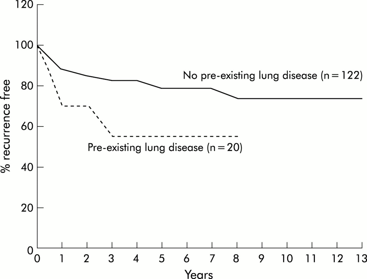
For patients who have not had a definitive surgical pleurodesis via a thoracotomy, a risk of recurrence should therefore be expected. While many studies have included details of the percentage of patients suffering a recurrence, very few have given much detail of the timing of these recurrences after the first episode, and few have characterised those most at risk. In one study a 54.2% recurrence rate was recorded with the majority occurring within 1 year of the first pneumothorax,78 and in another study 72% of the recurrences occurred within 2 years of the first episode.79
Cumulative freedom from recurrence data have been published by Lippert et al79 and stratified according to smoking history and underlying lung disease over a follow up period of up to 13 years. The shape of the curve (fig 2) does indeed imply that the biggest risk of recurrence is in the first year. One author has intimated that a further prospective trial he and colleagues are currently undertaking may provide a clearer month by month detail of recurrence rates.

Cumulative freedom from pneumothorax recurrence in relation to pre-existing lung disease (adapted with permission from Lippert et al79).
At present the recommended 6 week cut off seems to be arbitrary, with a significant fall in risk only appearing to occur after 1 year has elapsed. Furthermore, current advice does not take into account those with a higher risk of recurrence such as smokers, those with pre-existing lung disease, taller men, and possibly women.79,80 Thoracoscopic examination of the pleura does not permit any greater prediction of those at greatest risk of recurrence.79,81
In conclusion, a definitive surgical intervention makes the risk of recurrence of a pneumothorax negligible. Such patients may be able to fly 6 weeks after surgery and resolution of the pneumothorax in the absence of other contraindications. Careful medical assessment is required beforehand. For others the risk of a further pneumothorax is considerable for at least a year after the first episode. This risk is greatest for those with underlying lung disease and for continuing smokers.
While the likelihood of recurrence during flight is low and there is no evidence that air travel precipitates recurrence, the sequelae of recurrence at altitude may be significant. Recurrence of a pneumothorax while flying is likely to have more serious effects than a first episode, and recurrence in passengers with pre-existing lung disease is more likely to have serious consequences. Passengers may therefore choose to avoid this risk by delaying air travel for 1 year after a pneumothorax. This strategy should be given special consideration by those who smoke and/or have underlying lung disease.
Venous thromboembolic disease (VTE)
Fourteen papers were reviewed but the evidence is conflicting with many questions unanswered. BTS guidelines on suspected pulmonary thromboembolism list six major risk factors for VTE.82 Air travel is classified as one of several lesser risks. The evidence quoted in favour of an increased risk of air travel relates to long haul flights.83,84 Such reports are supported by others dating back over 20 years,85–88 and by more recent surveys.89–91 It is not possible from the published data to quantify the risk, and the underlying mechanisms have not been elucidated. Hypotheses include immobility, seated position, dehydration, and alcohol ingestion. Owing to delayed onset of symptoms and rapid dispersal of patients after a flight, many current reports are likely to underestimate the size of the problem.
In small studies evidence suggests that co-morbidity may increase the risk of VTE associated with air travel.89,90 Some studies suggest that previous VTE increases the risk of air travel associated recurrence,89–93 but the data are controversial. Further research is needed to determine whether delay in travel for those at risk is beneficial, and whether avoidance of alcohol and dehydration and upgrading reduce risk. Research is also required to examine the potential role of prophylactic low molecular weight heparin, full formal anticoagulation, and mechanical prophylactic methods including graded elastic compression hosiery and full leg pneumatic compression devices. The latter may be impractical on board an aeroplane and have not been studied in this context. However, they have been shown to have an additive effect in other at risk situations.94 A recent study suggests that symptomless deep vein thrombosis may occur in up to 10% of airline passengers, and that wearing elastic compression stockings during long haul flights is associated with a reduced incidence.95
The role of aspirin in this setting also requires investigation. A study of 13 356 patients undergoing surgery for hip fracture and 4088 patients undergoing elective arthroplasty showed that aspirin reduces the risk of pulmonary embolism and deep vein thrombosis by at least one third throughout a period of increased risk.96 The authors conclude that there is now good evidence for considering aspirin routinely in a wide range of groups at high risk of thromboembolism.
Thoracic surgery
There are few data available, but it is clear that the volume of gas in air spaces will increase by 30% at a cabin altitude of 2438 m (8000 ft). Postoperative complications such as sepsis or volume depletion should have resolved before patients undergo air travel. Severe headache precipitated by airline travel has been recorded 7 days after a spinal anaesthetic, presumed to be due to cabin pressure changes inducing a dural leak.97 North American guidelines13 highlight the fact that postoperative patients are in a state of increased oxygen consumption due to surgical trauma, possible sepsis, and increased adrenergic drive. Oxygen delivery may be reduced or fixed in patients who are elderly, volume depleted, anaemic, or who have cardiopulmonary disease. Reduced use of transfusions means that postoperative patients are now often more anaemic than previously.
Logistics of travel with oxygen
Berg et al98 have investigated the effects of oxygen supplementation in a group of 18 patients with severe COPD (mean FEV1 31% predicted). Baseline Pao2 at sea level was 9.47 kPa, which fell to 6.18 kPa when exposed to an altitude of 2438 m in a hypobaric chamber. The subjects were then given supplemental oxygen; 24% oxygen by Venturi mask increased Pao2 to 8.02 kPa, 28% oxygen by Venturi mask increased Pao2 to 8.55 kPa, and 4 l/min via nasal prongs increased Pao2 to 10.79 kPa. This suggests that, in patients with COPD, 24% and 28% oxygen via Venturi masks (and probably 2 l/min via nasal prongs) will improve hypoxaemia at 2438 m but will not fully correct it to sea level values. However, oxygen given at 4 l/min via nasal prongs will overcorrect hypoxaemia to produce values above sea level baseline values.
In practical terms, aircraft oxygen delivery systems are usually limited to 2 or 4 l/min. This is probably best delivered by nasal prongs as the simple oxygen masks provided by many airlines may allow some re-breathing and worsen carbon dioxide retention in susceptible subjects. Using 100% oxygen at a rate of 4 l/min via nasal prongs from a cylinder will produce a Pao2 at 2438 m (8000 ft) cabin altitude slightly higher than sea level Pao2 on air. Using 2 l/min via nasal prongs should correct the fall in oxygenation. Patients who require LTOT are not excluded from air travel, but no randomised controlled trials exist on which to base recommendations on the optimal flow rate.
The method of oxygen delivery depends upon the specific aircraft, but the supply is usually from cylinders. In some aircraft oxygen can also be tapped from the “ring main” of oxygen.99 Patients are not allowed to use their own oxygen equipment on the aircraft but can take an empty oxygen cylinder or oxygen concentrator as baggage. Charges may be made for both services, as well as a charge for supplemental oxygen. Regulations vary with each airline, which can decline the patient's request to travel.100 A comparative study of arranging in-flight oxygen on commercial air carriers was performed by members of the respiratory therapy department at the Cleveland Clinic Foundation in Cleveland, Ohio;101 76% of the 33 carriers contacted offered in-flight oxygen. There was significant variation in oxygen device and litre flow availability. Flow options varied from only two flow rates (36% of carriers) to a range of 1–15 l/min (one carrier). All carriers provided nasal cannulae, which was the only device available on 21 carriers. Charges varied considerably. Six carriers supplied oxygen free of charge while 18 carriers charged a fee ranging from $64 to $1500. Charges for an accompanying empty cylinder ranged from none to $250. Most carriers required 48–72 hours advance notice; one required one month's notice.
FUTURE DIRECTIONS
The justification for these recommendations is the significant number of in-flight medical incidents resulting from respiratory disease. The paucity of evidence on which to base them leads us to propose the following for patients with respiratory disease:
a prospective study to establish the predictive value of spirometric tests, equations, hypoxic challenge, and walk tests in different disease groups;
research to ascertain the effect of reduced humidity in the aircraft cabin;
comparison of the effects of long haul and short haul flights;
a prospective study to examine the risk of air travel for patients with diffuse parenchymal lung disease;
a prospective study to examine the risk of staying at altitude for patients with obstructive sleep apnoea;
a prospective study to clarify the benefit, if any, in delayed travel for those at risk where the risk will reduce with time—for example, after surgery or fracture.
In order to perform such studies it may be appropriate to establish a voluntary national reporting system to record in-flight respiratory incidents and collect airline data.
APPENDIX 1: Reviewers
Dr A G Arnold, Consultant Respiratory Physician, Castle Hill Hospital, North Humberside; Mrs R Barnes, Chief Executive, Cystic Fibrosis Trust; Miss A Bradley, Chief Executive, National Asthma Campaign; British Thoracic Society Standards of Care Committee; Dr M Britton, Chairman, British Lung Foundation & Breathe Easy; Dr J Coakley, Chairman, Intensive Care Society; Dr C Davidson, Home Mechanical Ventilation Group UK; Dr R J O Davies, Chairman, BTS Working Party on Pleural Disease; Dr D J C Flower, Consultant Occupational Physician, British Airways; Dr S A Goodwin, Airport Medical Services, Horley, Surrey; Professor D Peira Gray, President, Royal College of General Practitioners; Dr B Higgins, Chairman, BTS Standards of Care Committee; Dr S Hill, ARTP, Queen Elizabeth Hospital, Birmingham; Dr D Holland, Consultant Anaesthetist, Southmead Hospital, Bristol, and Medical Director & Adviser to CEGA Air Ambulance Limited, Chichester, West Sussex; Professor G Pasvol, Professor in Infection & Tropical Medicine, Imperial College Faculty of Medicine and Honorary Consultant in Infectious Disease, Northwick Park Hospital; Professor D Price, GP Airways Group; Professor S G Spiro, Respiratory Medicine Group, Royal College of Physicians, London; Dr H Swanton, President, British Cardiac Society; Mr M Winter, Sunrise Medical, Reading, Berkshire.
APPENDIX 2: Grading scheme for recommendations
Criteria for grading of recommendations are based on a paper by Petrie et al published on behalf of the Scottish Intercollegiate Guidelines Network102
APPENDIX 3: National referral centres with decompression chambers
-
RAF Centre for Aviation Medicine, RAF Henlow, Hitchin, Bedfordshire SG16 6DN. Tel 01462 851 515
-
Qinetiq Centre for Human Sciences, Building A50, Cody Technical Park, Farnborough, Hampshire GU14 OLX. Tel 01252 396 498 (general enquiries) or 01252 393 937 (decompression chamber)
-
HMS Sultan, Military Road, Gosport, Hampshire PO12 3BY. Tel 02392 542 934
APPENDIX 4: Major destinations exceeding 2438 m (8000 ft)
This is not an exhaustive list and passengers are recommended to contact the carrier if they suspect their destination may be at high altitude:
Bangda, Tibet: 15 548 ft
Bengdag, China: 14 100 ft
Bogota, Colomba: 8355 ft
La Paz, Bolivia: 13 310 ft
Lhasa, Tibet: 14 315 ft
Quito, Ecuador: 9222 ft
Telluride, USA: 9086 ft
APPENDIX 5: Sample MEDIF form (see 301 and 302)


APPENDIX 6: Conversion chart from feet to metres
Conversion chart from feet to metres
APPENDIX 7: Conversion algorithms for saturations to kPa and mm Hg
Conversion algorithm: saturations to kPa and mm Hg
APPENDIX 8: EXAMPLES OF EQUATIONS FOR PREDICTING HYPOXAEMIA
-
This relates Pao2 at altitude (Alt) to Pao2 at sea level (Ground)26:

-
This relates Pao2 Alt to Pao2 Ground and includes FEV1 in litres26:
-
This relates Pao2 Alt to Pao2 Ground and includes FEV1 as % predicted26:
-
This relates Pao2 Alt to Pao2 Ground and includes flight or destination altitude27:


Notes:
-
Thousands of feet should be entered as feet divided by 1000; 8000 feet would thus be entered in the equation as 8.0 not as 8000.
-
All these papers use mm Hg; 1 kPa = 7.5 mm Hg.
REFERENCES
Supplementary materials
- BTS recommendations
Managing passengers with respiratory disease planning air travelThe full report of the British Thoracic Society Standards of Care Committee regarding the fitness of passengers with respiratory disease to fly are published in the April 2002 issue of (Thorax 2002;57:289-304).
A summary for primary care is presented here.