


Abstract
The present article reviews the relevant stereological estimators for obtaining reliable quantitative structural data from the lungs. Stereological sampling achieves reliable, quantitative information either about the whole lung or complete lobes, whilst minimising the workload. Studies have used systematic random sampling, which has fixed and constant sampling probabilities on all blocks, sections and fields of view. For an estimation of total lung or lobe volume, the Cavalieri principle can be used, but it is not useful in estimating individual cell volume due to various effects from over- or underprojection. If the number of certain structures is required, two methods can be used: the disector and the fractionator.
The disector method is a three-dimensional stereological probe for sampling objects according to their number. However, it may be affected on tissue deformation and, therefore, the fractionator method is often the preferred sampling principle. In this method, a known and predetermined fraction of an object is sampled in one or more steps, with the final step estimating the number. Both methods can be performed in a physical and optical manner, therefore enabling cells and larger lung structure numbers (e.g. number of alveoli) to be estimated. Some estimators also require randomisation of orientation, so that all directions have an equal chance of being chosen. Using such isotropic sections, surface area, length, and diameter can be estimated on a Cavalieri set of sections.
Stereology can also illustrate the potential for transport between two compartments by analysing the barrier width. Estimating the individual volume of cells can be achieved by local stereology using a two-step procedure that first samples lung cells using the disector and then introduces individual volume estimation of the sampled cells.
The coefficient of error of most unbiased stereological estimators is a combination of variance from blocks, sections, fields of view, and noise due to random positioning of the probes. This can be decreased by increasing the number of units from the element causing the most variance. Overall, stereology provides lung scientists with efficient tools for estimating structural components correctly.
Quantitative structural investigations of lungs may reveal important information about the function and organisation of the lung being studied. Therefore, it is important that the various structural components can be measured correctly. Quantification of these components is also important when examining how lungs react to trauma, chemicals, disease and genetic engineering.
The problem that confronts the researcher is that a single lung structure can generate widely differing sections or projections, while several different lung structures may generate similar sections or projections. A two-dimensional section through a three-dimensional lung results in an irreversible loss of qualitative information and a reversible quantitative change of information. A section through a single lung capillary, which results in many unconnected profiles of the capillary, is an example of loss of qualitative information (that all capillary profiles represent just one capillary segment). The apparent thicknesses of the lung air–blood barrier in two dimensions may be larger than the true thicknesses in three dimensions, exemplifying a quantitative change of information. Two-dimensional projections of a three-dimensional lung result in a qualitative change of information and a quantitative increase of information, both of which are irreversible. In total projection of thick lung sections, false connections may appear between, for example, alveoli and capillaries, resulting in an irreversible change of qualitative information. In similar thick sections using projection, too many pneumocytes are seen, illustrating an irreversible increase in information.
The purpose of the present article is therefore to present the relevant stereological methods for use in bioscience with special emphasis on lung research.
SAMPLING
Instead of making prohibitively time-consuming three-dimensional reconstructions, stereology aims at minimising the workload using sampling, while still obtaining reliable, quantitative information either about the whole lung or complete lobes. The task of sampling has several problems due to the loss of information that three-dimensional lung tissue undergoes when viewed in two-dimensional sections. In this sense, it may be of interest to briefly mention the different sampling modalities in stereology, which ensure that the lung slices or structures of interest are sampled with the same probability. The lung should be envisaged as being cut into a number (N) of arbitrary slices. 1) Independent uniformly random sampling of the slices means that a random number between 1 and N is chosen and the corresponding slice is sampled. This continues until a fixed sample size (number = n) of slices is chosen. A new random number is chosen if the same random number is chosen twice. 2) Systematic uniformly random sampling (SURS) of 1/m, where m is the sampling period, of the slices is performed with a systematic and a random component. The systematic component is the decision to choose a sampling period of m. The random sampling component is to look up a random number between 1 and m. The first slice is decided by the random number, and from then on every mth slice is chosen from the ordered set of slices, arranged in a smooth order. This means that the smallest slices are placed at the ends of the set and those at the centre will be the biggest; slices in their natural sequence are often smooth. SURS is generally much more efficient than independent uniformly random sampling. 3) Cluster sampling of structures means that independent uniformly random sampling or SURS is used to sample clusters among the arbitrarily arranged clusters, with a probability, p (p = n/N in independent uniformly random sampling and p = 1/m in SURS). In the clusters sampled, the structures of interest are sampled by independent uniformly random sampling or SURS with probability q, making a total sampling fraction of p·q. The fractionator is an archetypical cluster sampling technique. 4) Stratified random sampling means that the lung is subdivided into strata (usually lobes). Each stratum is sampled using SURS or cluster sampling; total lung estimates are the sum of lobe estimates. Stratified sampling is advantageous when strata are quite different (which is not really the case in the lung); it adds to the efficiency in that the sampling intensity need not be the same in the strata.
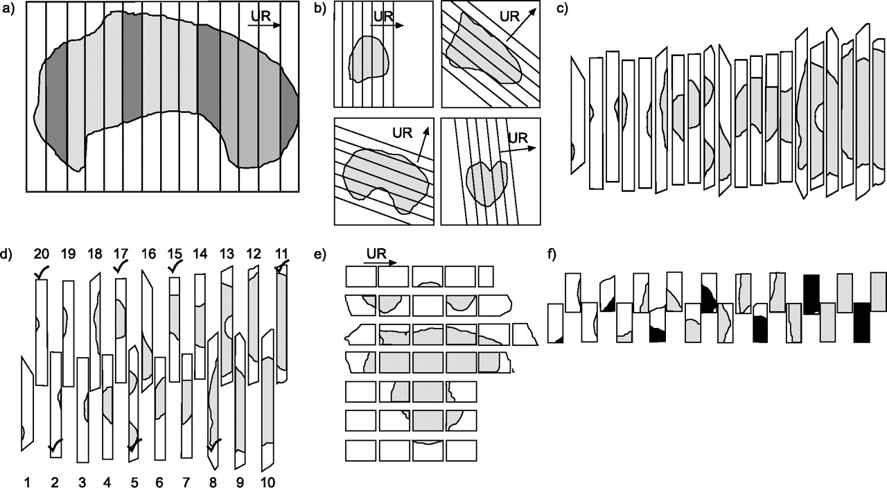
To date, state-of-the-art stereological lung studies have used uniformly random sampling, which is systematic for reasons of efficiency (fig. 1⇓), with fixed and constant sampling probabilities at the level of slices (constant thickness), fields of view (constant separation) and counting (same sampling intensity on all sections). This is the important ground rule in quantisation. The advantage is that the observer obtains accurate results regardless of inhomogeneity, which will only affect the precision of the data. The more inhomogeneous the lung structure, the more work is needed to obtain precise data. Inhomogeneity is known between lungs or lobes, etc. [1] and it is therefore beneficial to use both lungs in rodents, one lung in small mammals and a few lobes in large mammals. Typically, if a lobe happens to be small in a species, another lobe in the same lung may be big. Inhomogeneity can be reduced if both lobes are used and the workload is diminished. Similarly, if subsampling is performed, it is quite important to randomise between all lungs or lobes. For the same reason and the fact that the true story about the lung structure under study may never be known, it is never ideal to always take a particular subdivision of the lungs (left lung, upper left lobe, etc.). If freezing or one fixative can serve different research groups' needs for a particular study of lungs, the most practical way of handling the lungs is to slice them one or more times (multistage sampling) and then use SURS to obtain various sets of slices for the research groups.

Uniformly random (UR) and systematic uniformly random sampling (SURS) of a large lung or lobe. a) The specimen is embedded and sliced, optimally for isotropic uniformly random sectioning as shown in figures 2⇓ and 3⇓. b) All right-most cut surfaces are used for estimating total volume with the Cavalieri estimator; SURS is taken for cutting into bars, roughly parallel to the longest axis of each slice. c) All bars are sorted according to cut tissue area. d) Every second bar is pushed down to form a new row and all bars are relabelled as shown, starting with the smallest (this is the smooth fractionator). A SURS fraction (one-quarter) of the bars is sampled from the indicated sequence (tick marks in d). e) This is cut into blocks of a size that fits a glass slide. f) A SURS fraction (one-third) of all blocks is taken from a smooth arrangement (black tissue).
SAMPLING ANISOTROPIC STRUCTURES BY THE ORIENTATOR OR ISECTOR
Isotropy means that all directions have an equal chance of being chosen. There are three different ways of randomising orientation of section planes in stereology, as follows. 1) A design with completely random orientation is an isotropic design [2, 3], which is necessary if the stereological estimators require isotropic test planes. 2) A vertical design identifies or generates a vertical axis in the lung, which is rotated at random around this vertical axis, and stereological estimators requiring isotropic test planes can be used [4]. 3) When sections with an arbitrary orientation are sufficient, the randomisation of orientation can be ignored. The advantage of arbitrary and vertical sections is the possibility of keeping track of the architecture of the tissue being studied.
Estimations of the number of structures as well as connectivity of structures and the estimation of three-dimensional volume by point-counting, including the Cavalieri principle, may be performed on sections with arbitrary orientation. All other stereological methods require either isotropic test lines or isotropic test planes. Larger macroscopic lung structures are tubes and are anisotropic. Considering that the identification of regions in the lung is not a problem compared to, for example, the central nervous system, it is usually beneficial to choose an isotropic uniformly random (IUR) design (figs 2⇓ and 3⇓) for subsampling and estimation of various parameters. Alveoli and their substructures are essentially isotropic.

The orientator technique for producing isotropic uniformly random (IUR) slabs (and sections). a and b) The lung tissue is embedded in a block large enough for making the two cuts, without hitting the lung. First, the block is placed on the equiangular circle (c), with an edge parallel to the 1–1 direction, and a random number is selected. The block is cut perpendicular to the table in a direction parallel to that of the random number (15 in a). VA is the vertical axis around which the cut is uniform random. Secondly, the block is made to rest on the face just cut, with the edge of the previous resting face parallel to the 1–1 direction of the circle (d; cosine-weighted section planes). As shown, VA is now horizontal. A cut is made parallel to a new random direction (60 in b). The face just cut (▒) is IUR with respect to the specimen which may now be re-embedded and sliced into slabs parallel to the face (sections taken from the top of the slabs are IUR sections).
The isector technique for producing isotropic uniformly random (IUR) slabs (and sections). a) The lung lobe is cut into ∼15 slabs (— — —) and four slabs (———–) are sampled using systematic uniformly random sampling. b) Using a template with holes placed randomly over the slabs sampled, small lung lobe blocks are sampled uniformly random and embedded in spherical agar isectors. c) The isectors are embedded in the final embedding media and the central section will be isotropic, uniform and random. Note that several isectors can be embedded in a single block.
TOTAL VOLUME ESTIMATION USING THE CAVALIERI PRINCIPLE
The Cavalieri principle can be used for estimating the volume of any lung structure [5]. With a random start, the object is cut into slices of a known and fixed thickness. The volume is estimated by multiplying the distance between the slices by the total cut area of the structures under study. The cut area of the structures may be estimated by point-counting or using the two-dimensional nucleator [6]. The use of the Cavalieri principle requires the following: 1) that the position of the first slice is random; 2) that the slices are parallel; and 3) that the thickness of the slices is constant. The Cavalieri principle is typically used for efficient estimation of the total volume (reference volume) of lungs and lobes, but, due to various effects from over- or underprojection, it is not useful for estimating individual cell volume. However, the Cavalieri principle in combination with point-counting is ideal for estimating total volumes of various lung compartments. This can be extended down to the subcellular level and is known as the hierarchical design [7]. Point-counting at the subcellular level is illustrated in figure 4⇓. As with all size estimators, the Cavalieri principle is affected by tissue deformation, typically shrinkage.

An illustration of point-counting of lamellar bodies in a type 2 pneumocyte. All crosses are used to estimate the volume of lamellar bodies and encircled crosses (one in every 16) are used to estimate the reference volume, which is equal to the volume of the type 2 pneumocyte. Crosses hit an object if the object can be seen in the upper corner of the cross. Seven crosses hit lamellar bodies and six encircled crosses hit the cell. In this field of view, the volume fraction of mitochondria per tubular cell constitutes 7/(9·16) = 0.05, because 16 times as many crosses are used to estimate lamellar body volume as cell volume.
DISECTOR
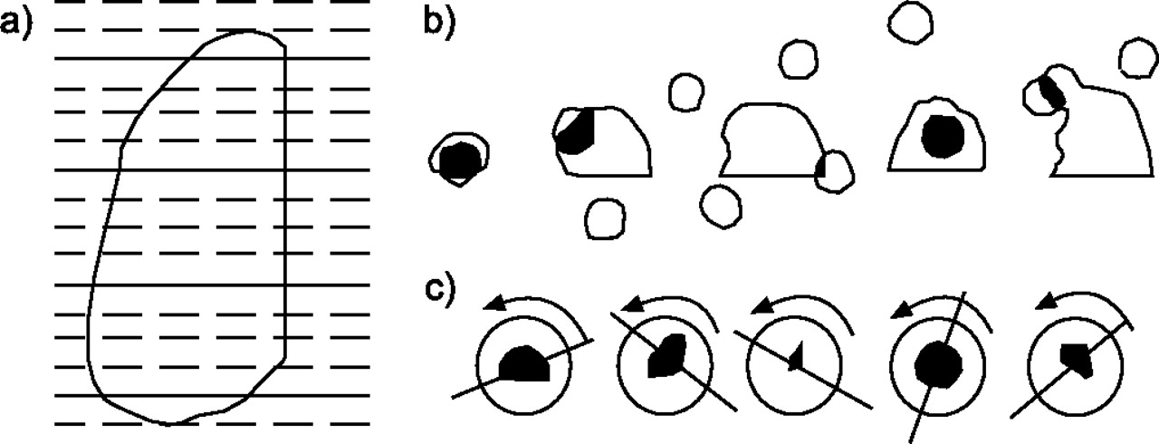
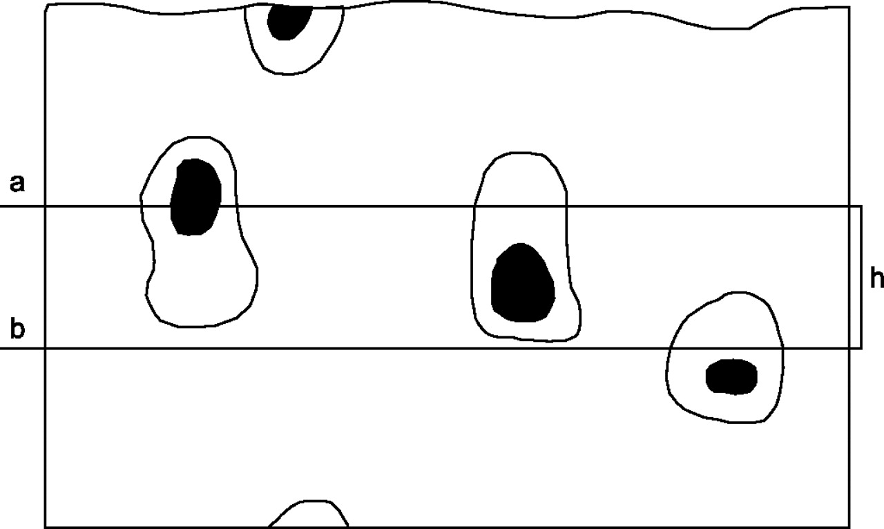
The disector is a three-dimensional stereological probe for sampling objects according to their number [8]. It consists of the following three elements: 1) a pair of physical or optical section planes separated by a known distance; 2) an integral test system with test points; and 3) an unbiased two-dimensional counting frame with a known area [9], as well as a counting rule. The objects are sampled by the disector if they are present in the counting frame in one section plane and not in the other plane. If the number of sampled lung structures is related to the volume between the two parallel section planes, it is possible to estimate the numerical density of the lung structures.
The disector is a volume probe and it is therefore important that the numerical density is transformed to a total quantity to avoid the “reference trap” [10]. The term reference trap refers to cases where wrong conclusions have been drawn from densities alone. The Cavalieri principle is usually used to estimate the reference volume. The disector is also dependent on tissue deformation, which is not the case for the fractionator and, therefore, the fractionator is usually preferred whenever applicable for number estimation. In practice, the disector should mainly be used in studies of number of subcellular studies, which will often be at the electron microscopic level. Here the disector can be used both as an ordinary physical disector (two physical sections) or as a double disector [11].
FRACTIONATOR
The fractionator is a sampling principle in which a known and predetermined fraction of an object is sampled, often in several steps of different sampling fractions [12]. In the final sample, the structural quantity (usually number) is estimated. The total number is estimated by multiplying the inverse sampling fraction by the number in the final sample. As with the disector, the fractionator can be performed in both a physical and an optical manner.
If the aim is to estimate the total number of a certain cell type in the mouse lung, the optical fractionator design would probably appear as follows. The lung is sliced and a fraction of the slices is taken using SURS after rearranging them in a smooth order. This constitutes the slice sampling fraction. Using a motorised stage, visualising the fields of view in a meandering fashion (SURS), the area sampling fraction is defined as the ratio between the two-dimensional unbiased counting frame and the step lengths in the x–y direction. The height sampling fraction is based on the ratio between the disector height and the section height, which is weighted with the number of cells counted per field of view [13]. The inverse of each these three fractions is then multiplied by the number of cells counted in this final sample to estimate the total number of cells.
If the aim is to count specifically stained pneumocytes in a lung, it is important to make a z-axis distribution in sections with a final thickness ≥25 µm. This distribution shows the number of cells in different heights of the section. By making this distribution of ∼300 cells, it is possible to check for full penetration of the stain, lost caps, section compression and shrinkage in order to decide whether the thick sections can be used for the optical fractionator and where to put the guard heights. In an appropriately sampled field of view, the microscope, equipped with oil objective lenses of a high numerical aperture, is focused from top to bottom and the microcator is used to measure the section height. When focusing on the bottom of the top guard height, all pneumocytes either above or in focus at this level are disregarded. From here, all pneumocytes coming into focus when focusing the microscope down to the lower guard height are counted. Pneumocytes in focus at this level are also counted. The two-dimensional counting frame is used to judge whether or not a pneumocyte is sampled in the x–y plane of the section. This optical counting principle is illustrated in figure 5⇓.
The optical fractionator is illustrated at the top using a thick section (∼30 µm) viewed edge-on. The pneumocyte cell nuclei are stained with standard histochemical stains and an antibody stains the cell membranes. In an appropriately sampled field of view, the microscope, equipped with an oil objective lens of a high numerical aperture, is focused a few micrometres down (a) in order to avoid distortion and unevenness of the section surface. All pneumocyte cell nuclei either above or in focus at level a are disregarded (two nuclei). From here, all nuclei coming into focus when focusing the microscope down to level b are counted. Nuclei in focus at level b are also counted. The height of the disector is h. If the whole height between a and b is used for counting, one pneumocyte will be counted here. The two-dimensional counting frame (not shown) is used to judge whether or not a nucleus is sampled in the x–y plane of the section. A microcator must be used to measure the height in the z-axis. A guard height is used above and below the optical disector to avoid ambiguities in identification of pneumocytes and to avoid the problems of lost caps (nuclei lost at either the top or bottom). A z-axis distribution decides the distance of the guard height.
If the aim is to estimate the number of alveoli [1, 14], which are bigger lung structures than cells, the physical fractionator is the method of choice. Due to their complexity, the number of alveoli or alveolar openings is counted by use of the Euler number using a modified ConnEulor counting rule [15]. This requires that there are no completely isolated cavities inside interalveolar septa and that the interalveolar septa generating the walls of the alveoli are one connected net, with no isolated septal pieces present. The section and area sampling fraction for a mouse lung would be carried out as previously described, but instead of cutting thick sections, two thin (4 µm for mouse lung) neighbouring sections, the reference section and the look-up section, are cut. On finding corresponding fields of view on the two sections, certain significant topological changes between the reference section and the look-up section are observed. A septal segment present in the reference section sampled by the two-dimensional counting frame that has disappeared in the look-up section is referred to as an “island”. Two septal segments directly neighbouring each other in a look-up section, which has been sampled as one uninterrupted septal segment in the counting frame in the reference section, are called a “bridge”. By taking into account the difference between the number of bridges and islands counted, and multiplying this number by the total inverse sampling fraction, the total number of alveoli for this lung can be estimated.
The physical fractionator and the Euler number have also been used in other organs [16] for estimating the number of microvessels. In this case, the topological event of a bridge occurs when two microvessel profiles merge into one large profile in the two-dimensional counting frame. An island occurs when one microvessel profile disappears from the two-dimensional counting frame to the look-up section. In addition, the rare topological event of a “hole” is also counted. A hole is an isolated part of nonlumen inside the lumen of a microvessel profile disappearing from the two-dimensional counting frame in the sampling section to the look-up section. These three microvessel events, bridges minus holes and islands, constitute the basis for the estimation of the Euler number and thereby the number of microvessels. The present authors have carried out pilot experiments in mice lungs using 2 µm-thick neighbouring paraffin sections and antibody stains of microvessels; however, due to the very small diameter of these microvessels, thinner sections may be needed.
Previously, the use of the physical fractionator required two identical projection microscopes in a dark room; however, computerised microscopes modified for stereology can nowadays show both the sampling section and look-up section in a split view on one monitor after automatically having found the corresponding fields of view. This saves quite some time compared with the manual method and makes the physical fractionator a must for larger structures that are not suitable for the optical fractionator. In addition, it makes the physical fractionator an attractive alternative for the optical fractionator when problems occur with penetration of stains or extreme collapse of section heights to much less than 25 µm.
SURFACE AREA, LENGTH, AND DIAMETER
Using IUR sections, total surface area and total length may, with great advantage, be estimated on a Cavalieri set of sections, i.e. seven to nine sections chosen by SURS. Surface area density is estimated by counting intersections on isotropic test lines and the specific surface is estimated by taking into consideration the volume of tissue investigated. Length density is estimated as that number of profiles sampled by isotropic two-dimensional counting frames monitored for tissue volume. Both length and surface area density are then converted to totals by multiplying them by the reference volume estimated by the Cavalieri principle.
Estimation of diameter requires that the cross-section of the tubes is circular. The diameter of a tube sampled by the two-dimensional isotropic counting frame is measured as the longest diameter of the tube profile perpendicular to the longest axis of the tube profile. Two diameters of figure of eight-shaped tube profiles are measured. Fuzzy tube profiles that represent grazing cuts should be discarded from diameter measurements because they will underestimate the diameter.
If the aim of a lung researcher is to sample bronchia (or vessels) with respect to a certain feature, the challenge is that the lung contains bronchia with many different diameters. One way to tackle this challenge is by the use of a magnifying glass to sample all visible bronchia with an isotropic two-dimensional unbiased counting frame and measure their diameter, sampling only those with, for example, a diameter ≥2 mm. In the next step, at the light microscopic level, all bronchia with a diameter <2 mm are sampled. It is then possible to make a diameter-weighted length distribution of all bronchia and measure other features of this proper sample of bronchia. Such an example has been published for brain microvessels [17].
There are many examples of generation-governed sampling of bronchia and vessels in the lung, perhaps because it is generally believed that this has biological and medical significance. This kind of sampling can never be performed uniformly and is not unbiased. It is therefore important that very cautious conclusions are drawn from such studies. If the researcher still decides to use generation-governed sampling, it may be performed on isotropic Cavalieri sections.
In asthma and other lung diseases, it is often quite relevant to estimate the correct three-dimensional layer thickness of various structures in lung tubes, e.g. bronchia. Ideally the tubes should be nonbranching and the correct layer thickness of any structure can be measured perpendicular to the longest axis of the tube profile where the tube wall is in focus.
BARRIER THICKNESS
As the lung is an organ in which transport of molecules between two biological compartments is a very important function, and the ease and potential of transport between the two compartments can be illustrated by the membrane/barrier width, the diffusion capacity of lung membranes has been studied for decades [18]. In stereological terms, membrane width estimation is nearly independent of the membrane thickness; this is in contrast to profile counts on sections, which are proportional to the height of the structure. In 1978, the first estimator of membrane width appeared, based on design-based sampling and uniformly oriented intercept lengths [19]. In the following year, the use of orthogonal intercept lengths further improved the efficiency of membrane-width estimation [20]. This latter method requires IUR sections.
If the aim is to estimate wall or barrier thickness in circular structures, e.g. vessels or bronchia, an even more efficient method for determining IUR sections exists. First, the circular structures are sampled by the two-dimensional counting frame. Secondly, the longest axis of the profiles is identified, and, finally, the shortest distance of the wall/barrier is measured in parallel with the longest diameter perpendicular to the longest axis. This distance is the correct three-dimensional thickness of the wall/barrier.
NUCLEATOR AND ROTATOR
When the task is to estimate the individual volume of lung cells, it is neither efficient nor accurate to use the Cavalieri principle at the light microscopic level due to effects from over- and underprojection. Instead, local stereology using a two-step procedure offers some interesting possibilities. The first step is to sample the lung cells with equal probability using the disector, i.e. the objects are selected according to their number irrespective of their size and shape. The second step introduces individual volume estimation of the sampled cells. This makes possible not only estimation of the average volume of the cell population but also the relative and absolute frequency distribution of the cell volumes. The most frequently used number-weighted local volume estimators comprise the nucleator [6] and the planar rotator (fig. 6⇓) [21]. Both the nucleator and rotator rely on a unique point, e.g. a nucleolus, as a reference from where the volume estimation is performed. This feature makes them ideal for optical sectioning in the optical fractionator. In order for the nucleator or rotator to work, it is important to realise that individual lung cell borders must be distinguishable on ordinary light microscopic sections. These two local volume estimators are independent of tissue deformation in the z-axis but are influenced by tissue deformation in the x–y-axes.

Local estimation of the number-weighted mean volume of a type 2 pneumocyte. In order to use the planar rotator, the section is isotropic (see figs 2⇑ and 3⇑). Isotropic, uniform, random and parallel test lines (drawn with full lines), a distance (d) apart, are applied to the sampled cell. The lattice of the test lines must be placed so its axis (arrows) passes through the sampled nucleus. For each test line crossing the cell height (h), linear intercepts from the arrow line to the cell border are measured on both sides of the arrow line. The number-weighted cell volume is estimated by squaring all intercepts at both sides of the arrow line and summing over all test lines. If a test line intersects the cell membrane twice, the length of this test line is calculated by adding up the squared lengths with alternating signs, with the intersection most distant from the arrow line being positive.
COEFFICIENT OF ERROR
The imprecision or variability (the coefficient of error (CE); see [22] for various ways to estimate CE) of almost all unbiased stereological estimators is a combination of variance from blocks, sections, fields of view, and “noise”. Noise is the independent noise due to random positioning of the stereological probes and is, for all practical purposes, related to the total count at the level of fields of view. The exception is the estimation of number of discrete particles like cells, where noise is the major uncertainty [23]. The independence of this noise means that there is no way to avoid it under uniform random sampling. It is likely that enough cells will always have to be counted for the desired precision. The block variation is usually most relevant for direct estimators for total content in a specimen, as in several fractionator designs [23]. For large specimens, a sufficient number of blocks are essential for counteracting the heterogeneity at the specimen level. In such cases, only one section is taken from each block. Within the sections or fields, the variability is with respect either to the density of what is being counted or to the previous field or section. If the major contributor to the variance is identified in the nested sampling designs (i.e. blocks, sections, or fields of view), increasing the number of units from this element might decrease the variance [24–26].
PERSPECTIVES
The introduction of the combined forces from stereology and image analysis, the proportionator [27], will provide the lung scientist with even more efficient tools to estimate various structural components correctly. This may, in turn, provide lung scientists with better possibilities of studying the effects of trauma, chemicals, disease, and genetic engineering on lung structure.
- © ERSJ Ltd
References


